ABC Family Arquivos Brasileiros de Cardiologia International Journal of Cardiovascular Sciences ABC Heart Failure & Cardiomyopathy ABC Imagem Cardiovascular
Português English
ABC Family
Português English
  • About
    • About ABC Family
    • Internal Regulation
    • Editorial Board
    • Brazilian Society of Cardiology (SBC)
  • Issues
  • SBC Guidelines
  • Content
    • Videos
    • Podcasts
    • News
  • About
    • About ABC Family
    • Internal Regulation
    • Editorial Board
    • Brazilian Society of Cardiology (SBC)
  • Issues
  • SBC Guidelines
  • Content
    • Videos
    • Podcasts
    • News

ABC Family

  • ABC Family
  • Arquivos Brasileiros de Cardiologia
  • International Journal of Cardiovascular Sciences
  • ABC Heart Failure & Cardiomyopathy
  • ABC Imagem Cardiovascular
Search in

No content found.

Journals
Ordering
Article types
Others

  1. Home
  2. Keyword: Three-Dimensional Imaging
Figure 1
ABCIC
Review Article

My Approach to Three-Dimensional Echocardiography in Mitral Stenosis: How and When?

Alexandre Costa Souza et al. Marcus Vinicius Silva Freire de Carvalho Marco André Moraes Sales

Abstract

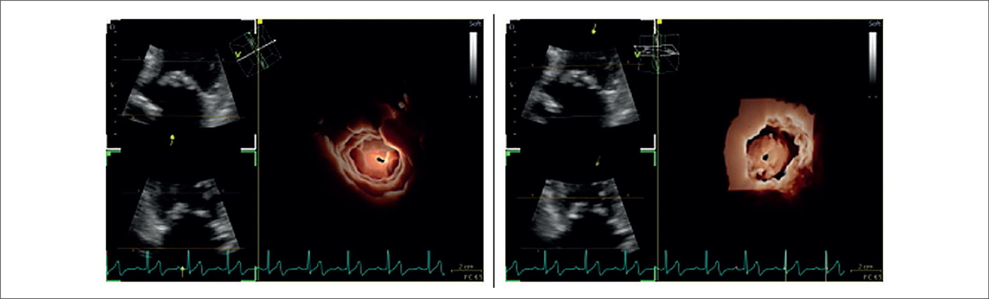
Three-dimensional echocardiography has become an essential tool in the assessment of mitral stenosis, allowing detailed anatomical analyses and reliable measurements, even in complex anatomies or extensive calcification. Based on daily clinical experience in an imaging laboratory, this article offers a practical guide for image acquisition and optimization, multiplanar reconstruction, and three-dimensional planimetry, focusing on technical adjustments, the use of tools such as rotation and cropping, and strategies for integrating the method into the routine workflow of cardiovascular imaging centers.

  • Mitral Valve Stenosis
  • Three-Dimensional Echocardiography
  • Three-Dimensional Imaging
Read Full Article
Creative Commons Attribution (CC BY) Creative Commons license
14/11/2025 DOI: 10.36660/abcimg.20250057i Views 2
Figure 1
ABCIC
Review Article

My Approach to 3-Dimensional Left Ventricular Strain

Rafael Bonafim Piveta et al. Miguel Osman Dias Aguiar Liria Lima Maria da Silva Marcelo Luiz Campos Vieira

Abstract

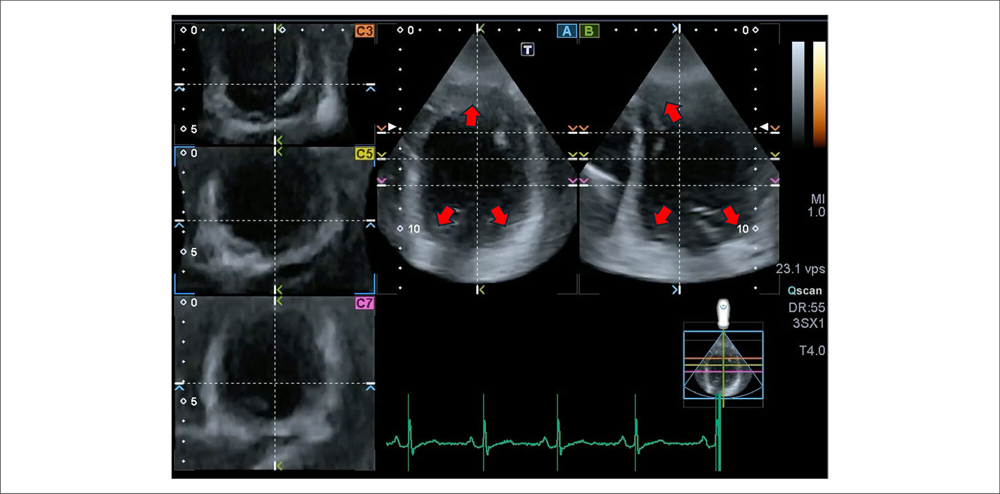
The role of strain analysis using 2-dimensional speckle tracking (2DST) has been well documented, and it represents an important complementary tool in the assessment of cardiac mechanics, adding value in the identification of incipient and subclinical myocardial injury in different clinical scenarios. A significant advance related to echocardiography was the development of 3-dimensional speckle tracking (3DST), which has the potential to overcome many limitations intrinsic to 2-dimensional technology, offering additional parameters of myocardial deformation (such as area strain) and a more comprehensive quantification of the geometry and function of the left ventricular myocardium, based on a single image acquisition. Among its main limitations, the low temporal and spatial resolution stands out, in addition to the dependence on high-quality images and a well-trained operator. Although it is a relatively recent technique that is still under development, several experimental studies and some clinical investigations have already demonstrated the reproducibility and potential applicability of 3DST. The objective of this article is to add information about adequate analysis of 3-dimensional left ventricular strain and explore its main points of vulnerability, discussing important variables to increase the accuracy and reproducibility of this technology.

  • Echocardiography
  • Heart Ventricles
  • Three-Dimensional Imaging
Read Full Article
Creative Commons Attribution (CC BY) Creative Commons license
28/05/2024 DOI: 10.36660/abcimg.20240028i Views 2
Figure 1
ABC
Image

Spatio-Temporal Image Correlation: Three-Dimensional Imaging for Fetal Cardiac Screening and Congenital Heart Disease Assessment

André Malho et al. Renato Silveira Ximenes Nathalie Jeanne Bravo-Valenzuela Edward Araujo Júnior

Heart defects are among the most common congenital malformations, occurring in 4 to 12 per 1,000 live births, depending on the population studied. Congenital heart disease (CHD) is an important cause of mortality related to birth defects. Therefore, prenatal diagnosis of CHD is crucial to improve the prognosis associated with this disease. In this scenario, the development of prenatal cardiac ultrasound screening programs serves as crucial tools that have increased the early detection of CHD. These programs achieve this by adding the ventricular outflow tract and superior mediastinum views (three vessels – 3V and 3V with trachea) to the standard four-chamber (4C) view when scanning the cardiac ultrasound.

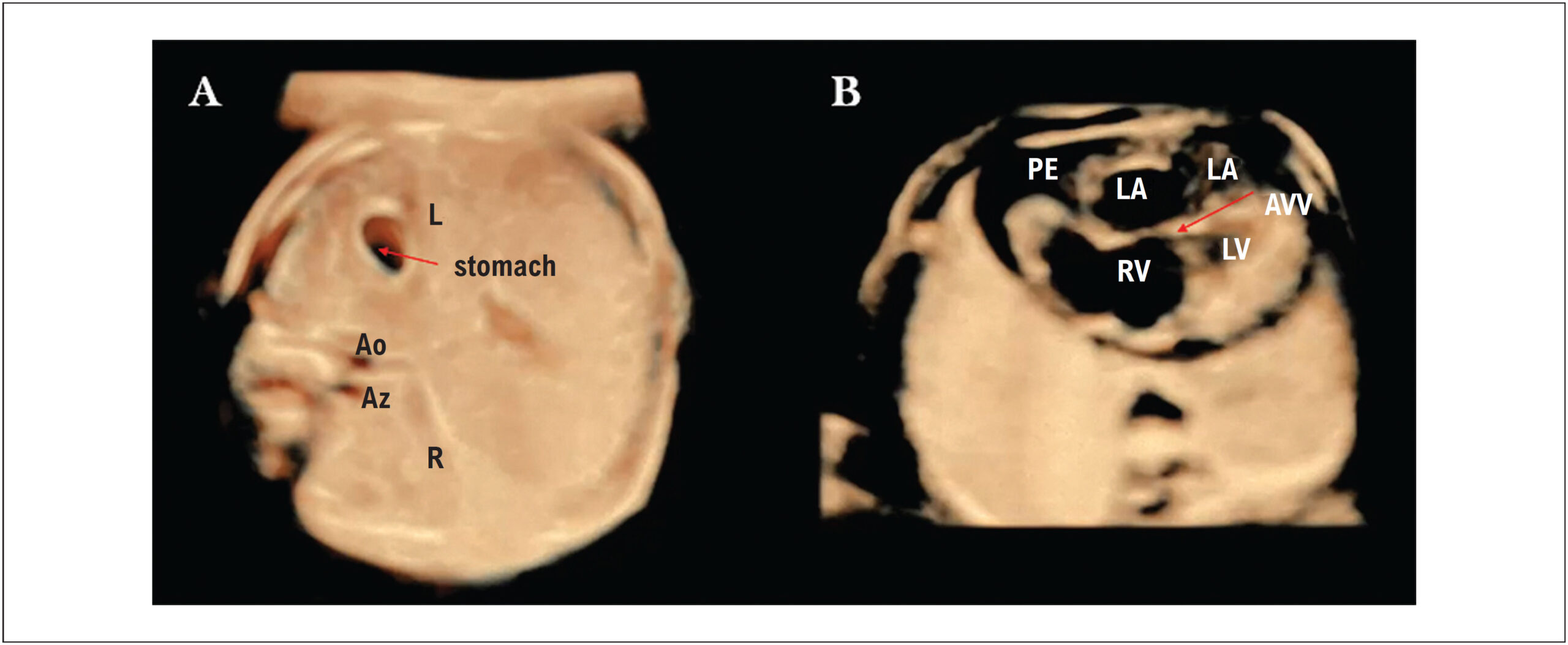
The development of advanced imaging technologies, such as three- and four-dimensional (3D/4D) ultrasound and Spatio-Temporal Image Correlation (STIC), has improved the quality of ultrasound images of the fetal heart with a positive impact on the diagnosis of cardiac malformations. The 4D-STIC technology allows the capture of thousands of cardiac images using a 3D volumetric transducer by a single 7.5 to 15-second slow-motion scan of the 4C view of the fetal heart. The acquired 3D/4D volumes contain a “block” of information of the complete cardiac cycle that can be stored for later off-line navigation of the fetal heart, allowing detailed analysis of cardiac anatomy by the sonographer, or sent via Internet link for analysis by experts in tertiary centers.

[…]

  • Congenital Heart Defects
  • fetal heart
  • Three-Dimensional Imaging
  • Ultrasonography
Read Full Article
Creative Commons Attribution (CC BY) Creative Commons license
29/04/2024 DOI: 10.36660/abc.20230580i Views 1

How to cite this article

EndNote RIS

Electronic Document Format (Vancouver)

Souza AC, Carvalho MVSF, Sales MAM. My Approach to Three-Dimensional Echocardiography in Mitral Stenosis: How and When?. ABC Imagem Cardiovascular 2025;38(3):e20250057.

Electronic Document Format (ABNT)

Souza, Alexandre Costa; Carvalho, Marcus Vinicius Silva Freire de; Sales, Marco André Moraes. My Approach to Three-Dimensional Echocardiography in Mitral Stenosis: How and When?. ABC Imagem Cardiovascular, v. 38, n. 3, e20250057, Nov. 2025.

Electronic Document Format (APA)

Souza, A. C., Carvalho, M. V. S. F. , & Sales, M. A. M. (2025). My Approach to Three-Dimensional Echocardiography in Mitral Stenosis: How and When?. ABC Imagem Cardiovascular, 38(3), e20250057.

Electronic Document Format (ISO)

Souza, Alexandre Costa and Carvalho, Marcus Vinicius Silva Freire de and Sales, Marco André Moraes. My Approach to Three-Dimensional Echocardiography in Mitral Stenosis: How and When?. ABC Imagem Cardiovascular [online]. 2025, vol. 38, n. 3, [cited 2026-10-14], e20250057. Available from: <https://journals.cardiol.br/en/article/my-approach-to-three-dimensional-echocardiography-in-mitral-stenosis-how-and-when/>. ISSN 2318-8219

EndNote RIS

Electronic Document Format (Vancouver)

Piveta RB, Aguiar MOD, Silva LLM, Vieira MLC. My Approach to 3-Dimensional Left Ventricular Strain. ABC Imagem Cardiovascular 2024;37(2):e20240028.

Electronic Document Format (ABNT)

Piveta, Rafael Bonafim; Aguiar, Miguel Osman Dias; Silva, Liria Lima Maria da; Vieira, Marcelo Luiz Campos. My Approach to 3-Dimensional Left Ventricular Strain. ABC Imagem Cardiovascular, v. 37, n. 2, e20240028, May. 2024.

Electronic Document Format (APA)

Piveta, R. B., Aguiar, M. O. D., Silva, L. L. M. , & Vieira, M. L. C. (2024). My Approach to 3-Dimensional Left Ventricular Strain. ABC Imagem Cardiovascular, 37(2), e20240028.

Electronic Document Format (ISO)

Piveta, Rafael Bonafim and Aguiar, Miguel Osman Dias and Silva, Liria Lima Maria da and Vieira, Marcelo Luiz Campos. My Approach to 3-Dimensional Left Ventricular Strain. ABC Imagem Cardiovascular [online]. 2024, vol. 37, n. 2, [cited 2026-10-14], e20240028. Available from: <https://journals.cardiol.br/en/article/my-approach-to-3-dimensional-left-ventricular-strain/>. ISSN 2318-8219

EndNote RIS

Electronic Document Format (Vancouver)

Malho A, Ximenes RS, Bravo-Valenzuela NJ, Araujo Júnior E. Spatio-Temporal Image Correlation: Three-Dimensional Imaging for Fetal Cardiac Screening and Congenital Heart Disease Assessment. Arquivos Brasileiros de Cardiologia 2024;121(4):e20230580.

Electronic Document Format (ABNT)

Malho, André; Ximenes, Renato Silveira; Bravo-Valenzuela, Nathalie Jeanne; Araujo Júnior, Edward. Spatio-Temporal Image Correlation: Three-Dimensional Imaging for Fetal Cardiac Screening and Congenital Heart Disease Assessment. Arquivos Brasileiros de Cardiologia, v. 121, n. 4, e20230580, Apr. 2024.

Electronic Document Format (APA)

Malho, A., Ximenes, R. S., Bravo-Valenzuela, N. J., & Araujo Júnior, E. (2024). Spatio-Temporal Image Correlation: Three-Dimensional Imaging for Fetal Cardiac Screening and Congenital Heart Disease Assessment. Arquivos Brasileiros de Cardiologia, 121(4), e20230580.

Electronic Document Format (ISO)

Malho, André and Ximenes, Renato Silveira and Bravo-Valenzuela, Nathalie Jeanne and Araujo Júnior, Edward. Spatio-Temporal Image Correlation: Three-Dimensional Imaging for Fetal Cardiac Screening and Congenital Heart Disease Assessment. Arquivos Brasileiros de Cardiologia [online]. 2024, vol. 121, n. 4, [cited 2026-10-14], e20230580. Available from: <https://journals.cardiol.br/en/article/spatio-temporal-image-correlation-three-dimensional-imaging-for-fetal-cardiac-screening-and-congenital-heart-disease-assessment/>. ISSN 0066-782X

  • About ABC Family
  • Internal Regulation
  • Editorial Board
  • Brazilian Society of Cardiology (SBC)

Issues

SBC Guidelines

  • Videos
  • Podcasts
  • News