| ABCIC
ABC Family Arquivos Brasileiros de Cardiologia International Journal of Cardiovascular Sciences ABC Heart Failure & Cardiomyopathy ABC Imagem Cardiovascular
Português English
ABC Family | ABCIC
Português English
  • About
    • About the Journal
    • Internal Regulation
    • Editors Section
    • Editorial Board
    • SBC Award
  • Instructions to Authors
    • Guidelines for Publication
    • Editorial Policy
    • Ethics Policy
    • Policy on the Use of Artificial Intelligence
    • Mandatory Publication Form
  • Issues
  • SBC Guidelines
  • About
    • About the Journal
    • Internal Regulation
    • Editors Section
    • Editorial Board
    • SBC Award
  • Instructions to Authors
    • Guidelines for Publication
    • Editorial Policy
    • Ethics Policy
    • Policy on the Use of Artificial Intelligence
    • Mandatory Publication Form
  • Issues
  • SBC Guidelines

ABC Family

  • ABC Family
  • Arquivos Brasileiros de Cardiologia
  • International Journal of Cardiovascular Sciences
  • ABC Heart Failure & Cardiomyopathy
  • ABC Imagem Cardiovascular
Search in

No content found.

Journals
Ordering
Article types
Others

  1. Home
  2. ABCIC
  3. Issues
  4. 2026, Volume 39, Number 1
Erratum

ERRATUM: Recalibrating the Barometer: Echocardiography in Diastolic Dysfunction and the Era of New Algorithms

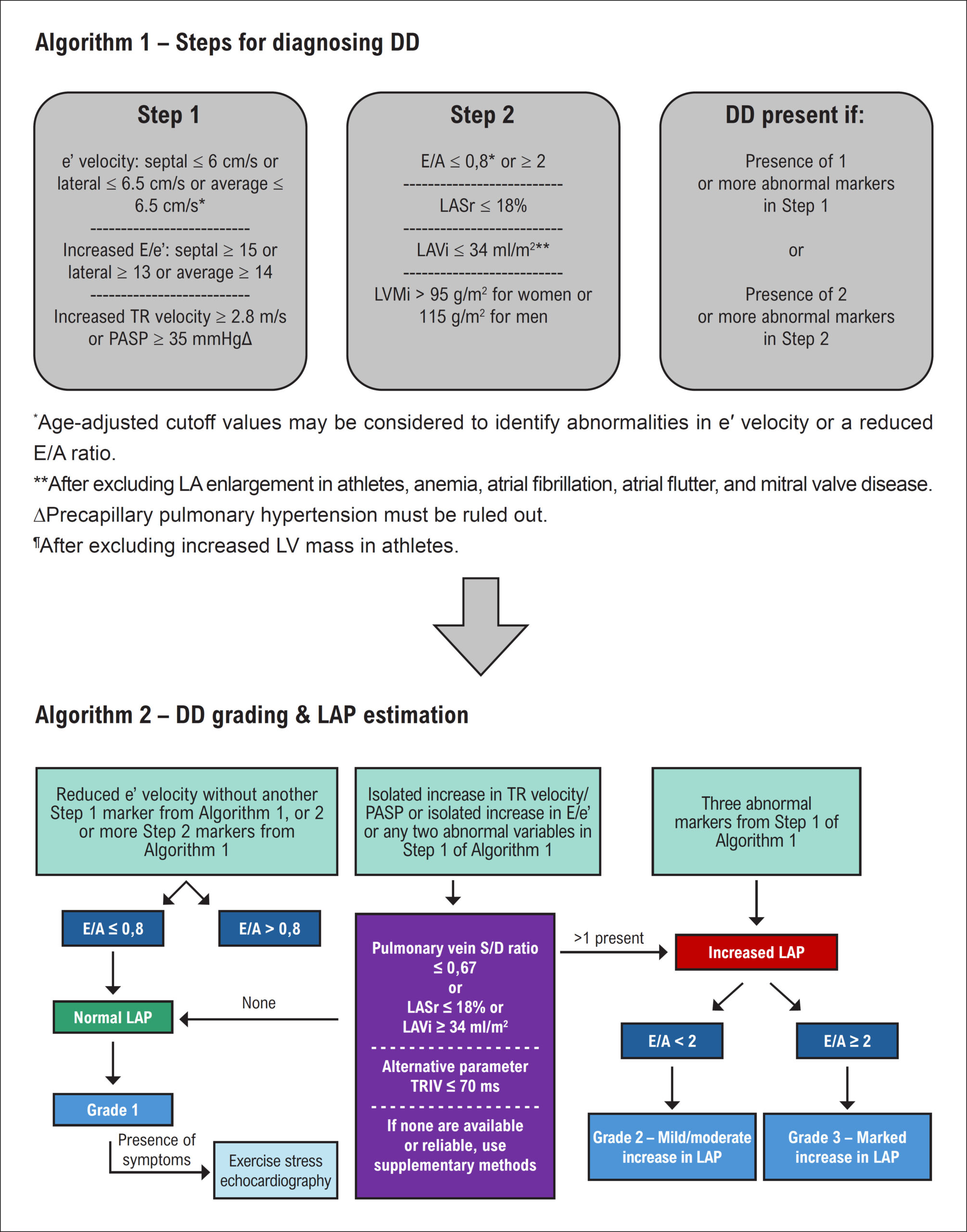
In the Editorial “Recalibrating the Barometer: Echocardiography in Diastolic Dysfunction and the Era of New Algorithms,” DOI: https://doi.org/10.36660/abcimg.20260004i, published in the journal Arquivos Brasileiros de Cardiologia: Imagem Cardiovascular, Arq Bras Cardiol: Imagem cardiovasc. 2026;39(1):e20260004, on page 2, in Figure 1, replace the line “LAVi ≤ 34 mL/m2” with “LAVi ≥ 34 mL/m2”.

Editorial Team

[…]

Read Full Article
Creative Commons Attribution (CC BY) Creative Commons license
24/08/2026 DOI: 10.36660/abcimg.20260055i Views 3
Figure 1
Review Article

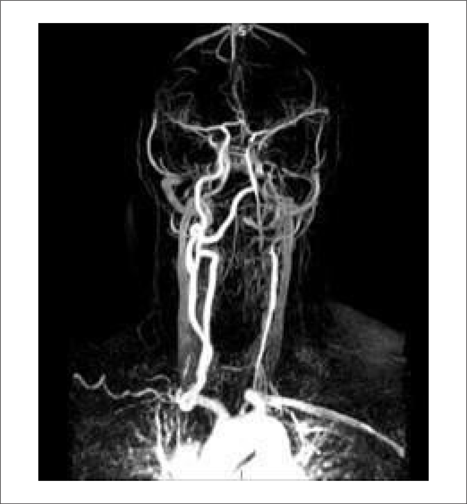
Neurological Manifestations of Takayasu Arteritis: A Case Report and Literature Review

Amanda Antunes Arantes Rolim et al. Tainá Cândida de Almeida Gontijo Carneiro Flávia de Campos Dilson Palhares Ferreira

Abstract

Takayasu arteritis (TA) is a rare large-vessel vasculitis that primarily involves the aorta and its major branches and predominantly affects women of reproductive age. We report the case of a woman who experienced an ischemic stroke at age 20 and a transient ischemic attack at age 53, with TA diagnosed only after the second cerebrovascular event. Although ischemic stroke is an uncommon initial manifestation of TA, early recognition and timely management are essential to prevent further complications and improve long-term outcomes.

  • Neurologic Manifestations
  • Stroke
  • Takayasu Arteritis
Read Full Article
Creative Commons Attribution (CC BY) Creative Commons license
25/05/2026 DOI: 10.36660/abcimg.20250110i
Figure 1
Case Report

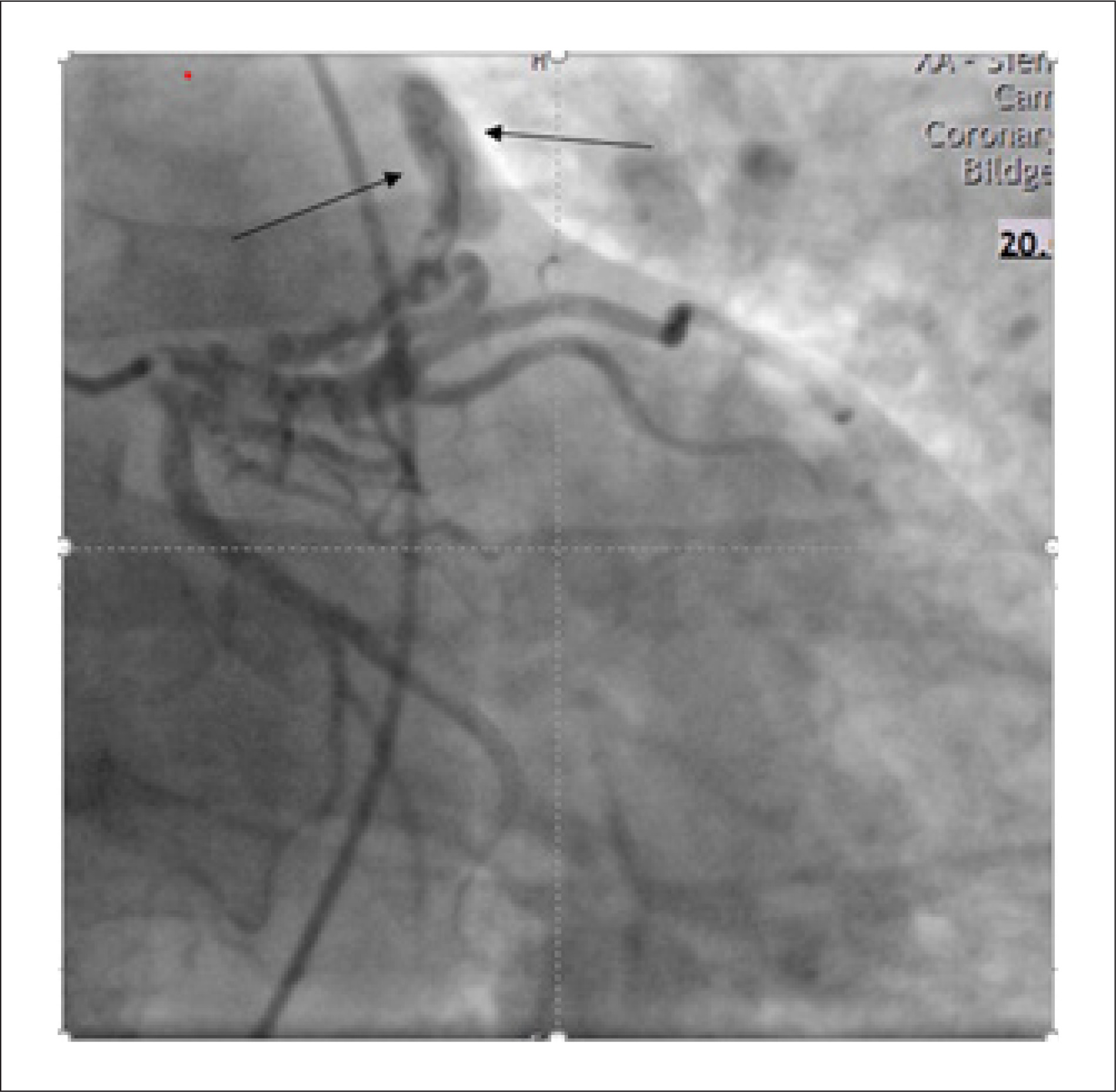
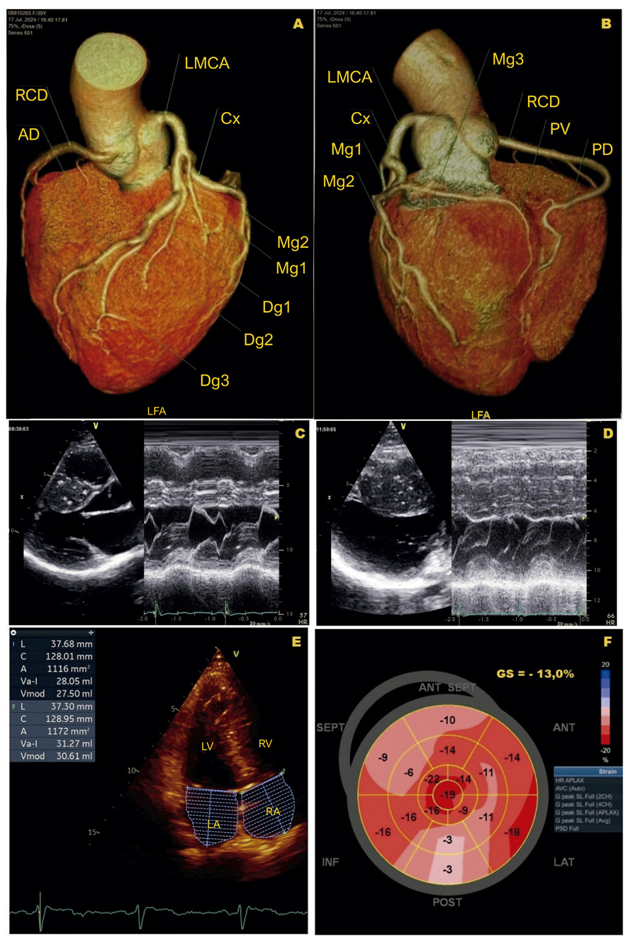
Coexistence of Partial Anomalous Pulmonary Venous Connection and Coronary Artery Fistulas: A Rare Case Report

Mourad Haj Abdo et al. Hussain Latsh Mathias Wagner George König Sebastian Barth

Abstract

Background

Dyspnoea is a common clinical symptom that frequently prompts hospital admission and is associated with significant morbidity. While it most often results from prevalent cardiopulmonary conditions, rare congenital cardiovascular anomalies can also manifest with dyspnoea. Partial Anomalous Pulmonary Venous Connection (PAPVC) and Coronary Artery Fistulas (CAFs) are uncommon congenital malformations of the cardiovascular system, and their simultaneous presence is exceedingly rare. Early recognition of such anomalies is critical to avoid progressive hemodynamic compromise and to guide appropriate management strategies.

Case Presentation

We present the case of a 55-year-old man who experienced an acute onset of dyspnoea lasting approximately two hours. Initial clinical assessment and routine investigations—including physical examination, echocardiography, electrocardiography, and right and left heart catheterization—raised suspicion of an underlying cardiac abnormality, prompting further evaluation. Subsequent cardiac Magnetic Resonance Imaging (MRI) and Multi-Detector Computed Tomography (MDCT) revealed the presence of a Partial Anomalous Pulmonary Venous Connection (PAPVC) accompanied by Coronary Artery Fistulas (CAFs). Given the non-complex characteristics of the shunt in this case, a shared decision was reached with the patient to proceed with conservative management.

Discussion

PAPVC and Coronary Artery Fistulas CAFs are rare entities that should be considered in the differential diagnosis. However, recommending their evaluation as initial diagnostic hypotheses may result in unnecessary investigations.

  • Coronary Vessels
  • Differential Diagnosis
  • Echocardiography
Read Full Article
Creative Commons Attribution (CC BY) Creative Commons license
25/05/2026 DOI: 10.36660/abcimg.20250030i Views 1
Correlation Between Venous Excess Ultrasound and N-Terminal Pro-B-Type Natriuretic Peptide Levels in Patients With Acute Decompensated Heart Failure
Original Article

Correlation Between Venous Excess Ultrasound and N-Terminal Pro-B-Type Natriuretic Peptide Levels in Patients With Acute Decompensated Heart Failure

Marcella Pereira Flores et al. Alexandre Costa Souza Marcus Vinicius Silva Freire de Carvalho Rodrigo Morel Vieira de Melo … Lívia Rodrigues Sampaio Cavalcante Natália Duarte Barroso Yuri Xavier de Carvalho Raisa Mainarte Franco Barros Clara Talita Silva Lobo Adriano Chaves de Almeida Filho

Abstract

Background:

The Venous Excess Ultrasound (VExUS) score has been proposed as an ultrasonographic method for assessing systemic venous congestion in patients with acute decompensated heart failure (ADHF). However, the relationship between VExUS and the biomarker N-terminal pro-B-type natriuretic peptide (NT-proBNP) remains unclear in literature.

Objectives:

To evaluate the correlation between the VExUS score and serum NT-proBNP levels in patients with ADHF.

Methods:

This retrospective observational study included 117 patients hospitalized with ADHF. Systemic venous congestion was assessed using the VExUS score, and serum NT-proBNP levels were obtained from laboratory records. Comparisons between groups were performed using the Kruskal-Wallis test, followed by Dunn’s test for multiple comparisons. The strength of association between variables was analyzed using Spearman’s correlation coefficient.

Results:

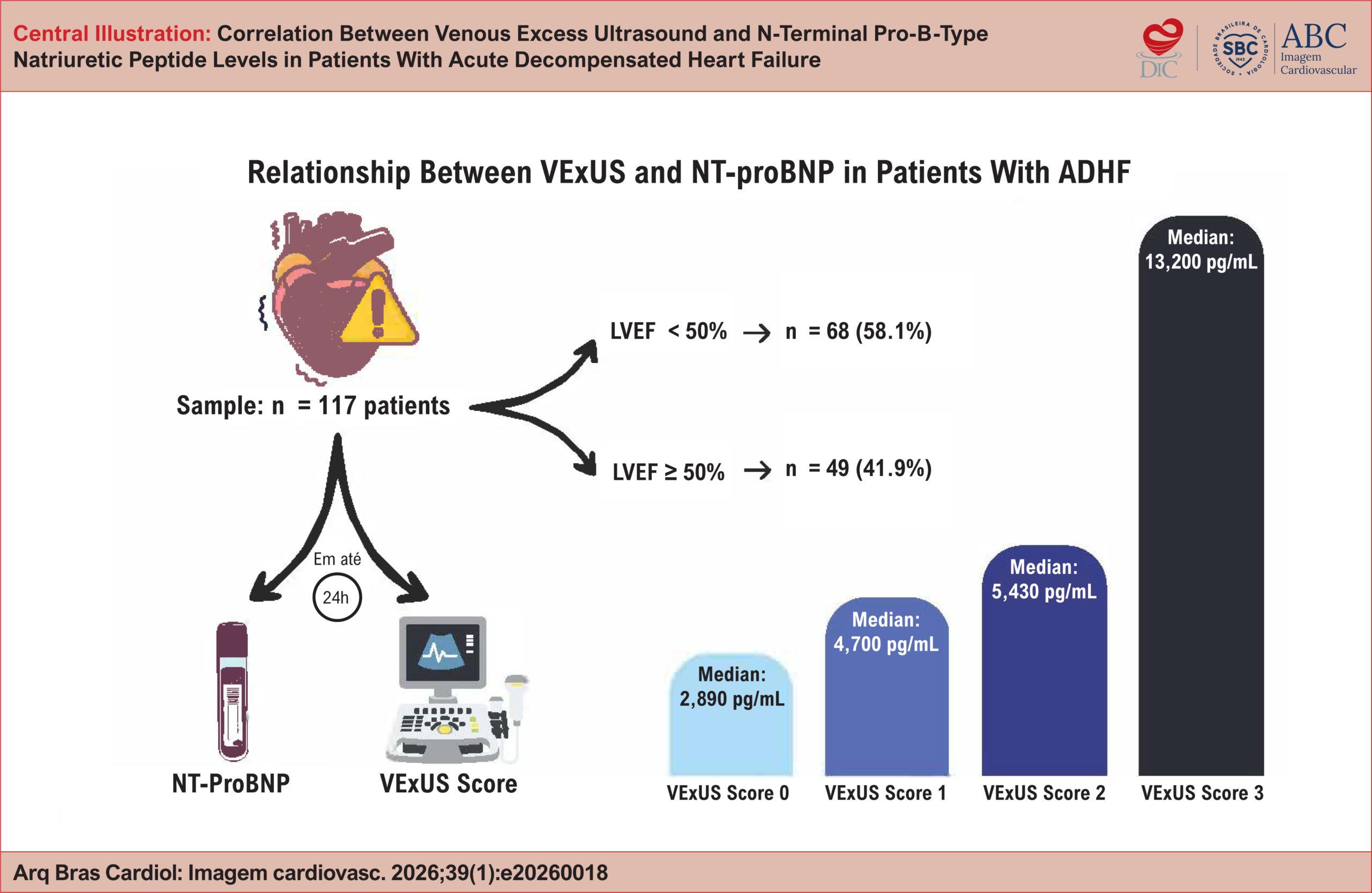
NT-proBNP levels increased progressively with increasing VExUS severity, with median values of 2,890 pg/mL (VExUS 0), 4,700 pg/mL (VExUS 1), 5,430 pg/mL (VExUS 2), and 13,200 pg/mL (VExUS 3). Statistical analysis demonstrated a significant difference between groups (Kruskal-Wallis: χ2 = 39.18; p < 0.0001). Dunn's test indicated that patients with VExUS 3 had significantly higher NT-proBNP levels compared with the other groups (p < 0.01). A moderate positive correlation was observed between the variables (Spearman's coefficient ρ = 0.567; p < 0.0001).

Conclusion:

The results indicate that the VExUS score is associated with NT-proBNP levels and may be integrated into clinical reasoning when assessing venous congestion in patients with ADHF.

  • Heart Failure
  • Inpatients
  • Ultrasonics
Read Full Article
Creative Commons Attribution (CC BY) Creative Commons license
25/05/2026 DOI: 10.36660/abcimg.20260018i Views 1
Step-by-Step Approach to the Evaluation of Constrictive Pericarditis
Review Article

Step-by-Step Approach to the Evaluation of Constrictive Pericarditis

Aline Travessa et al. Paulo Henrique Pereira Louise Moutinho Machado Natalia Sousa Esteves

Abstract

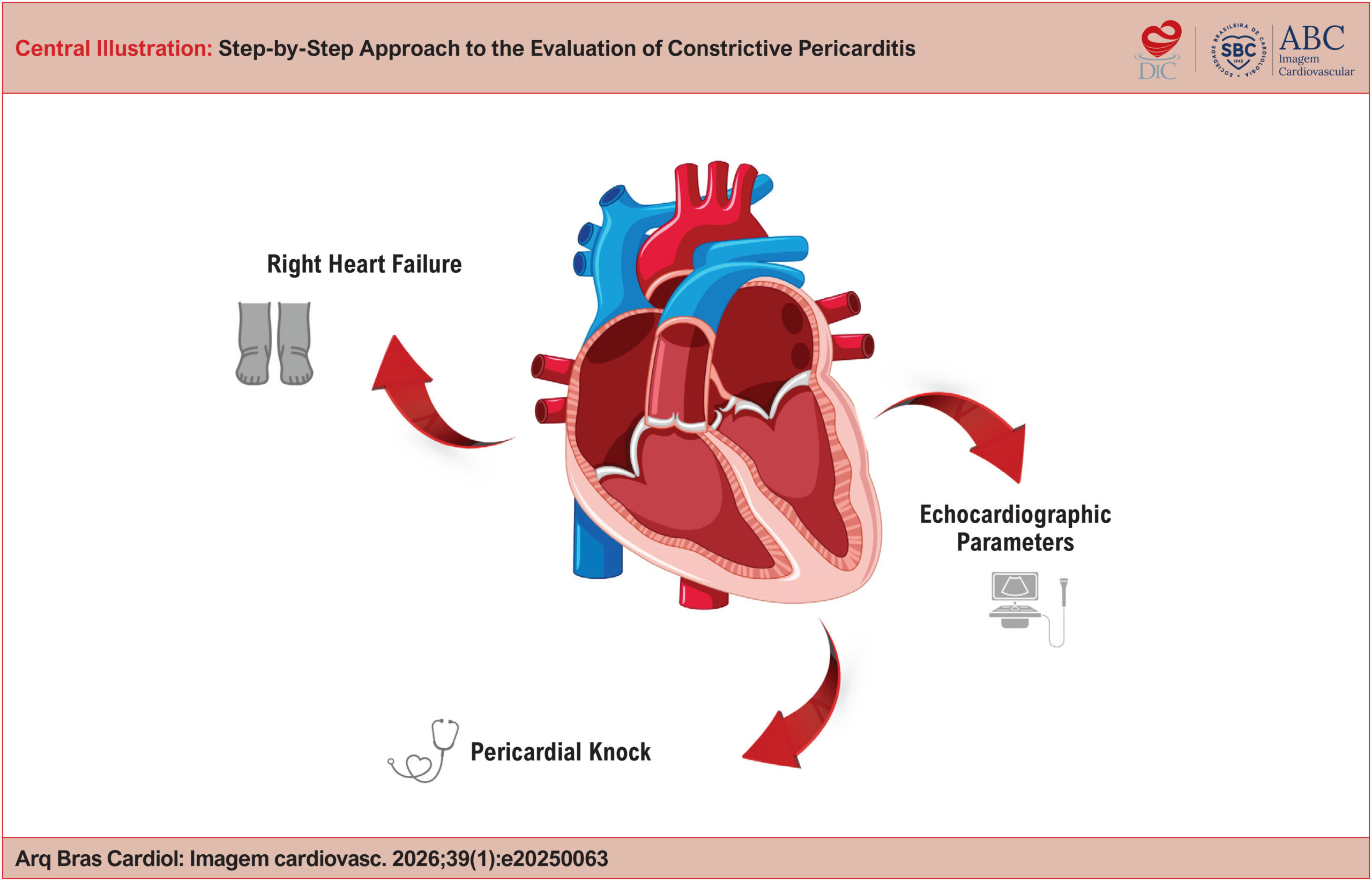
Constrictive Pericarditis (CP) is an uncommon but potentially curable condition, typically presenting with right-sided heart failure and impaired ventricular filling. Diagnosis can be challenging and requires a structured approach that integrates various cardiovascular imaging modalities. This article offers a practical guide, based on evidence and clinical experience, for the step-by-step recognition of Constrictive Pericarditis, highlighting echocardiographic findings.

  • Doppler Echocardiography
  • Echocardiography
  • Pericarditis
Read Full Article
Creative Commons Attribution (CC BY) Creative Commons license
25/05/2026 DOI: 10.36660/abcimg.20250063i
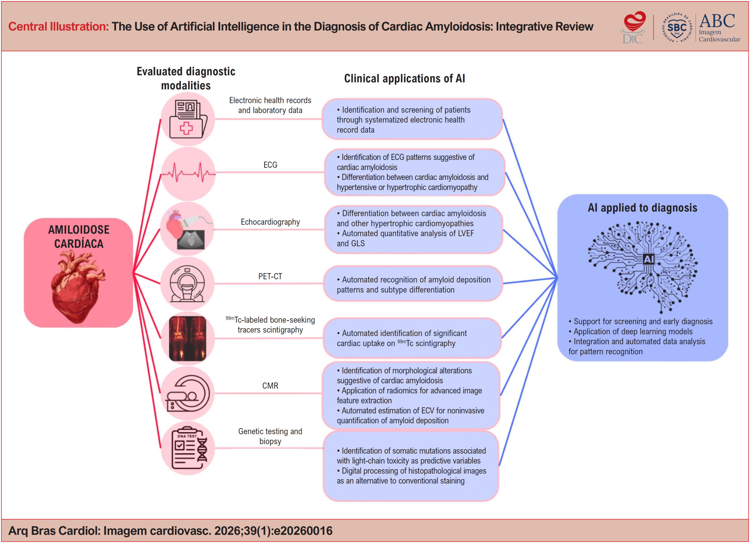
The Use of Artificial Intelligence in the Diagnosis of Cardiac Amyloidosis: Integrative Review
Review Article

The Use of Artificial Intelligence in the Diagnosis of Cardiac Amyloidosis: Integrative Review

Nilson Batista Lemos et al. Gabriela Aparecida Moreira Araújo Marcelo Dantas Tavares de Melo

Abstract

Fundamento:

Cardiac amyloidosis is a rare form of infiltrative cardiomyopathy characterized by the deposition of proteins in the myocardium, resulting in increased wall thickness, impaired ventricular function, and possible progression to heart failure. Diagnosis is challenging due to the low prevalence of the disease and the nonspecific nature of its clinical manifestations. The application of artificial intelligence (AI) to the analysis of medical tests emerges as a promising strategy for early detection, more accurate diagnosis, and timely initiation of treatment.

Methods:

An integrative literature review was conducted on the use of AI in the diagnosis of cardiac amyloidosis. Articles published between 2019 and 2024 were searched in the PubMed, Scopus, Web of Science, Embase, and Cochrane Library databases.

Results:

Of the 420 articles initially identified, 21 met the eligibility criteria and were included in the final analysis. A predominance of retrospective observational studies applying machine learning models was observed. Among the diagnostic modalities evaluated in association with AI, electrocardiography and echocardiography were the most frequently studied tests.

Conclusion:

AI demonstrates high potential to improve the screening and diagnosis of cardiac amyloidosis when applied to the analysis of clinical and imaging tests. The findings of this review indicate that AI may accelerate the diagnostic process, reduce the need for invasive procedures, and optimize the use of health care resources. However, to expand its integration into clinical practice and enhance its generalizability, further model refinement and validation in more diverse populations are required.

  • Amyloidosis
  • Artificial Intelligence
  • Diagnosis
Read Full Article
Creative Commons Attribution (CC BY) Creative Commons license
25/05/2026 DOI: 10.36660/abcimg.20260016i Views 1
Figure 1
Editorial

Nuclear Cardiology: From Consolidation to Integration

Simone Cristina Soares Brandão

Introduction

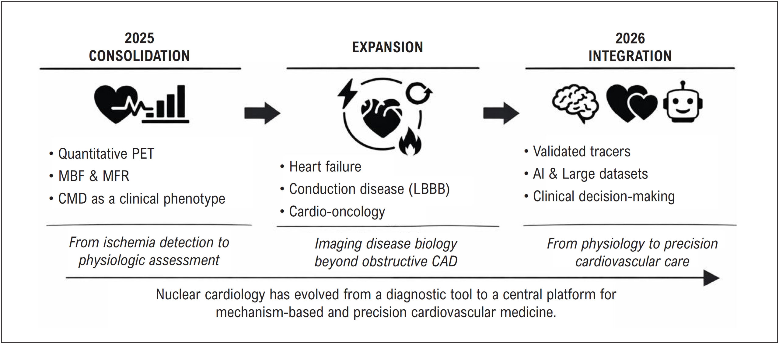
Over the past decade, nuclear cardiology has undergone a profound transformation.– What was once primarily a diagnostic tool focused on ischemia detection has evolved into a comprehensive platform for physiological understanding, risk stratification, and precision cardiovascular care.,- In this context, the field has shifted from simply identifying disease to elucidating its underlying mechanisms, echoing Marie Curie’s insight that “nothing in life is to be feared, it is only to be understood.”

The year 2025 did not stand out for isolated disruptive discoveries, but rather for something equally important: clinical consolidation.,, During this period, previously emerging concepts matured into actionable clinical paradigms, allowing the field to move from promise to practice.,

[…]

  • Inflammation
  • Positron-Emission Tomography
  • Precision Medicine
Read Full Article
Creative Commons Attribution (CC BY) Creative Commons license
25/05/2026 DOI: 10.36660/abcimg.20260007i Views 1
Cardiovascular Complications in Patients with COVID-19 and Their Relationship with Mortality: An Echocardiographic Perspective
Original Article

Cardiovascular Complications in Patients with COVID-19 and Their Relationship with Mortality: An Echocardiographic Perspective

João Henrique Andrade de Almeida et al. Gustavo Miranda de Azevedo Ferreira Marina de Azevedo Martins Andrade Heloísa Marceliano Nunes Igor Brasil Costa

Abstract

Background:

Echocardiography plays a fundamental role in the diagnosis of cardiovascular diseases related to SARS-CoV-2 infection. Despite prior investigation in the literature, studies focusing on cardiovascular manifestations among patients from the Brazilian Legal Amazon region remain scarce.

Objectives:

To identify cardiovascular complications of COVID-19 in patients admitted to a hospital in the North Region of Brazil and their potential association with mortality.

Methods:

We conducted a retrospective cohort study including 25 medical records of adults diagnosed with COVID-19. These patients were admitted to a hospital in Belém, Pará, in Northern Brazil, between March 2020 and December 2020. Demographic and clinical characteristics, as well as echocardiographic findings, were extracted from the medical records. Statistical analyses were performed adopting a significance level of α < 0.05. The project was approved by the Research Ethics Committee (approval number: 5.540.025).

Results:

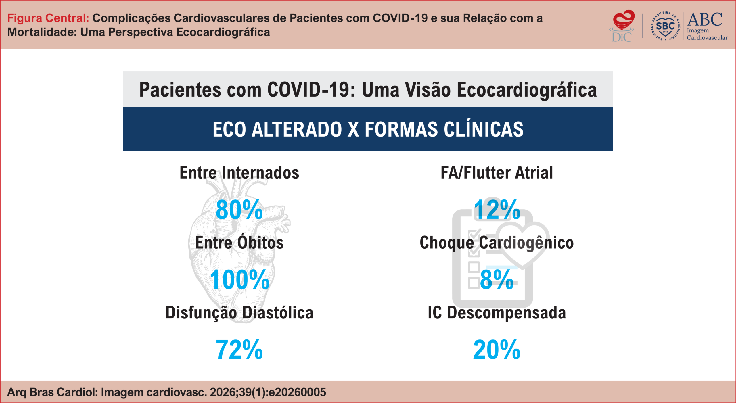
Among the 25 patients, 15 (60%) were male, with a mean age of 69.6 ± 14.4 years. Hypertension (n = 23; 92%) was the most prevalent cardiovascular risk factor, followed by diabetes (n = 11; 44%) and obesity (n = 8; 32%). We detected echocardiographic abnormalities in 20 (80%) individuals, with diastolic dysfunction being the most frequent (n = 18; 72%). All 7 patients who died showed abnormal echocardiography (p > 0.05). Atrial fibrillation or flutter (n = 5; 20%), decompensated heart failure (n = 3; 12%), and cardiogenic shock (n = 2; 8%) were the most common types of cardiovascular involvement.

Conclusions:

An increasing trend in COVID-19 mortality was observed among patients with cardiovascular complications. Nevertheless, the statistical power was limited by the small sample size.

  • Cardiovascular Diseases
  • COVID-19
  • Echocardiography
  • Mortality
Read Full Article
Creative Commons Attribution (CC BY) Creative Commons license
25/05/2026 DOI: 10.36660/abcimg.20260005i Views 3
Editorial

Hypertrophic Cardiomyopathy: Standardization of Echocardiographic Assessment in an Era of New Therapies

Marcelo Goulart Paiva

Hypertrophic cardiomyopathy (HCM) has become the most common inherited myocardial disease, with an estimated global prevalence between 1:200 and 1:500. Despite its relatively high frequency in the general population, the disease remains significantly underdiagnosed. Only about 15% of affected individuals are clinically identified, due mainly to the wide variability of phenotypes and clinical manifestations., Slightly more than half of patients may develop progressive symptoms or experience adverse events throughout their lifetime. Early identification, risk stratification, and cardiovascular therapies and interventions have reduced mortality rates to < 1.0% per year.

The pathophysiology of HCM is based on myocardial hypertrophy in the absence of secondary causes, associated with hypercontractility and diastolic dysfunction, resulting from abnormal myosin activation. Approximately 75% of patients present with left ventricular outflow tract (LVOT) obstruction at rest or after provocative maneuvers. In the absence of an obstructive pattern, the disease course is usually favorable, oligosymptomatic, or asymptomatic, with a minority progressing to advanced stages.–

[…]

  • Echocardiography
  • Hypertrophic Cardiomyopathy
  • Left Ventricular Outflow Obstruction
Read Full Article
Creative Commons Attribution (CC BY) Creative Commons license
11/05/2026 DOI: 10.36660/abcimg.20260027i Views 2
Editorial

Phenotyping of Congestion in Decompensated Heart Failure: Life-Saving Accuracy

Amanda Fernandes

Diagnosing heart failure (HF) in patients with multiple comorbidities continues to pose a challenge, especially when overlaps with pulmonary disease, obesity, or atypical clinical presentation. In this context, complementary diagnostic tools, such as NT-proBNP and venous excess ultrasound (VExUS) score, enable earlier and more accurate assessment of congestion, reducing adverse effects and fostering timely implementation of guideline-directed medical therapy.

Management of decompensated HF becomes even more complex when there is subclinical fluid retention, a frequently underestimated condition that is associated with higher rates of hospital readmission and a comparable mortality risk to that observed in patients with clinically evident edema., In these scenarios, VExUS has become a central tool in the diagnostic and therapeutic arsenal, both in differentiating the etiology of dyspnea and in managing patients with overt congestion and cardiorenal syndrome, by allowing for safer and more individualized management of diuresis, minimizing the risk of kidney injury.

[…]

  • Heart Failure
  • Natriuretic Peptides
  • ultrasound
Read Full Article
Creative Commons Attribution (CC BY) Creative Commons license
11/05/2026 DOI: 10.36660/abcimg.20260019i
Figure 1
Case Report

Aneurysm of the Suprahepatic Inferior Vena Cava: A Case Report

Gabriella Ghattas Mariano et al. Calina Araujo Thaines Gabrielle Silva Desani Letícia de Castro Gouvêa … Paulo Vítor Cabral Covilo Rogério de Paula Garcia Caravante

Introduction

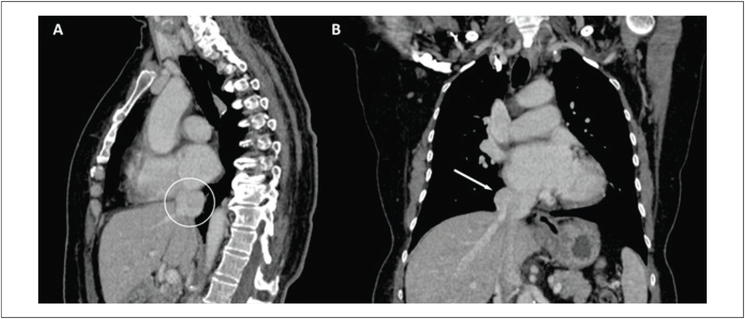
Aneurysms of the inferior vena cava (IVC) are a rare occurrence in the cardiovascular system and can present with a variety of signs and symptoms. In many cases, patients remain asymptomatic, which makes diagnosis difficult. Venous aneurysms are defined as an abnormal and persistent dilation of a vein in a specific area, with a diameter at least twice that considered normal. An IVC aneurysm is a specific type of venous aneurysm. This case report is relevant because this condition is rare; by 2021, only around 70 cases had been described in literature.

Aneurysms may arise due to fragility in the vessel wall and can be triggered by various factors, including hypertension, trauma, infection, and genetic conditions. Smoking, atherosclerosis, and chronic obstructive pulmonary disease are also considered risk factors for aneurysm development. Although they can occur in different regions of the body, they most frequently affect the cerebral arteries, the aorta, and the peripheral arteries.

[…]

  • Aneurysm
  • Cardiovascular Diseases
  • Case Reports
  • Inferior vena cava
Read Full Article
Creative Commons Attribution (CC BY) Creative Commons license
11/05/2026 DOI: 10.36660/abcimg.20250055i Views 1
Figure 1
Editorial

Recalibrating the Barometer: Echocardiography in Diastolic Dysfunction and the Era of New Algorithms

Maria Estefania Bosco Otto et al. Jorge Eduardo Assef Gustavo Nishida

Diastolic dysfunction (DD) remains a diagnostic challenge, not for lack of available parameters, but because uncertainty emerges when complex physiology is reduced to static labels in echocardiographic reports. In clinical practice, physicians are often less concerned with the specific grade of DD and more interested in its prognostic implications and in whether increased filling pressures may explain dyspnea, guide further investigation, and support therapeutic decisions. It is precisely in this variable, mean left atrial pressure (MLAP) and left ventricular (LV) filling pressure (LVFP), that echocardiography must be most pragmatic, minimizing the number of “indeterminate” reports and offering an operational conclusion grounded in integrated physiology.–

The structured reasoning that supports this goal began with the 2009 American Society of Echocardiography/European Association of Echocardiography guideline on the evaluation of LV diastolic function, a landmark document that organized modern thinking on diastole, established a shared language for key pathophysiological mechanisms, and systematized the interpretation of echocardiographic parameters. Beyond its conceptual value, it reinforced a critical principle for echocardiography laboratories: the assessment of diastole must translate into a clinically meaningful message, particularly when heart failure with preserved ejection fraction (HFpEF) is suspected.

[…]

  • Diastolic Heart Failure
  • Echocardiography
  • Left Ventricular Dysfunction
Read Full Article
Creative Commons Attribution (CC BY) Creative Commons license
20/04/2026 DOI: 10.36660/abcimg.20260004i Views 2
Comparison of Cardiac Structural Changes After Surgical and Transcatheter Atrial Septal Defect Closure With Color Doppler Echocardiography
Original Article

Comparison of Cardiac Structural Changes After Surgical and Transcatheter Atrial Septal Defect Closure With Color Doppler Echocardiography

Tuğçe Akın et al. Zeynep Bilge Yılmaz Dere Yılmaz Yozgat Halil Türkoğlu Murat Ugurlucan

ABSTRACT

Background:

Surgical and transcatheter techniques represent the two principal approaches for atrial septal defect (ASD) closure. Although both are widely used, comparative evidence regarding their mid-term effects on cardiac remodeling and right ventricular (RV) function remains limited.

Objectives:

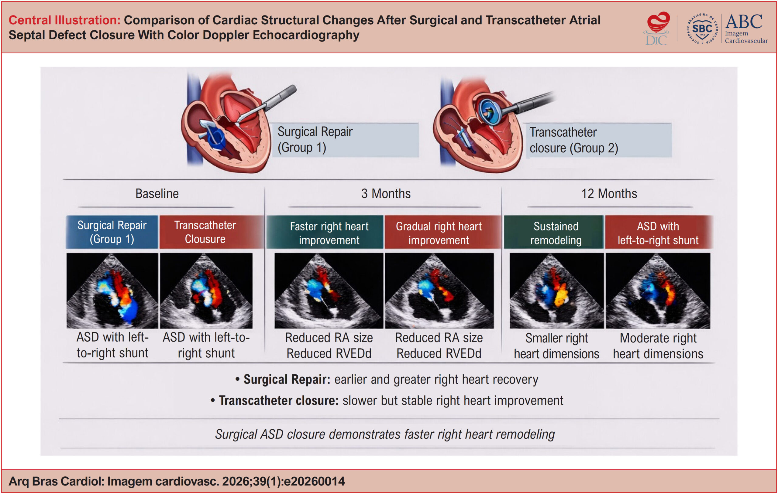
To compare mid-term cardiac structural remodeling and right ventricular functional recovery after surgical versus transcatheter ASD closure in pediatric patients using serial color Doppler echocardiographic assessment. Additionally, to determine whether either technique leads to faster or greater improvement in right heart morphology and function.

Methods:

We retrospectively evaluated 69 pediatric patients who underwent ASD closure at a single center. A total of 39 patients underwent surgical repair (Group 1), and 30 patients underwent transcatheter closure (Group 2). Transthoracic color Doppler echocardiography was performed before the procedure and at 3 and 12 months after intervention. Measures of atrial and ventricular morphology and function were analyzed.

Results:

At 3 months, the surgical group showed significantly greater improvement in right atrium (RA) major axis, RA volume, interventricular septal thickness in diastole, interventricular septal thickness in systole, and RV end-diastolic diameter (RVEDd) compared with the transcatheter group (all p < 0.05). At 12 months, surgical repair remained superior regarding improvement in RA major axis, RA volume, and RVEDd (all p < 0.05). Residual shunt was identified in only one patient in each group at 12 months.

Conclusions:

urgical ASD closure was associated with earlier and more consistent recovery of right atrial and ventricular geometry and function compared with transcatheter closure. These findings indicate that surgical closure may offer advantages for selected patients, particularly in relation to right heart remodeling during the first postoperative year.

  • Atrial Heart Septal Defects
  • Echocardiography
  • Operative Surgical Procedures
Read Full Article
Creative Commons Attribution (CC BY) Creative Commons license
20/04/2026 DOI: 10.36660/abcimg.20260014i Views 1
My Approach to VExUS Assessment Using Transesophageal Echocardiography: A Step-by-Step Performance Guide
Review Article

My Approach to VExUS Assessment Using Transesophageal Echocardiography: A Step-by-Step Performance Guide

Angelo Antunes Salgado et al. Marcos Paulo Lacerda Bernardo Marcelo Ramalho Fernandes

ABSTRACT

Monitoring systemic venous congestion has become essential in the management of critically ill patients, enabling accurate diagnosis, severity grading, and prognostic stratification. Literature shows that congestion is strongly associated with the development of acute kidney injury and increased mortality when compared with optimized volume states.

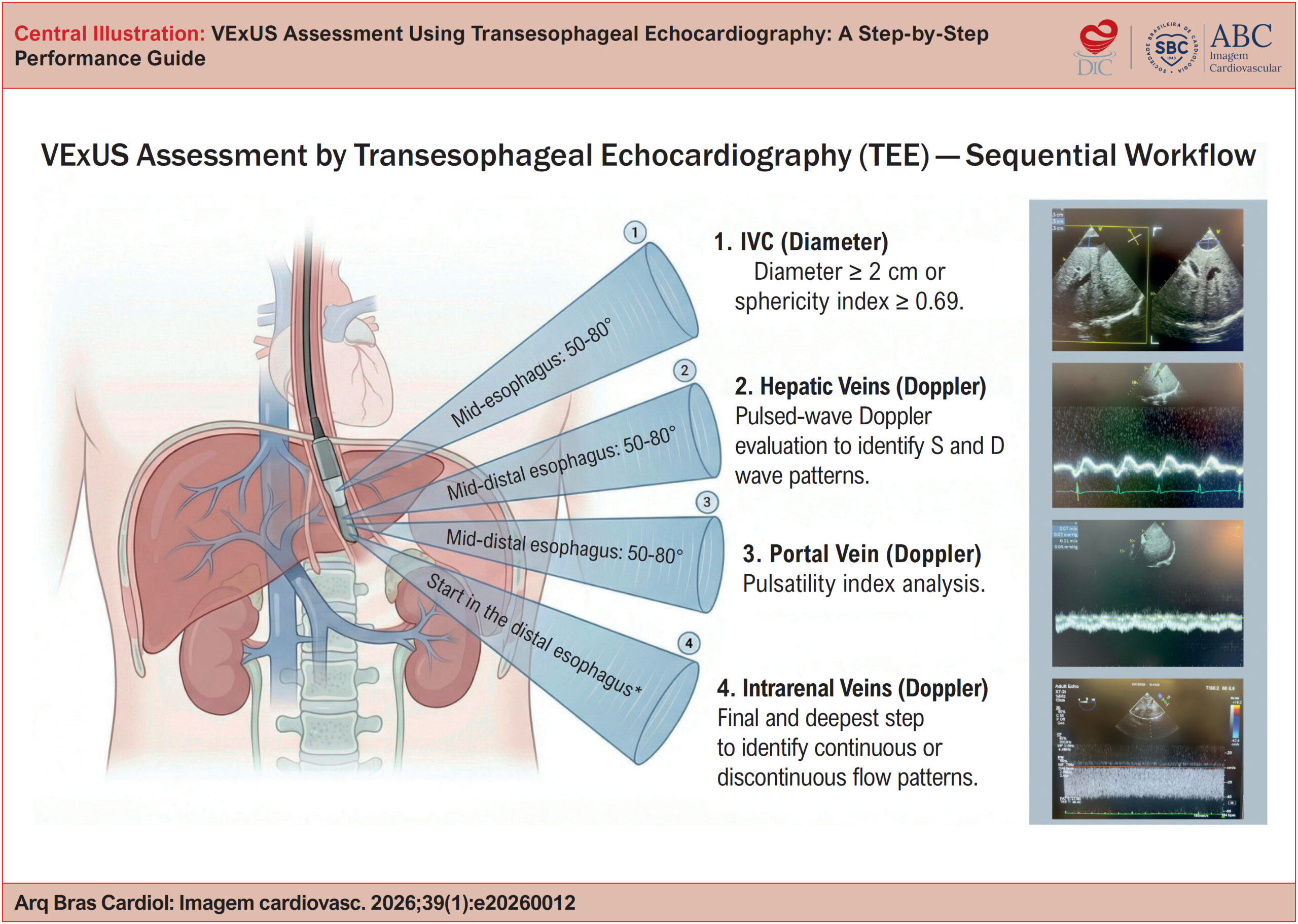
In this setting, intraoperative Transesophageal Echocardiography (TEE) has emerged as an advanced and versatile tool. In addition to allowing detailed assessment of cardiac function, TEE is effective in assessing intravascular volume status and predicting fluid responsiveness through dynamic measurement of stroke volume and the degree of systemic congestion. TEE also enables direct visualization of abdominal vessels, such as the hepatic, portal, and intrarenal veins, thereby facilitating the identification of pathological pulsatile flow patterns even in patients with limited transthoracic acoustic windows.

The integration of protocols such as VExUS (or its modified version, mVExUS) allows for a personalized approach focused on “perfusion without congestion.” This review outlines the practical application of VExUS assessment using TEE, its technical limitations, and how to use it to guide hemodynamic resuscitation, minimizing organ injury and optimizing clinical outcomes.

  • Operative Surgical Procedures
  • Prognosis
  • Transesophageal echocardiography
Read Full Article
Creative Commons Attribution (CC BY) Creative Commons license
20/04/2026 DOI: 10.36660/abcimg.20260012i Views 1
Editorial

Echocardiography with Ultrasound Enhancement Agents and the Diagnostic Challenge of Cardiac Masses: Solid Evidence for a Complex Clinical Problem

Rafael Bonafim Piveta et al. Miguel Osman Dias Aguiar

The proper characterization of intracardiac masses continues to be one of the most relevant challenges in contemporary cardiovascular imaging. Thrombi, benign tumors, and malignant neoplasms share some similar morphological characteristics in conventional echocardiography, but they entail radically different approaches, prognoses, and therapeutic urgencies. Despite significant advances in cardiovascular imaging diagnostic techniques, critical decisions, such as whether to anticoagulate or operate, investigate or observe, treat urgently or monitor, are still frequently challenging in clinical practice. Echocardiography, although indispensable as an initial method, often fails to differentiate thrombi, benign tumors, and malignant neoplasms in a significant number of patients. Given this scenario, it is legitimate to question: why does echocardiography with ultrasound enhancing agents (UEAs), available for decades, still play a secondary role in many diagnostic algorithms?

The meta-analysis, “Diagnostic performance of contrast-enhanced echocardiography in differentiating cardiac masses”, presents important arguments for this context. Using methodological rigor, aligned with PRISMA-DTA and Cochrane Handbook recommendations,, the authors demonstrate that echocardiography with UEAs has shown exceptional diagnostic performance in two of the most critical dilemmas in clinical practice: differentiating tumors from thrombi and distinguishing benign from malignant tumors.

[…]

  • cardiac masses
  • contrast
  • Echocardiography
Read Full Article
Creative Commons Attribution (CC BY) Creative Commons license
20/04/2026 DOI: 10.36660/abcimg.20260009i Views 1
Cardiac Sarcoidosis: The Role of Multimodal Imaging
Review Article

Cardiac Sarcoidosis: The Role of Multimodal Imaging

Lara Cristiane Terra Ferreira Carreira et al. Lívia Carreira Adriana Soares Xavier de Brito

ABSTRACT

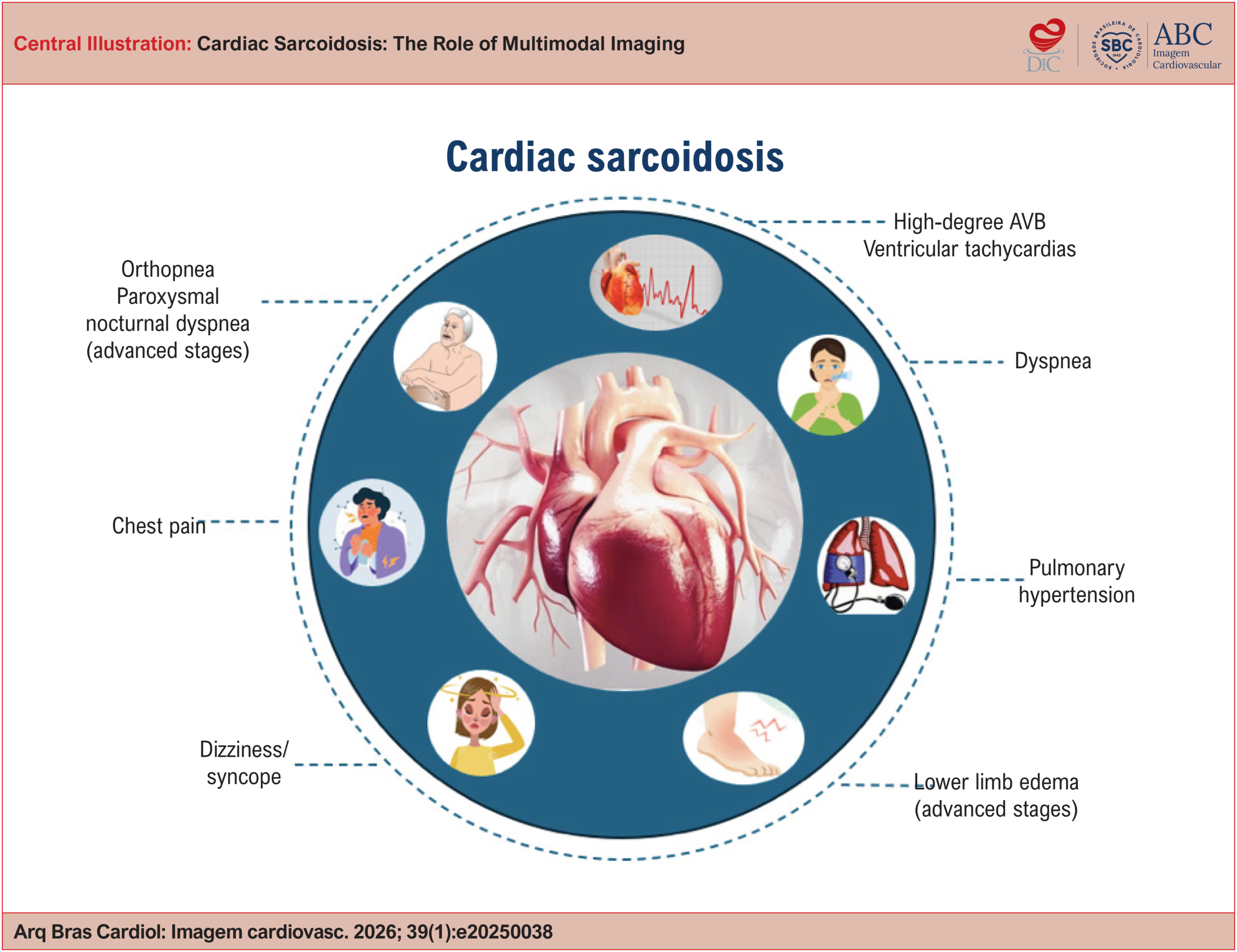
Cardiac sarcoidosis (CS) is a potentially severe manifestation of systemic sarcoidosis, associated with advanced atrioventricular block, ventricular arrhythmias, heart failure, and sudden death. Diagnosis remains challenging due to phenotypic variability and the limitations of conventional diagnostic methods. Advances in imaging techniques, especially the combination of positron emission tomography/computed tomography (PET/CT) using 18F-FDG and cardiac magnetic resonance imaging (CMR), have revolutionized the diagnostic approach and therapeutic monitoring of CS. This article reviews current concepts of CS and its diagnosis, with a focus on the role of PET/CT, the importance of appropriate patient preparation, and integration with CMR.

  • Fluorodeoxyglucose F18
  • Magnetic Resonance Imaging
  • Multimodal Imaging
  • Positron-Emission Tomography
  • Sarcoidosis
Read Full Article
Creative Commons Attribution (CC BY) Creative Commons license
20/04/2026 DOI: 10.36660/abcimg.20250038i Views 1
My Approach to Coronary Flow Assessment With Transthoracic Echocardiography
Review Article

My Approach to Coronary Flow Assessment With Transthoracic Echocardiography

José Maria Del Castillo et al. Issam Shehadeh

Abstract

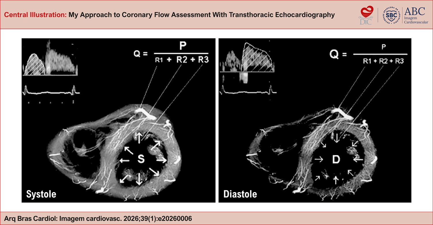
The assessment of coronary flow reserve is an extremely important step in stress echocardiography, both within and outside the context of coronary artery disease. For some, it is seen as an impossible parameter; for others, it is essential. In any case, everything begins with the proper visualization of the coronary arteries at rest. In this article, we will address theoretical and practical concepts for incorporating coronary flow study into the routine of the echocardiographer.

  • Coronary Artery Disease
  • Doppler ultrasonography
  • Ecocardiografia sob Estresse
Read Full Article
Creative Commons Attribution (CC BY) Creative Commons license
17/04/2026 DOI: 10.36660/abcimg.20260006i Views 1
Figure 1
Brief Communication

Left Ventricular Outflow Tract Velocity-Time Integral (LVOT VTI) as a Marker of Cardiac Performance: Mortality Data of the ELSA-Brasil Cohort

Mariana de Castro Lopes et al. Altair Ivory Heidemann Jr Eduardo G. Pianca Bruce B. Duncan … Murilo Foppa Angela B.S Santos

Transthoracic Echocardiography (TTE) is a well-established tool for assessing cardiac function and hemodynamics. While Left Ventricular (LV) systolic and diastolic function are commonly used metrics, Cardiac Output (CO) remains central for evaluating hemodynamic status, especially in critical care settings. TTE-derived parameters, such as Left Ventricular Outflow Tract Velocity-Time Integral (LVOT VTI) and LVOT area, when combined with body habitus and heart rate, provide noninvasive estimates of cardiac output. However, the accuracy of CO calculations may decrease when multiple covariates are included, with LVOT diameter being a major source of error. In contrast, isolated LVOT VTI may represent a simpler and more reliable surrogate of cardiac performance.

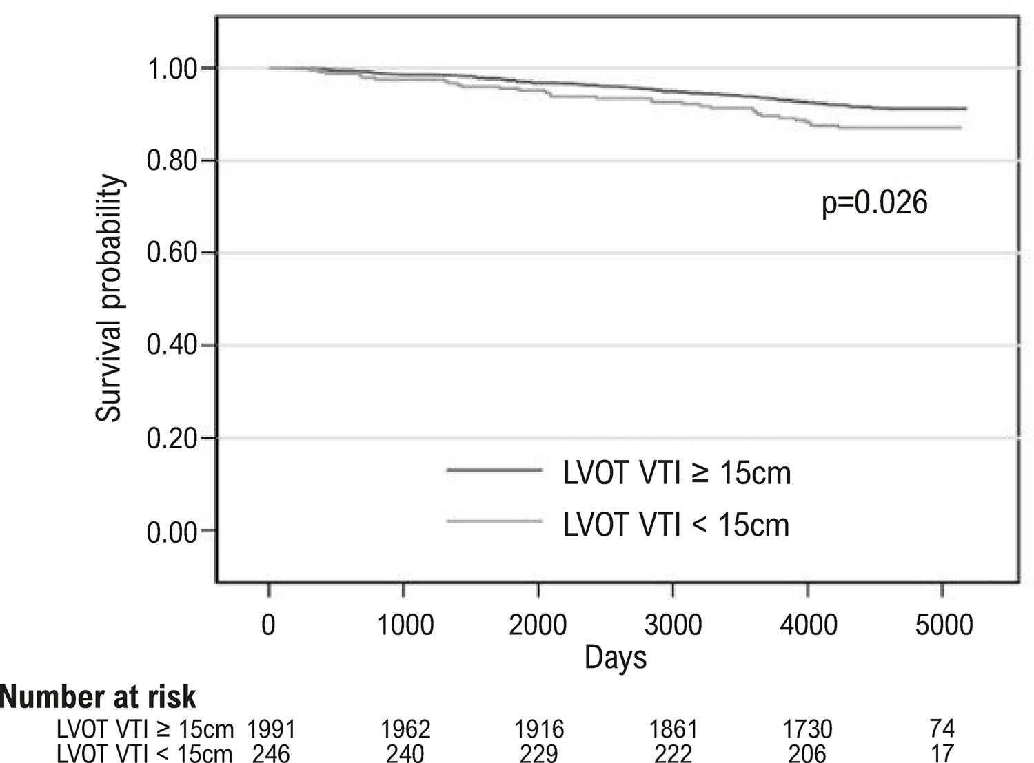
Among patients with Heart Failure with reduced Ejection Fraction (HFrEF), LVOT VTI values below 12 cm or 8 cm have been associated with worse cardiovascular outcomes, with a progressive increase in risk as VTI decreases., Similarly, in intermediate to high-risk pulmonary embolism, LVOT VTI ≤ 15 cm has been associated with higher in-hospital mortality, cardiopulmonary arrest, shock, and need for reperfusion therapy. In secondary mitral regurgitation, LVOT VTI ≤ 17 cm predicts both cardiovascular and all-cause mortality. And in ambulatory adults with stable Coronary Artery Disease (CAD), LVOT VTI ≤ 18 cm was associated with heart failure hospitalization and mortality. Despite these findings, there is limited data to evaluate the usefulness of LVOT VTI in free-dwelling adults.

[…]

  • Echocardiography
  • Healthy Volunteers
  • Mortality
  • Risk Assessment
Read Full Article
Creative Commons Attribution (CC BY) Creative Commons license
17/04/2026 DOI: 10.36660/abcimg.20260013i Views 2
Development and Validation of a Predictive Model for Atrial Functional Mitral Regurgitation
Original Article

Development and Validation of a Predictive Model for Atrial Functional Mitral Regurgitation

Alexandre Costa Souza et al. Bruna de Mattos Ivo Junqueira Stephanie de Azevedo Drubi Priscila Pinheiro … Laila Caroline Gomes Pedro Henrique Correia Filgueiras Ricardo André Sales Pereira Guedes Marco André Moraes Sales Yuri Xavier de Carvalho Carolina Thê Macêdo

Abstract

Background

Identifying atrial etiology in patients with mitral regurgitation remains challenging because the diagnosis is often established by exclusion. The use of a multivariable model may enhance diagnostic accuracy in the context of atrial functional mitral regurgitation.

Objective

To develop and validate a multivariable logistic regression model based on clinical and echocardiographic characteristics to predict atrial functional mitral regurgitation.

Methods

This cross-sectional study included patients with significant mitral regurgitation diagnosed by transesophageal echocardiography. The dataset was randomly divided into a training set (70%) and a validation set (30%). Statistical analyses were performed using a significance level of 5%.

Results

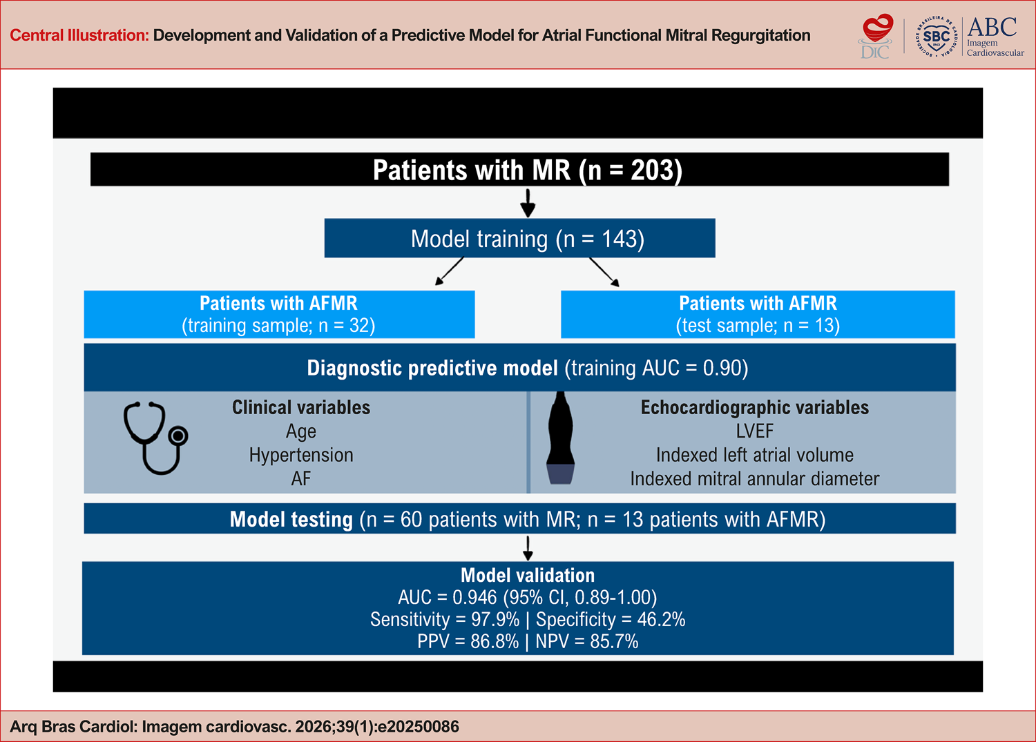
A total of 203 patients were included. The median age was 79 years in the atrial group and 72 years in the non-atrial group (p = 0.0022). Receiver operating characteristic curve analysis demonstrated good discriminative performance, with an area under the curve of 0.896 (95% CI, 0.845-0.947) in the training set. In the validation set, the model achieved an area under the curve of 0.946 (95% CI, 0.89-1.00), which indicates high predictive accuracy. Model calibration assessed by the Hosmer-Lemeshow test (chi-square test = 5.197; df = 8; p = 0.736) demonstrated good agreement between predicted and observed outcomes.

Conclusion

A multivariable model was derived and validated as a useful tool for predicting atrial etiology in patients with mitral regurgitation, potentially reducing diagnostic variability in clinical practice.

  • Echocardiography
  • Logistic Models
  • Mitral Valve Insufficiency
  • Statistical Models
Read Full Article
Creative Commons Attribution (CC BY) Creative Commons license
17/04/2026 DOI: 10.36660/abcimg.20250086i Views 3
Diagnostic Performance Of Contrast-Enhanced Echocardiography In Differentiating Cardiac Masses: A Systematic Review And Meta-analysis
Original Article

Diagnostic Performance Of Contrast-Enhanced Echocardiography In Differentiating Cardiac Masses: A Systematic Review And Meta-analysis

João Guilherme G. Pedrosa et al. Felizardo José Leandro Pereira Antonio Lacerda Cavalcanti Neto Renata Ramos Stropp … Giordano Persuhn Rolim de Moura Marcelo Tavares Sadrak Lyon Dantas Pontes Alex dos Santos Felix

Abstract

Background

Conventional echocardiography often struggles to differentiate intracardiac masses, particularly in patients with poor acoustic windows. Contrast-enhanced Echocardiography (CEE) overcomes this limitation by visualizing perfusion patterns — distinguishing avascular thrombi from vascularized tumors. We aimed to synthesize existing evidence to evaluate the diagnostic accuracy of CEE.

Objectives

To evaluate the diagnostic accuracy of CEE for differentiating cardiac masses in adults, using histopathology as reference and reporting AUC, sensitivity, specificity, PPV, and NPV.

Methods

Systematic searches of PubMed, Web of Science, Cochrane Library, and EMBASE were performed on August 10, 2025. Studies meeting PICOTT criteria were included; extracted data included sensitivity, specificity, AUC, and 2×2 tables. Pooled estimates were obtained using standard bivariate and SROC models for diagnostic meta-analysis. Statistical significance set at P < 0.05.

Results

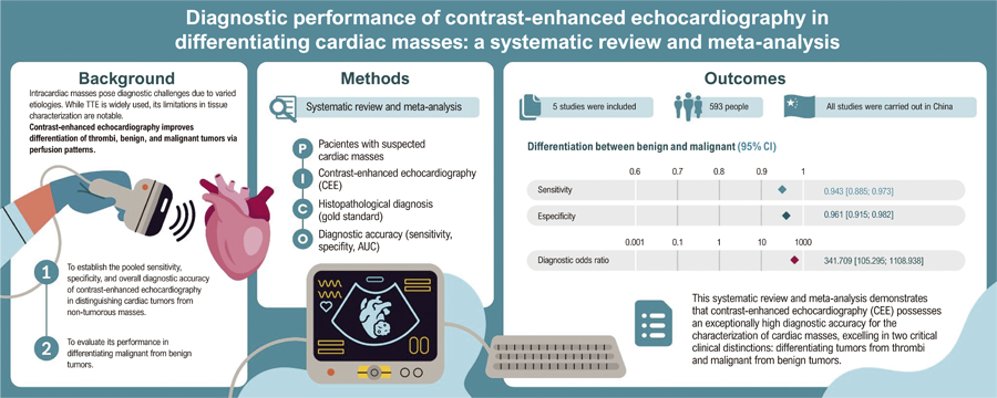
Five prospective cohort studies (total n = 381 patients) were included. For tumor vs non-tumor, pooled sensitivity = 100% and specificity = 100% (95% CI 99.5–100%; I2 = 0%; heterogeneity P = 0.985), diagnostic odds ratio (DOR) = 3,890.65, AUC = 0.989. For malignant vs benign tumors, pooled sensitivity = 94.3% (95% CI 88.5–97.3%; I2 = 0%; P = 0.681), specificity = 96.1% (95% CI 91.5–98.2%; I2 = 0%; P = 0.970), DOR = 341.71, SROC AUC = 0.976.

Conclusions

CEE showed very high diagnostic accuracy in the available prospective series. However, the small number of studies and limited sample sizes warrant cautious interpretation; larger prospective multicenter studies with standardized CEE protocols are needed to confirm these results.

  • Cardiac Neoplasms
  • Contrast Media
  • Echocardiography
  • Meta-Analysis
  • systematic review
Read Full Article
Creative Commons Attribution (CC BY) Creative Commons license
17/04/2026 DOI: 10.36660/abcimg.20250082i Views 1
Figure 1
Case Report

Hemodynamic Impact of Hypertrophic Cardiomyopathy at Rest and During Supine Bicycle Exercise: Additional Value of Postprandial Assessment

Marília Esther Benevides Abreu et al. Tereza Cristina Pinheiro Diógenes Isadora Sucupira Machado Chagas Humberto Mororó Xerex José Sebastião De Abreu

Abstract

A patient with severe, symptomatic hypertrophic cardiomyopathy (HCM) without a significant left ventricular outflow tract (LVOT) gradient at rest requires further evaluation. During exercise echocardiography (EE) performed on a supine bicycle, a latent or underestimated dynamic obstruction may be identified in real time, with postprandial assessment being particularly relevant.

  • Exercise
  • Exercise Test
  • Hypertrophic Cardiomyopathy
  • Postprandial
Read Full Article
Creative Commons Attribution (CC BY) Creative Commons license
17/04/2026 DOI: 10.36660/abcimg.20250049i
Editorial

Functional Mitral Regurgitation of Atrial Origin: Search for Diagnostic Criteria

Minna Moreira Dias Romano

Identifying the various mechanisms of Mitral Regurgitation (MR) is essential for understanding the disease, prognosis, and planning structural therapeutic approaches. Recently, several studies on valvular heart disease have recognized functional MR. This condition is associated with the impact of cardiac geometry on mitral valve function, rather than primary valvular injury such as rheumatic, degenerative, infectious (endocarditis), or congenital diseases.

Furthermore, within the group of patients with functional MR, a subgroup has been identified that does not present Left Ventricular (LV) dilation or dysfunction. In this subgroup, the likely mechanism of MR is chronic enlargement of the Left Atrium (LA) and the mitral annulus, commonly associated with atrial fibrillation. Preliminary studies suggest associated left atrial dysfunction, which can be measured through tissue mechanics analysis. These factors likely contribute to the reduction of the coaptation surface of the mitral leaflet edges.

[…]

  • Atrial Fibrillation
  • Echocardiography
  • Mitral Valve Insufficiency
Read Full Article
Creative Commons Attribution (CC BY) Creative Commons license
10/04/2026 DOI: 10.36660/abcimg.20260008i Views 2
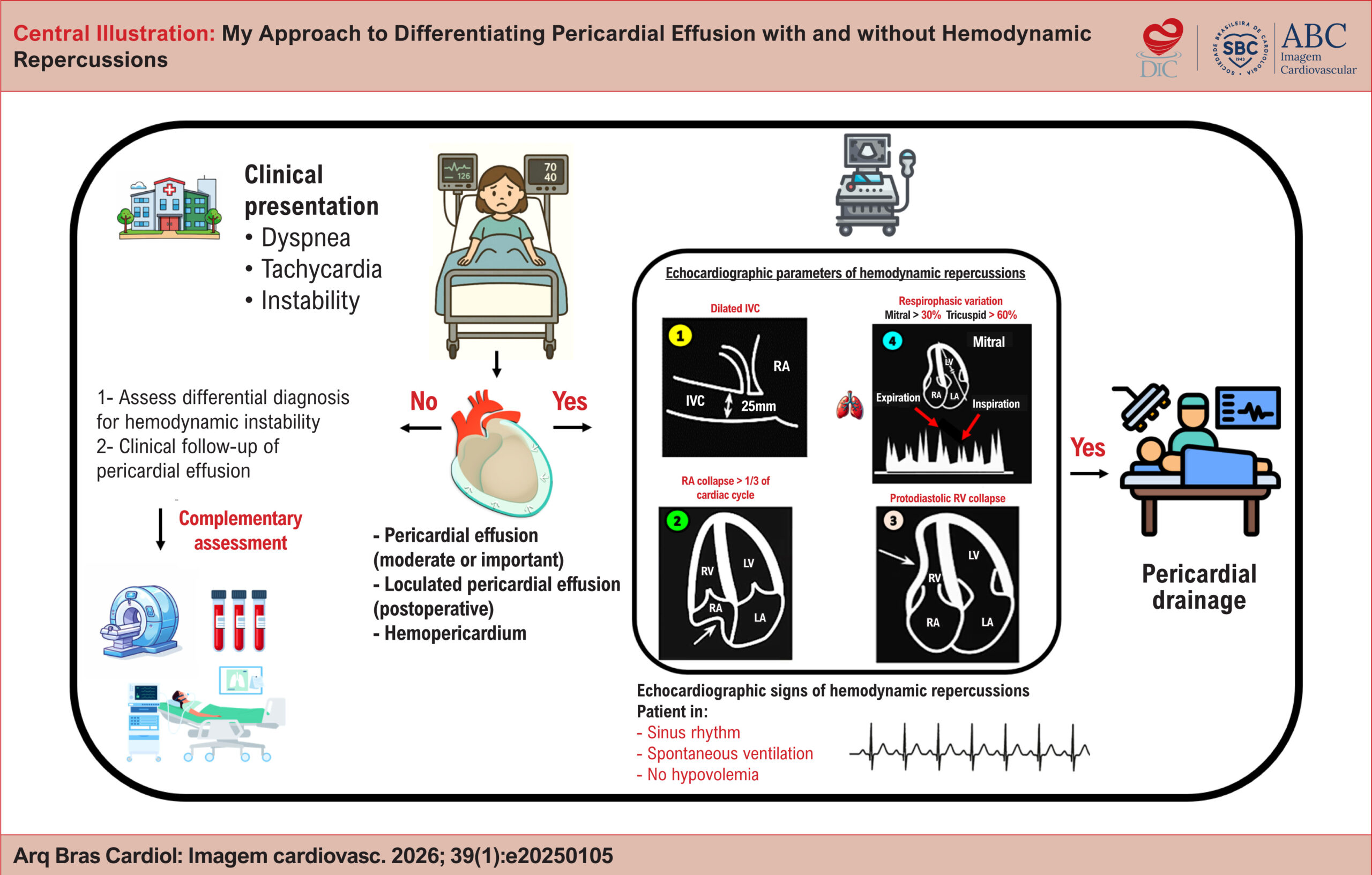
My Approach to Differentiating Pericardial Effusion with and without Hemodynamic Repercussions
Review Article

My Approach to Differentiating Pericardial Effusion with and without Hemodynamic Repercussions

Helder Moura Gomes et al. Halsted Alarcão Gomes Pereira da Silva

Abstract

Pericardial effusion is a common finding in cardiology practice. It is frequently identified in outpatient and inpatient follow-up examinations, especially during the postoperative period of heart surgery. In clinically stable patients, proper assessment can allow for early detection of signs of clinical deterioration. In patients in shock, careful analysis of pericardial effusion can confirm or rule out this condition as the main cause of hemodynamic instability. Precise identification of the location, anatomical characterization of severity, and analysis of hemodynamic repercussions by means of Doppler are essential elements to guide medical management. In addition to technical assessment, precautions when describing findings in imaging reports are equally important. This is because the hemodynamic repercussions observed on echocardiography do not always correspond to the patient’s clinical severity.

  • Cardiac Tamponade
  • Echocardiography
  • Inferior vena cava
  • Pericardial Effusion
Read Full Article
Creative Commons Attribution (CC BY) Creative Commons license
10/04/2026 DOI: 10.36660/abcimg.20250105i
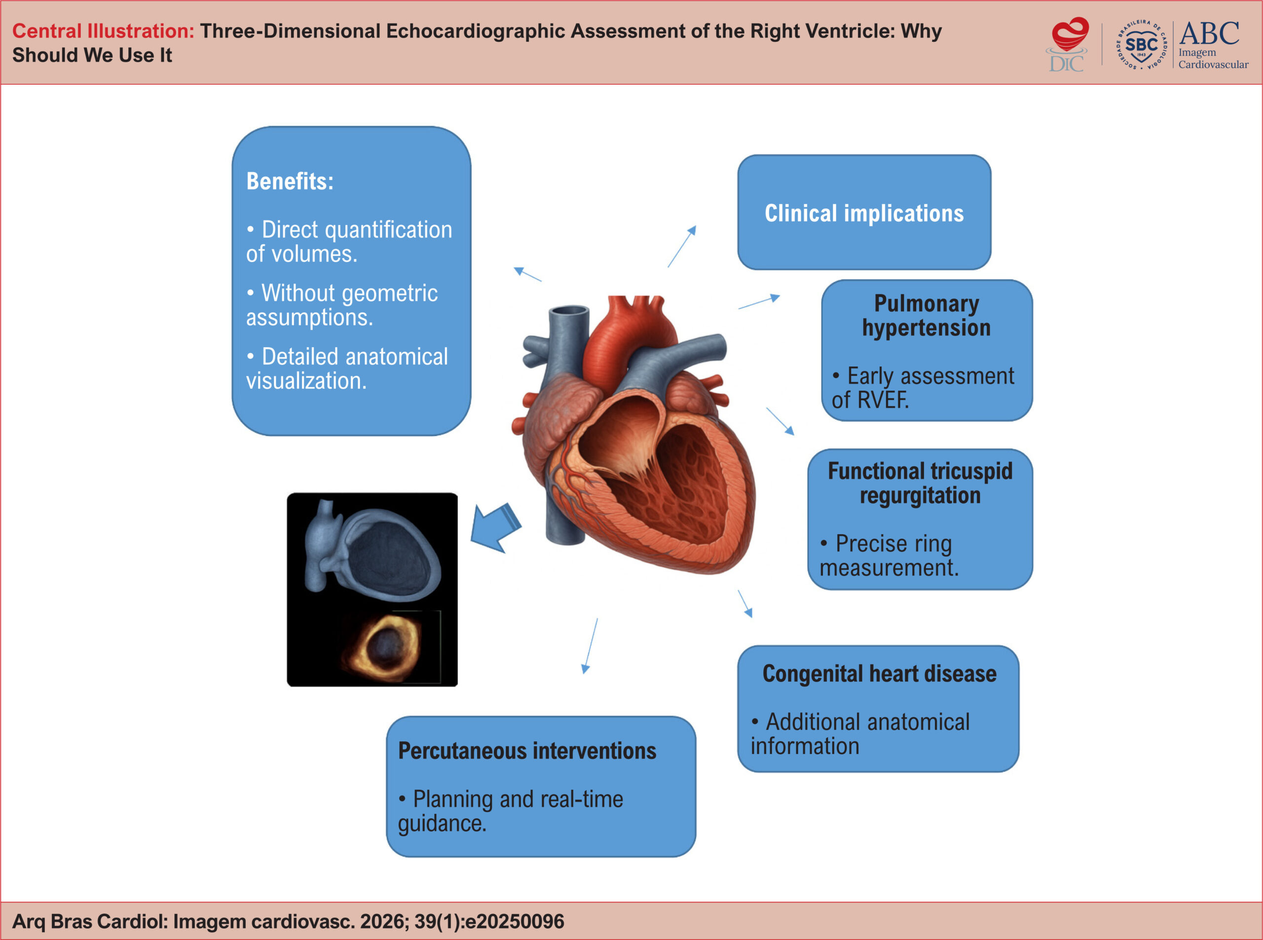
Three-Dimensional Echocardiographic Assessment of the Right Ventricle: Why Should We Use It
Review Article

Three-Dimensional Echocardiographic Assessment of the Right Ventricle: Why Should We Use It

Tiago R. Politi et al. Rodrigo B.M. Barretto João Cesar Nunes Sbano David Costa de Souza Le Bihan Wilson Mathias Jr.

Abstract

Right ventricular (RV) assessment using two-dimensional (2D) echocardiography has historically faced significant challenges due to the chamber’s complex and unique geometry and its thoracic orientation. In this context, three-dimensional (3D) echocardiography has emerged as a promising tool to overcome and illuminate these limitations, enabling accurate quantification of volumes and ejection fraction without relying on geometric assumptions. As a result, the routine incorporation of 3D echocardiography into RV evaluation may redefine diagnostic and prognostic paradigms, fostering a more precise and personalized approach in modern cardiology. To consolidate and highlight this technique, this review article explores the technical principles of 3D echocardiography for RV assessment, discusses its advantages over conventional 2D imaging, examines its validation against cardiac magnetic resonance (CMR), and reviews key clinical applications, including pulmonary hypertension, functional tricuspid regurgitation, congenital heart disease, and right-sided heart failure. Additionally, the article outlines current limitations of the technique, future perspectives, and practical recommendations based on contemporary literature.

  • Cardiac Magnetic Resonance Imaging
  • Ejection Fraction
  • Right ventricle
  • Three-Dimensional Echocardiography
Read Full Article
Creative Commons Attribution (CC BY) Creative Commons license
10/04/2026 DOI: 10.36660/abcimg.20250096i Views 1

Could not load more articles. Please try again.

How to cite this article

EndNote RIS

Electronic Document Format (Vancouver)

. ERRATUM: Recalibrating the Barometer: Echocardiography in Diastolic Dysfunction and the Era of New Algorithms. ABC Imagem Cardiovascular 2026;39(1):e20260055.

Electronic Document Format (ABNT)

. ERRATUM: Recalibrating the Barometer: Echocardiography in Diastolic Dysfunction and the Era of New Algorithms. ABC Imagem Cardiovascular, v. 39, n. 1, e20260055, Aug. 2026.

Electronic Document Format (APA)

(2026). ERRATUM: Recalibrating the Barometer: Echocardiography in Diastolic Dysfunction and the Era of New Algorithms. ABC Imagem Cardiovascular, 39(1), e20260055.

Electronic Document Format (ISO)

. ERRATUM: Recalibrating the Barometer: Echocardiography in Diastolic Dysfunction and the Era of New Algorithms. ABC Imagem Cardiovascular [online]. 2026, vol. 39, n. 1, [cited 2026-10-14], e20260055. Available from: <https://journals.cardiol.br/en/article/erratum-recalibrating-the-barometer-echocardiography-in-diastolic-dysfunction-and-the-era-of-new-algorithms/>. ISSN 2318-8219

EndNote RIS

Electronic Document Format (Vancouver)

Rolim AAA, Carneiro TCAG, Campos F, Ferreira DP. Neurological Manifestations of Takayasu Arteritis: A Case Report and Literature Review. ABC Imagem Cardiovascular 2026;39(1):e202500110.

Electronic Document Format (ABNT)

Rolim, Amanda Antunes Arantes; Carneiro, Tainá Cândida de Almeida Gontijo; Campos, Flávia de; Ferreira, Dilson Palhares. Neurological Manifestations of Takayasu Arteritis: A Case Report and Literature Review. ABC Imagem Cardiovascular, v. 39, n. 1, e202500110, May. 2026.

Electronic Document Format (APA)

Rolim, A. A. A., Carneiro, T. C. A. G., Campos, F. , & Ferreira, D. P. (2026). Neurological Manifestations of Takayasu Arteritis: A Case Report and Literature Review. ABC Imagem Cardiovascular, 39(1), e202500110.

Electronic Document Format (ISO)

Rolim, Amanda Antunes Arantes and Carneiro, Tainá Cândida de Almeida Gontijo and Campos, Flávia de and Ferreira, Dilson Palhares. Neurological Manifestations of Takayasu Arteritis: A Case Report and Literature Review. ABC Imagem Cardiovascular [online]. 2026, vol. 39, n. 1, [cited 2026-10-14], e202500110. Available from: <https://journals.cardiol.br/en/article/neurological-manifestations-of-takayasu-arteritis-a-case-report-and-literature-review/>. ISSN 2318-8219

EndNote RIS

Electronic Document Format (Vancouver)

Abdo MH, Latsh H, Wagner M, König G, Barth S. Coexistence of Partial Anomalous Pulmonary Venous Connection and Coronary Artery Fistulas: A Rare Case Report. ABC Imagem Cardiovascular 2026;39(1):e20250030.

Electronic Document Format (ABNT)

Abdo, Mourad Haj; Latsh, Hussain; Wagner, Mathias; König, George; Barth, Sebastian. Coexistence of Partial Anomalous Pulmonary Venous Connection and Coronary Artery Fistulas: A Rare Case Report. ABC Imagem Cardiovascular, v. 39, n. 1, e20250030, May. 2026.

Electronic Document Format (APA)

Abdo, M. H., Latsh, H., Wagner, M., König, G., & Barth, S. (2026). Coexistence of Partial Anomalous Pulmonary Venous Connection and Coronary Artery Fistulas: A Rare Case Report. ABC Imagem Cardiovascular, 39(1), e20250030.

Electronic Document Format (ISO)

Abdo, Mourad Haj and Latsh, Hussain and Wagner, Mathias and König, George and Barth, Sebastian. Coexistence of Partial Anomalous Pulmonary Venous Connection and Coronary Artery Fistulas: A Rare Case Report. ABC Imagem Cardiovascular [online]. 2026, vol. 39, n. 1, [cited 2026-10-14], e20250030. Available from: <https://journals.cardiol.br/en/article/coexistence-of-partial-anomalous-pulmonary-venous-connection-and-coronary-artery-fistulas-a-rare-case-report/>. ISSN 2318-8219

EndNote RIS

Electronic Document Format (Vancouver)

Flores MP, Souza AC, Carvalho MVSF, Melo RMV, Cavalcante LRS, Barroso ND, Carvalho YX, et al. Correlation Between Venous Excess Ultrasound and N-Terminal Pro-B-Type Natriuretic Peptide Levels in Patients With Acute Decompensated Heart Failure. ABC Imagem Cardiovascular 2026;39(1):e20260018.

Electronic Document Format (ABNT)

Flores, Marcella Pereira; Souza, Alexandre Costa; Carvalho, Marcus Vinicius Silva Freire de; Melo, Rodrigo Morel Vieira de; Cavalcante, Lívia Rodrigues Sampaio; Barroso, Natália Duarte; Carvalho, Yuri Xavier de; Barros, Raisa Mainarte Franco; Lobo, Clara Talita Silva; Almeida Filho, Adriano Chaves de. Correlation Between Venous Excess Ultrasound and N-Terminal Pro-B-Type Natriuretic Peptide Levels in Patients With Acute Decompensated Heart Failure. ABC Imagem Cardiovascular, v. 39, n. 1, e20260018, May. 2026.

Electronic Document Format (APA)

Flores, M. P., Souza, A. C., Carvalho, M. V. S. F. , Melo, R. M. V. , Cavalcante, L. R. S., Barroso, N. D., Carvalho, Y. X. , Barros, R. M. F., Lobo, C. T. S., & Almeida Filho, A. C. (2026). Correlation Between Venous Excess Ultrasound and N-Terminal Pro-B-Type Natriuretic Peptide Levels in Patients With Acute Decompensated Heart Failure. ABC Imagem Cardiovascular, 39(1), e20260018.

Electronic Document Format (ISO)

Flores, Marcella Pereira and Souza, Alexandre Costa and Carvalho, Marcus Vinicius Silva Freire de and Melo, Rodrigo Morel Vieira de and Cavalcante, Lívia Rodrigues Sampaio and Barroso, Natália Duarte and Carvalho, Yuri Xavier de and Barros, Raisa Mainarte Franco and Lobo, Clara Talita Silva and Almeida Filho, Adriano Chaves de. Correlation Between Venous Excess Ultrasound and N-Terminal Pro-B-Type Natriuretic Peptide Levels in Patients With Acute Decompensated Heart Failure. ABC Imagem Cardiovascular [online]. 2026, vol. 39, n. 1, [cited 2026-10-14], e20260018. Available from: <https://journals.cardiol.br/en/article/correlation-between-venous-excess-ultrasound-and-n-terminal-pro-b-type-natriuretic-peptide-levels-in-patients-with-acute-decompensated-heart-failure/>. ISSN 2318-8219

EndNote RIS

Electronic Document Format (Vancouver)

Travessa A, Pereira PH, Machado LM, Esteves NS. Step-by-Step Approach to the Evaluation of Constrictive Pericarditis. ABC Imagem Cardiovascular 2026;39(1):e20250063.

Electronic Document Format (ABNT)

Travessa, Aline; Pereira, Paulo Henrique; Machado, Louise Moutinho; Esteves, Natalia Sousa. Step-by-Step Approach to the Evaluation of Constrictive Pericarditis. ABC Imagem Cardiovascular, v. 39, n. 1, e20250063, May. 2026.

Electronic Document Format (APA)

Travessa, A., Pereira, P. H., Machado, L. M., & Esteves, N. S. (2026). Step-by-Step Approach to the Evaluation of Constrictive Pericarditis. ABC Imagem Cardiovascular, 39(1), e20250063.

Electronic Document Format (ISO)

Travessa, Aline and Pereira, Paulo Henrique and Machado, Louise Moutinho and Esteves, Natalia Sousa. Step-by-Step Approach to the Evaluation of Constrictive Pericarditis. ABC Imagem Cardiovascular [online]. 2026, vol. 39, n. 1, [cited 2026-10-14], e20250063. Available from: <https://journals.cardiol.br/en/article/step-by-step-approach-to-the-evaluation-of-constrictive-pericarditis/>. ISSN 2318-8219

EndNote RIS

Electronic Document Format (Vancouver)

Lemos NB, Araújo GAM, Melo MDT. The Use of Artificial Intelligence in the Diagnosis of Cardiac Amyloidosis: Integrative Review. ABC Imagem Cardiovascular 2026;39(1):e20260016.

Electronic Document Format (ABNT)

Lemos, Nilson Batista; Araújo, Gabriela Aparecida Moreira; Melo, Marcelo Dantas Tavares de. The Use of Artificial Intelligence in the Diagnosis of Cardiac Amyloidosis: Integrative Review. ABC Imagem Cardiovascular, v. 39, n. 1, e20260016, May. 2026.

Electronic Document Format (APA)

Lemos, N. B., Araújo, G. A. M., & Melo, M. D. T. (2026). The Use of Artificial Intelligence in the Diagnosis of Cardiac Amyloidosis: Integrative Review. ABC Imagem Cardiovascular, 39(1), e20260016.

Electronic Document Format (ISO)

Lemos, Nilson Batista and Araújo, Gabriela Aparecida Moreira and Melo, Marcelo Dantas Tavares de. The Use of Artificial Intelligence in the Diagnosis of Cardiac Amyloidosis: Integrative Review. ABC Imagem Cardiovascular [online]. 2026, vol. 39, n. 1, [cited 2026-10-14], e20260016. Available from: <https://journals.cardiol.br/en/article/the-use-of-artificial-intelligence-in-the-diagnosis-of-cardiac-amyloidosis-integrative-review/>. ISSN 2318-8219

EndNote RIS

Electronic Document Format (Vancouver)

Brandão SCS. Nuclear Cardiology: From Consolidation to Integration. ABC Imagem Cardiovascular 2026;39(1):e20260007.

Electronic Document Format (ABNT)

Brandão, Simone Cristina Soares. Nuclear Cardiology: From Consolidation to Integration. ABC Imagem Cardiovascular, v. 39, n. 1, e20260007, May. 2026.

Electronic Document Format (APA)

Brandão, S. C. S. (2026). Nuclear Cardiology: From Consolidation to Integration. ABC Imagem Cardiovascular, 39(1), e20260007.

Electronic Document Format (ISO)

Brandão, Simone Cristina Soares. Nuclear Cardiology: From Consolidation to Integration. ABC Imagem Cardiovascular [online]. 2026, vol. 39, n. 1, [cited 2026-10-14], e20260007. Available from: <https://journals.cardiol.br/en/article/nuclear-cardiology-from-consolidation-to-integration/>. ISSN 2318-8219

EndNote RIS

Electronic Document Format (Vancouver)

Almeida JHA, Ferreira GMA, Andrade MAM, Nunes HM, Costa IB. Cardiovascular Complications in Patients with COVID-19 and Their Relationship with Mortality: An Echocardiographic Perspective. ABC Imagem Cardiovascular 2026;39(1):e20260005.

Electronic Document Format (ABNT)

Almeida, João Henrique Andrade de; Ferreira, Gustavo Miranda de Azevedo; Andrade, Marina de Azevedo Martins; Nunes, Heloísa Marceliano; Costa, Igor Brasil. Cardiovascular Complications in Patients with COVID-19 and Their Relationship with Mortality: An Echocardiographic Perspective. ABC Imagem Cardiovascular, v. 39, n. 1, e20260005, May. 2026.

Electronic Document Format (APA)

Almeida, J. H. A. , Ferreira, G. M. A., Andrade, M. A. M., Nunes, H. M., & Costa, I. B. (2026). Cardiovascular Complications in Patients with COVID-19 and Their Relationship with Mortality: An Echocardiographic Perspective. ABC Imagem Cardiovascular, 39(1), e20260005.

Electronic Document Format (ISO)

Almeida, João Henrique Andrade de and Ferreira, Gustavo Miranda de Azevedo and Andrade, Marina de Azevedo Martins and Nunes, Heloísa Marceliano and Costa, Igor Brasil. Cardiovascular Complications in Patients with COVID-19 and Their Relationship with Mortality: An Echocardiographic Perspective. ABC Imagem Cardiovascular [online]. 2026, vol. 39, n. 1, [cited 2026-10-14], e20260005. Available from: <https://journals.cardiol.br/en/article/cardiovascular-complications-in-patients-with-covid-19-and-their-relationship-with-mortality-an-echocardiographic-perspective/>. ISSN 2318-8219

EndNote RIS

Electronic Document Format (Vancouver)

Paiva MG. Hypertrophic Cardiomyopathy: Standardization of Echocardiographic Assessment in an Era of New Therapies. ABC Imagem Cardiovascular 2026;39(1):e20260027.

Electronic Document Format (ABNT)

Paiva, Marcelo Goulart. Hypertrophic Cardiomyopathy: Standardization of Echocardiographic Assessment in an Era of New Therapies. ABC Imagem Cardiovascular, v. 39, n. 1, e20260027, May. 2026.

Electronic Document Format (APA)

Paiva, M. G. (2026). Hypertrophic Cardiomyopathy: Standardization of Echocardiographic Assessment in an Era of New Therapies. ABC Imagem Cardiovascular, 39(1), e20260027.

Electronic Document Format (ISO)

Paiva, Marcelo Goulart. Hypertrophic Cardiomyopathy: Standardization of Echocardiographic Assessment in an Era of New Therapies. ABC Imagem Cardiovascular [online]. 2026, vol. 39, n. 1, [cited 2026-10-14], e20260027. Available from: <https://journals.cardiol.br/en/article/hypertrophic-cardiomyopathy-standardization-of-echocardiographic-assessment-in-an-era-of-new-therapies/>. ISSN 2318-8219

EndNote RIS

Electronic Document Format (Vancouver)

Fernandes A. Phenotyping of Congestion in Decompensated Heart Failure: Life-Saving Accuracy. ABC Imagem Cardiovascular 2026;39(1):e20260019.

Electronic Document Format (ABNT)

Fernandes, Amanda. Phenotyping of Congestion in Decompensated Heart Failure: Life-Saving Accuracy. ABC Imagem Cardiovascular, v. 39, n. 1, e20260019, May. 2026.

Electronic Document Format (APA)

Fernandes, A. (2026). Phenotyping of Congestion in Decompensated Heart Failure: Life-Saving Accuracy. ABC Imagem Cardiovascular, 39(1), e20260019.

Electronic Document Format (ISO)

Fernandes, Amanda. Phenotyping of Congestion in Decompensated Heart Failure: Life-Saving Accuracy. ABC Imagem Cardiovascular [online]. 2026, vol. 39, n. 1, [cited 2026-10-14], e20260019. Available from: <https://journals.cardiol.br/en/article/phenotyping-of-congestion-in-decompensated-heart-failure-life-saving-accuracy/>. ISSN 2318-8219

EndNote RIS

Electronic Document Format (Vancouver)

Mariano GG, Thaines CA, Desani GS, Gouvêa LC, Covilo PVC, Caravante RPG. Aneurysm of the Suprahepatic Inferior Vena Cava: A Case Report. ABC Imagem Cardiovascular 2026;39(1):e20250055.

Electronic Document Format (ABNT)

Mariano, Gabriella Ghattas; Thaines, Calina Araujo; Desani, Gabrielle Silva; Gouvêa, Letícia de Castro; Covilo, Paulo Vítor Cabral; Caravante, Rogério de Paula Garcia. Aneurysm of the Suprahepatic Inferior Vena Cava: A Case Report. ABC Imagem Cardiovascular, v. 39, n. 1, e20250055, May. 2026.

Electronic Document Format (APA)

Mariano, G. G., Thaines, C. A., Desani, G. S., Gouvêa, L. C., Covilo, P. V. C., & Caravante, R. P. G. (2026). Aneurysm of the Suprahepatic Inferior Vena Cava: A Case Report. ABC Imagem Cardiovascular, 39(1), e20250055.

Electronic Document Format (ISO)

Mariano, Gabriella Ghattas and Thaines, Calina Araujo and Desani, Gabrielle Silva and Gouvêa, Letícia de Castro and Covilo, Paulo Vítor Cabral and Caravante, Rogério de Paula Garcia. Aneurysm of the Suprahepatic Inferior Vena Cava: A Case Report. ABC Imagem Cardiovascular [online]. 2026, vol. 39, n. 1, [cited 2026-10-14], e20250055. Available from: <https://journals.cardiol.br/en/article/aneurysm-of-the-suprahepatic-inferior-vena-cava-a-case-report/>. ISSN 2318-8219

EndNote RIS

Electronic Document Format (Vancouver)

Otto MEB, Assef JE, Nishida G. Recalibrating the Barometer: Echocardiography in Diastolic Dysfunction and the Era of New Algorithms. ABC Imagem Cardiovascular 2026;39(1):e20260004.

Electronic Document Format (ABNT)

Otto, Maria Estefania Bosco; Assef, Jorge Eduardo; Nishida, Gustavo. Recalibrating the Barometer: Echocardiography in Diastolic Dysfunction and the Era of New Algorithms. ABC Imagem Cardiovascular, v. 39, n. 1, e20260004, Apr. 2026.

Electronic Document Format (APA)

Otto, M. E. B., Assef, J. E., & Nishida, G. (2026). Recalibrating the Barometer: Echocardiography in Diastolic Dysfunction and the Era of New Algorithms. ABC Imagem Cardiovascular, 39(1), e20260004.

Electronic Document Format (ISO)

Otto, Maria Estefania Bosco and Assef, Jorge Eduardo and Nishida, Gustavo. Recalibrating the Barometer: Echocardiography in Diastolic Dysfunction and the Era of New Algorithms. ABC Imagem Cardiovascular [online]. 2026, vol. 39, n. 1, [cited 2026-10-14], e20260004. Available from: <https://journals.cardiol.br/en/article/recalibrating-the-barometer-echocardiography-in-diastolic-dysfunction-and-the-era-of-new-algorithms/>. ISSN 2318-8219

EndNote RIS

Electronic Document Format (Vancouver)

Akın T, Dere ZBY, Yozgat Y, Türkoğlu H, Ugurlucan M. Comparison of Cardiac Structural Changes After Surgical and Transcatheter Atrial Septal Defect Closure With Color Doppler Echocardiography. ABC Imagem Cardiovascular 2026;39(1):e20260014.

Electronic Document Format (ABNT)

Akın, Tuğçe; Dere, Zeynep Bilge Yılmaz; Yozgat, Yılmaz; Türkoğlu, Halil; Ugurlucan, Murat. Comparison of Cardiac Structural Changes After Surgical and Transcatheter Atrial Septal Defect Closure With Color Doppler Echocardiography. ABC Imagem Cardiovascular, v. 39, n. 1, e20260014, Apr. 2026.

Electronic Document Format (APA)

Akın, T., Dere, Z. B. Y., Yozgat, Y., Türkoğlu, H., & Ugurlucan, M. (2026). Comparison of Cardiac Structural Changes After Surgical and Transcatheter Atrial Septal Defect Closure With Color Doppler Echocardiography. ABC Imagem Cardiovascular, 39(1), e20260014.

Electronic Document Format (ISO)

Akın, Tuğçe and Dere, Zeynep Bilge Yılmaz and Yozgat, Yılmaz and Türkoğlu, Halil and Ugurlucan, Murat. Comparison of Cardiac Structural Changes After Surgical and Transcatheter Atrial Septal Defect Closure With Color Doppler Echocardiography. ABC Imagem Cardiovascular [online]. 2026, vol. 39, n. 1, [cited 2026-10-14], e20260014. Available from: <https://journals.cardiol.br/en/article/comparison-of-cardiac-structural-changes-after-surgical-and-transcatheter-atrial-septal-defect-closure-with-color-doppler-echocardiography/>. ISSN 2318-8219

EndNote RIS

Electronic Document Format (Vancouver)

Salgado AA, Bernardo MPL, Fernandes MR. My Approach to VExUS Assessment Using Transesophageal Echocardiography: A Step-by-Step Performance Guide. ABC Imagem Cardiovascular 2026;39(1):e20260012.

Electronic Document Format (ABNT)

Salgado, Angelo Antunes; Bernardo, Marcos Paulo Lacerda; Fernandes, Marcelo Ramalho. My Approach to VExUS Assessment Using Transesophageal Echocardiography: A Step-by-Step Performance Guide. ABC Imagem Cardiovascular, v. 39, n. 1, e20260012, Apr. 2026.

Electronic Document Format (APA)

Salgado, A. A., Bernardo, M. P. L., & Fernandes, M. R. (2026). My Approach to VExUS Assessment Using Transesophageal Echocardiography: A Step-by-Step Performance Guide. ABC Imagem Cardiovascular, 39(1), e20260012.

Electronic Document Format (ISO)

Salgado, Angelo Antunes and Bernardo, Marcos Paulo Lacerda and Fernandes, Marcelo Ramalho. My Approach to VExUS Assessment Using Transesophageal Echocardiography: A Step-by-Step Performance Guide. ABC Imagem Cardiovascular [online]. 2026, vol. 39, n. 1, [cited 2026-10-14], e20260012. Available from: <https://journals.cardiol.br/en/article/my-approach-to-vexus-assessment-using-transesophageal-echocardiography-a-step-by-step-performance-guide/>. ISSN 2318-8219

EndNote RIS

Electronic Document Format (Vancouver)

Piveta RB, Aguiar MOD. Echocardiography with Ultrasound Enhancement Agents and the Diagnostic Challenge of Cardiac Masses: Solid Evidence for a Complex Clinical Problem. ABC Imagem Cardiovascular 2026;39(1):e20260009.

Electronic Document Format (ABNT)

Piveta, Rafael Bonafim; Aguiar, Miguel Osman Dias. Echocardiography with Ultrasound Enhancement Agents and the Diagnostic Challenge of Cardiac Masses: Solid Evidence for a Complex Clinical Problem. ABC Imagem Cardiovascular, v. 39, n. 1, e20260009, Apr. 2026.

Electronic Document Format (APA)

Piveta, R. B., & Aguiar, M. O. D. (2026). Echocardiography with Ultrasound Enhancement Agents and the Diagnostic Challenge of Cardiac Masses: Solid Evidence for a Complex Clinical Problem. ABC Imagem Cardiovascular, 39(1), e20260009.

Electronic Document Format (ISO)

Piveta, Rafael Bonafim and Aguiar, Miguel Osman Dias. Echocardiography with Ultrasound Enhancement Agents and the Diagnostic Challenge of Cardiac Masses: Solid Evidence for a Complex Clinical Problem. ABC Imagem Cardiovascular [online]. 2026, vol. 39, n. 1, [cited 2026-10-14], e20260009. Available from: <https://journals.cardiol.br/en/article/echocardiography-with-ultrasound-enhancement-agents-and-the-diagnostic-challenge-of-cardiac-masses-solid-evidence-for-a-complex-clinical-problem/>. ISSN 2318-8219

EndNote RIS

Electronic Document Format (Vancouver)

Carreira LCTF, Carreira L, Brito ASX. Cardiac Sarcoidosis: The Role of Multimodal Imaging. ABC Imagem Cardiovascular 2026;39(1):e20250038.

Electronic Document Format (ABNT)

Carreira, Lara Cristiane Terra Ferreira; Carreira, Lívia; Brito, Adriana Soares Xavier de. Cardiac Sarcoidosis: The Role of Multimodal Imaging. ABC Imagem Cardiovascular, v. 39, n. 1, e20250038, Apr. 2026.

Electronic Document Format (APA)

Carreira, L. C. T. F., Carreira, L., & Brito, A. S. X. (2026). Cardiac Sarcoidosis: The Role of Multimodal Imaging. ABC Imagem Cardiovascular, 39(1), e20250038.

Electronic Document Format (ISO)

Carreira, Lara Cristiane Terra Ferreira and Carreira, Lívia and Brito, Adriana Soares Xavier de. Cardiac Sarcoidosis: The Role of Multimodal Imaging. ABC Imagem Cardiovascular [online]. 2026, vol. 39, n. 1, [cited 2026-10-14], e20250038. Available from: <https://journals.cardiol.br/en/article/cardiac-sarcoidosis-the-role-of-multimodal-imaging/>. ISSN 2318-8219

EndNote RIS

Electronic Document Format (Vancouver)

Castillo JMD, Shehadeh I. My Approach to Coronary Flow Assessment With Transthoracic Echocardiography. ABC Imagem Cardiovascular 2026;39(1):e20260006.

Electronic Document Format (ABNT)

Castillo, José Maria Del; Shehadeh, Issam. My Approach to Coronary Flow Assessment With Transthoracic Echocardiography. ABC Imagem Cardiovascular, v. 39, n. 1, e20260006, Apr. 2026.

Electronic Document Format (APA)

Castillo, J. M. D., & Shehadeh, I. (2026). My Approach to Coronary Flow Assessment With Transthoracic Echocardiography. ABC Imagem Cardiovascular, 39(1), e20260006.

Electronic Document Format (ISO)

Castillo, José Maria Del and Shehadeh, Issam. My Approach to Coronary Flow Assessment With Transthoracic Echocardiography. ABC Imagem Cardiovascular [online]. 2026, vol. 39, n. 1, [cited 2026-10-14], e20260006. Available from: <https://journals.cardiol.br/en/article/my-approach-to-coronary-flow-assessment-with-transthoracic-echocardiography/>. ISSN 2318-8219

EndNote RIS

Electronic Document Format (Vancouver)

Lopes MC, Heidemann Jr AI, Pianca EG, Duncan BB, Foppa M, Santos AB. Left Ventricular Outflow Tract Velocity-Time Integral (LVOT VTI) as a Marker of Cardiac Performance: Mortality Data of the ELSA-Brasil Cohort. ABC Imagem Cardiovascular 2026;39(1):e20260013.

Electronic Document Format (ABNT)

Lopes, Mariana de Castro; Heidemann Jr, Altair Ivory; Pianca, Eduardo G.; Duncan, Bruce B.; Foppa, Murilo; Santos, Angela B.S. Left Ventricular Outflow Tract Velocity-Time Integral (LVOT VTI) as a Marker of Cardiac Performance: Mortality Data of the ELSA-Brasil Cohort. ABC Imagem Cardiovascular, v. 39, n. 1, e20260013, Apr. 2026.

Electronic Document Format (APA)

Lopes, M. C., Heidemann Jr, A. I., Pianca, E. G., Duncan, B. B., Foppa, M., & Santos, A. B. (2026). Left Ventricular Outflow Tract Velocity-Time Integral (LVOT VTI) as a Marker of Cardiac Performance: Mortality Data of the ELSA-Brasil Cohort. ABC Imagem Cardiovascular, 39(1), e20260013.

Electronic Document Format (ISO)

Lopes, Mariana de Castro and Heidemann Jr, Altair Ivory and Pianca, Eduardo G. and Duncan, Bruce B. and Foppa, Murilo and Santos, Angela B.S. Left Ventricular Outflow Tract Velocity-Time Integral (LVOT VTI) as a Marker of Cardiac Performance: Mortality Data of the ELSA-Brasil Cohort. ABC Imagem Cardiovascular [online]. 2026, vol. 39, n. 1, [cited 2026-10-14], e20260013. Available from: <https://journals.cardiol.br/en/article/left-ventricular-outflow-tract-velocity-time-integral-lvot-vti-as-a-marker-of-cardiac-performance-mortality-data-of-the-elsa-brasil-cohort/>. ISSN 2318-8219

EndNote RIS

Electronic Document Format (Vancouver)

Souza AC, Junqueira BMI, Drubi SA, Pinheiro P, Gomes LC, Filgueiras PHC, Guedes RASP, et al. Development and Validation of a Predictive Model for Atrial Functional Mitral Regurgitation. ABC Imagem Cardiovascular 2026;39(1):e20250086.

Electronic Document Format (ABNT)

Souza, Alexandre Costa; Junqueira, Bruna de Mattos Ivo; Drubi, Stephanie de Azevedo; Pinheiro, Priscila; Gomes, Laila Caroline; Filgueiras, Pedro Henrique Correia; Guedes, Ricardo André Sales Pereira; Sales, Marco André Moraes; Carvalho, Yuri Xavier de; Macêdo, Carolina Thê. Development and Validation of a Predictive Model for Atrial Functional Mitral Regurgitation. ABC Imagem Cardiovascular, v. 39, n. 1, e20250086, Apr. 2026.

Electronic Document Format (APA)

Souza, A. C., Junqueira, B. M. I., Drubi, S. A., Pinheiro, P., Gomes, L. C., Filgueiras, P. H. C., Guedes, R. A. S. P., Sales, M. A. M., Carvalho, Y. X. , & Macêdo, C. T. (2026). Development and Validation of a Predictive Model for Atrial Functional Mitral Regurgitation. ABC Imagem Cardiovascular, 39(1), e20250086.

Electronic Document Format (ISO)

Souza, Alexandre Costa and Junqueira, Bruna de Mattos Ivo and Drubi, Stephanie de Azevedo and Pinheiro, Priscila and Gomes, Laila Caroline and Filgueiras, Pedro Henrique Correia and Guedes, Ricardo André Sales Pereira and Sales, Marco André Moraes and Carvalho, Yuri Xavier de and Macêdo, Carolina Thê. Development and Validation of a Predictive Model for Atrial Functional Mitral Regurgitation. ABC Imagem Cardiovascular [online]. 2026, vol. 39, n. 1, [cited 2026-10-14], e20250086. Available from: <https://journals.cardiol.br/en/article/development-and-validation-of-a-predictive-model-for-atrial-functional-mitral-regurgitation/>. ISSN 2318-8219

EndNote RIS

Electronic Document Format (Vancouver)

Pedrosa JGG, Pereira FJL, Cavalcanti Neto AL, Stropp RR, Moura GPR, Tavares M, Pontes SLD, et al. Diagnostic Performance Of Contrast-Enhanced Echocardiography In Differentiating Cardiac Masses: A Systematic Review And Meta-analysis. ABC Imagem Cardiovascular 2026;39(1):e20250082.

Electronic Document Format (ABNT)

Pedrosa, João Guilherme G.; Pereira, Felizardo José Leandro; Cavalcanti Neto, Antonio Lacerda; Stropp, Renata Ramos; Moura, Giordano Persuhn Rolim de; Tavares, Marcelo; Pontes, Sadrak Lyon Dantas; Felix, Alex dos Santos. Diagnostic Performance Of Contrast-Enhanced Echocardiography In Differentiating Cardiac Masses: A Systematic Review And Meta-analysis. ABC Imagem Cardiovascular, v. 39, n. 1, e20250082, Apr. 2026.

Electronic Document Format (APA)

Pedrosa, J. G. G., Pereira, F. J. L., Cavalcanti Neto, A. L., Stropp, R. R., Moura, G. P. R. , Tavares, M., Pontes, S. L. D., & Felix, A. S. (2026). Diagnostic Performance Of Contrast-Enhanced Echocardiography In Differentiating Cardiac Masses: A Systematic Review And Meta-analysis. ABC Imagem Cardiovascular, 39(1), e20250082.

Electronic Document Format (ISO)

Pedrosa, João Guilherme G. and Pereira, Felizardo José Leandro and Cavalcanti Neto, Antonio Lacerda and Stropp, Renata Ramos and Moura, Giordano Persuhn Rolim de and Tavares, Marcelo and Pontes, Sadrak Lyon Dantas and Felix, Alex dos Santos. Diagnostic Performance Of Contrast-Enhanced Echocardiography In Differentiating Cardiac Masses: A Systematic Review And Meta-analysis. ABC Imagem Cardiovascular [online]. 2026, vol. 39, n. 1, [cited 2026-10-14], e20250082. Available from: <https://journals.cardiol.br/en/article/diagnostic-performance-of-contrast-enhanced-echocardiography-in-differentiating-cardiac-masses-a-systematic-review-and-meta-analysis/>. ISSN 2318-8219

EndNote RIS

Electronic Document Format (Vancouver)

Abreu MEB, Diógenes TCP, Chagas ISM, Xerex HM, Abreu JSD. Hemodynamic Impact of Hypertrophic Cardiomyopathy at Rest and During Supine Bicycle Exercise: Additional Value of Postprandial Assessment. ABC Imagem Cardiovascular 2026;39(1):e20250049.

Electronic Document Format (ABNT)

Abreu, Marília Esther Benevides; Diógenes, Tereza Cristina Pinheiro; Chagas, Isadora Sucupira Machado; Xerex, Humberto Mororó; Abreu, José Sebastião De. Hemodynamic Impact of Hypertrophic Cardiomyopathy at Rest and During Supine Bicycle Exercise: Additional Value of Postprandial Assessment. ABC Imagem Cardiovascular, v. 39, n. 1, e20250049, Apr. 2026.

Electronic Document Format (APA)

Abreu, M. E. B., Diógenes, T. C. P., Chagas, I. S. M., Xerex, H. M., & Abreu, J. S. D. (2026). Hemodynamic Impact of Hypertrophic Cardiomyopathy at Rest and During Supine Bicycle Exercise: Additional Value of Postprandial Assessment. ABC Imagem Cardiovascular, 39(1), e20250049.

Electronic Document Format (ISO)

Abreu, Marília Esther Benevides and Diógenes, Tereza Cristina Pinheiro and Chagas, Isadora Sucupira Machado and Xerex, Humberto Mororó and Abreu, José Sebastião De. Hemodynamic Impact of Hypertrophic Cardiomyopathy at Rest and During Supine Bicycle Exercise: Additional Value of Postprandial Assessment. ABC Imagem Cardiovascular [online]. 2026, vol. 39, n. 1, [cited 2026-10-14], e20250049. Available from: <https://journals.cardiol.br/en/article/hemodynamic-impact-of-hypertrophic-cardiomyopathy-at-rest-and-during-supine-bicycle-exercise-additional-value-of-postprandial-assessment/>. ISSN 2318-8219

EndNote RIS

Electronic Document Format (Vancouver)

Romano MMD. Functional Mitral Regurgitation of Atrial Origin: Search for Diagnostic Criteria. ABC Imagem Cardiovascular 2026;39(1):e20260008.

Electronic Document Format (ABNT)

Romano, Minna Moreira Dias. Functional Mitral Regurgitation of Atrial Origin: Search for Diagnostic Criteria. ABC Imagem Cardiovascular, v. 39, n. 1, e20260008, Apr. 2026.

Electronic Document Format (APA)

Romano, M. M. D. (2026). Functional Mitral Regurgitation of Atrial Origin: Search for Diagnostic Criteria. ABC Imagem Cardiovascular, 39(1), e20260008.

Electronic Document Format (ISO)

Romano, Minna Moreira Dias. Functional Mitral Regurgitation of Atrial Origin: Search for Diagnostic Criteria. ABC Imagem Cardiovascular [online]. 2026, vol. 39, n. 1, [cited 2026-10-14], e20260008. Available from: <https://journals.cardiol.br/en/article/functional-mitral-regurgitation-of-atrial-origin-search-for-diagnostic-criteria/>. ISSN 2318-8219

EndNote RIS

Electronic Document Format (Vancouver)

Gomes HM, Silva HAGP. My Approach to Differentiating Pericardial Effusion with and without Hemodynamic Repercussions. ABC Imagem Cardiovascular 2026;39(1):e20250105.

Electronic Document Format (ABNT)

Gomes, Helder Moura; Silva, Halsted Alarcão Gomes Pereira da. My Approach to Differentiating Pericardial Effusion with and without Hemodynamic Repercussions. ABC Imagem Cardiovascular, v. 39, n. 1, e20250105, Apr. 2026.

Electronic Document Format (APA)

Gomes, H. M., & Silva, H. A. G. P. (2026). My Approach to Differentiating Pericardial Effusion with and without Hemodynamic Repercussions. ABC Imagem Cardiovascular, 39(1), e20250105.

Electronic Document Format (ISO)

Gomes, Helder Moura and Silva, Halsted Alarcão Gomes Pereira da. My Approach to Differentiating Pericardial Effusion with and without Hemodynamic Repercussions. ABC Imagem Cardiovascular [online]. 2026, vol. 39, n. 1, [cited 2026-10-14], e20250105. Available from: <https://journals.cardiol.br/en/article/my-approach-to-differentiating-pericardial-effusion-with-and-without-hemodynamic-repercussions/>. ISSN 2318-8219

EndNote RIS

Electronic Document Format (Vancouver)

Politi TR, Barretto RB, Sbano JCN, Bihan DCSL, Mathias Jr. W. Three-Dimensional Echocardiographic Assessment of the Right Ventricle: Why Should We Use It. ABC Imagem Cardiovascular 2026;39(1):e20250096.

Electronic Document Format (ABNT)

Politi, Tiago R.; Barretto, Rodrigo B.M.; Sbano, João Cesar Nunes; Bihan, David Costa de Souza Le; Mathias Jr., Wilson. Three-Dimensional Echocardiographic Assessment of the Right Ventricle: Why Should We Use It. ABC Imagem Cardiovascular, v. 39, n. 1, e20250096, Apr. 2026.

Electronic Document Format (APA)

Politi, T. R., Barretto, R. B., Sbano, J. C. N., Bihan, D. C. S. L., & Mathias Jr., W. (2026). Three-Dimensional Echocardiographic Assessment of the Right Ventricle: Why Should We Use It. ABC Imagem Cardiovascular, 39(1), e20250096.

Electronic Document Format (ISO)

Politi, Tiago R. and Barretto, Rodrigo B.M. and Sbano, João Cesar Nunes and Bihan, David Costa de Souza Le and Mathias Jr., Wilson. Three-Dimensional Echocardiographic Assessment of the Right Ventricle: Why Should We Use It. ABC Imagem Cardiovascular [online]. 2026, vol. 39, n. 1, [cited 2026-10-14], e20250096. Available from: <https://journals.cardiol.br/en/article/three-dimensional-echocardiographic-assessment-of-the-right-ventricle-why-should-we-use-it/>. ISSN 2318-8219

Sede – São Paulo
Rua Barata Ribeiro, 380 cj 54 Bela Vista
São Paulo, Brasil – CEP: 01308-000
Tel.: +55 (11) 3120-3363
Telefax: +55 (11) 3259-2988
E-mail: depeco@cardiol.br

Sede – Rio de Janeiro
Av. Marechal Câmara, 160
3º andar – Sala: 330 – Centro
Rio de Janeiro – CEP: 20020-907
Tel.: +55 (21) 3478-2700
ou: +55 (21) 99386-3961
E-mail: abcimaging@cardiol.br

  • About the Journal
  • Internal Regulation
  • Editors Section
  • Editorial Board
  • SBC Award

  • Guidelines for Publication
  • Editorial Policy
  • Ethics Policy
  • Policy on the Use of Artificial Intelligence
  • Mandatory Publication Form

Issues

SBC Guidelines