Three-Dimensional Echocardiographic Assessment of the Right Ventricle: Why Should We Use It
Abstract
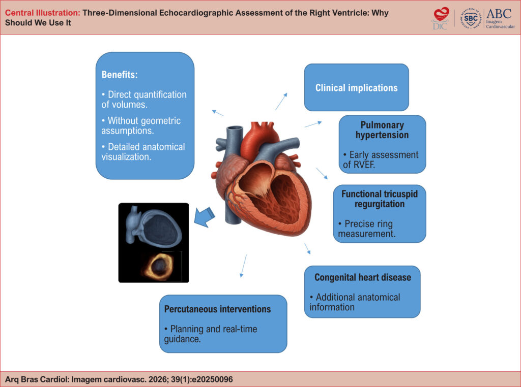
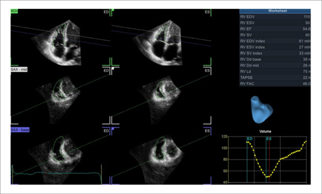
Right ventricular (RV) assessment using two-dimensional (2D) echocardiography has historically faced significant challenges due to the chamber’s complex and unique geometry and its thoracic orientation. In this context, three-dimensional (3D) echocardiography has emerged as a promising tool to overcome and illuminate these limitations, enabling accurate quantification of volumes and ejection fraction without relying on geometric assumptions. As a result, the routine incorporation of 3D echocardiography into RV evaluation may redefine diagnostic and prognostic paradigms, fostering a more precise and personalized approach in modern cardiology. To consolidate and highlight this technique, this review article explores the technical principles of 3D echocardiography for RV assessment, discusses its advantages over conventional 2D imaging, examines its validation against cardiac magnetic resonance (CMR), and reviews key clinical applications, including pulmonary hypertension, functional tricuspid regurgitation, congenital heart disease, and right-sided heart failure. Additionally, the article outlines current limitations of the technique, future perspectives, and practical recommendations based on contemporary literature.
Images
Image gallery fullscreen viewer
Comments are moderated by the editorial team before publication. Discussions should be evidence-based and use scientific language. Conflicts of interest should be declared.