ABC Family Arquivos Brasileiros de Cardiologia International Journal of Cardiovascular Sciences ABC Heart Failure & Cardiomyopathy ABC Imagem Cardiovascular
Português English
ABC Family
Português English
  • About
    • About ABC Family
    • Internal Regulation
    • Editorial Board
    • Brazilian Society of Cardiology (SBC)
  • Issues
  • SBC Guidelines
  • Content
    • Videos
    • Podcasts
    • News
  • About
    • About ABC Family
    • Internal Regulation
    • Editorial Board
    • Brazilian Society of Cardiology (SBC)
  • Issues
  • SBC Guidelines
  • Content
    • Videos
    • Podcasts
    • News

ABC Family

  • ABC Family
  • Arquivos Brasileiros de Cardiologia
  • International Journal of Cardiovascular Sciences
  • ABC Heart Failure & Cardiomyopathy
  • ABC Imagem Cardiovascular
Search in

No content found.

Journals
Ordering
Article types
Others

  1. Home
  2. Keyword: Computed Tomography
ABC
Editorial

The “Value of 0”: Is There an Added Value in Using the Calcium Score in the Stratification of Symptomatic Individuals with a Zero Score?

Catarina Ferreira

Coronary calcium score (CCS) is a good marker of cardiovascular risk, with an independent prognostic value that is incremental over traditional clinical risk factors.– It is performed using non-contrast computed tomography with very low radiation exposure to patients.

Its use is now recommended in selected asymptomatic individuals to refine risk assessment and help initiate or prolong preventive pharmacotherapies.

[…]

  • Cardiac Imaging Techniques
  • Chest Pain
  • Computed Tomography
  • Coronary Calcium
  • Coronary Disease
  • X-Ray
Read Full Article
Creative Commons Attribution (CC BY) Creative Commons license
07/04/2023 DOI: 10.36660/abc.20230105 Views 2
Figure 1
ABCHF
Review Article

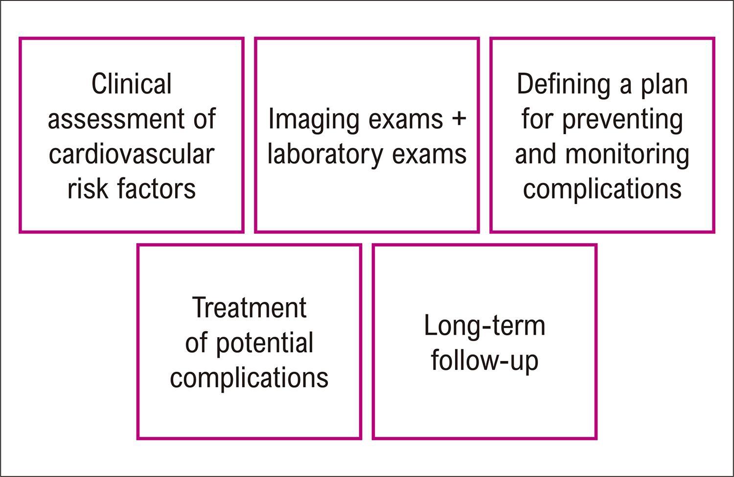
Critical Analysis and Applicability of Imaging Methods in Monitoring and Diagnosing Ventricular Dysfunction in Patients with Cancer

Claudio Tinoco Mesquita et al. Marcelo Dantas Tavares de Melo Ariane Binoti Pacheco Leal André Luiz Cerqueira de Almeida

Abstract

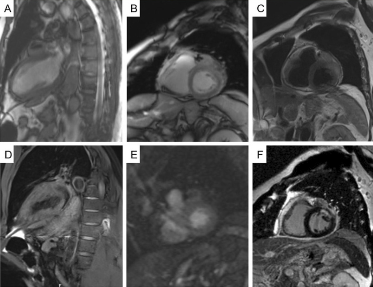
Cardio-oncology is a subspecialty of cardiology that has become necessary as a consequence of the favorable impact of cancer treatment, which increases patients’ survival rates, but makes them more prone to the cardiovascular side effects of cancer therapies in the short and long term. The presence of predisposing factors such as pre-existing cardiovascular disease, cardiovascular risk factors, genetic predisposition, previous antineoplastic therapies, and increased patient age is associated with a higher risk of cardiotoxicity in cancer treatment and may aggravate its complications. The use of imaging methods is fundamental in the management and detection of complications in cancer treatment. Echocardiography is considered the standard method for assessing left ventricular function and should be used in all patients. Magnetic resonance imaging is the best alternative for evaluation in patients with other associated conditions, especially advanced coronary disease, and in cases where it is difficult to obtain adequate echocardiographic images. Nuclear medicine offers options for patients for whom the use of echocardiography and magnetic resonance imaging is limited and for patients whose clinical and laboratory assessments conflict. Judicious use of imaging techniques leads to better patient outcomes during cancer treatment.

  • Cardiotoxicity
  • Computed Tomography
  • Echocardiography
  • Heart Failure
  • Magnetic Resonance
  • Scintigraphy
Read Full Article
Creative Commons Attribution (CC BY) Creative Commons license
18/12/2022 DOI: 10.36660/abchf.20220087 Views 2
Figure 1
ABC
Original Article

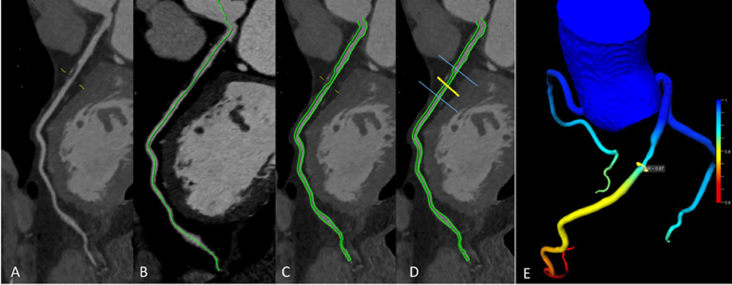
Diagnostic Performance of a Machine Learning-Based CT-Derived FFR in Detecting Flow-Limiting Stenosis

Thamara Carvalho Morais et al. Antonildes Nascimento Assunção-Jr Roberto Nery Dantas Júnior Carla Franco Grego da Silva … Caroline Bastida de Paula Roberto Almeida Torres Tiago Augusto Magalhães César Higa Nomura Luiz Francisco Rodrigues de Ávila José Rodrigues Parga Filho

Abstract

Background

The non-invasive quantification of the fractional flow reserve (FFRCT) using a more recent version of an artificial intelligence-based software and latest generation CT scanner (384 slices) may show high performance to detect coronary ischemia.

Objectives

To evaluate the diagnostic performance of FFRCT for the detection of significant coronary artery disease (CAD) in contrast to invasive FFR (iFFR) using previous generation CT scanners (128 and 256- detector rows).

Methods

Retrospective study with patients referred to coronary artery CT angiography (CTA) and catheterization (iFFR) procedures. Siemens Somatom Definition Flash (256-detector rows) and AS+ (128-detector rows) CT scanners were used to acquire the images. The FFRCT and the minimal lumen area (MLA) were evaluated using a dedicated software (cFFR version 3.0.0, Siemens Healthineers, Forchheim, Germany). Obstructive CAD was defined as CTA lumen reduction ≥ 50%, and flow-limiting stenosis as iFFR ≤0.8. All reported P values are two-tailed, and when <0.05, they were considered statistically significant.

Results

Ninety-three consecutive patients (152 vessels) were included. There was good agreement between FFRCT and iFFR, with minimal FFRCT overestimation (bias: -0.02; limits of agreement:0.14-0.09). Different CT scanners did not modify the association between FFRCT and FFRi (p for interaction=0.73). The performance of FFRCT was significantly superior compared to the visual classification of coronary stenosis (AUC 0.93vs.0.61, p<0.001) and to MLA (AUC 0.93vs.0.75, p<0.001), reducing the number of false-positive cases. The optimal cut-off point for FFRCT using a Youden index was 0.85 (87% Sensitivity, 86% Specificity, 73% PPV, 94% NPV), with a reduction of false-positives.

Conclusion

Machine learning-based FFRCT using previous generation CT scanners (128 and 256-detector rows) shows good diagnostic performance for the detection of CAD, and can be used to reduce the number of invasive procedures.

  • Computed Tomography
  • Coronary Artery Disease
  • Machine Learning
  • Myocardial Fractional Flow Reserve
  • Myocardial Ischemic
Read Full Article
Creative Commons Attribution (CC BY) Creative Commons license
14/06/2021 DOI: 10.36660/abc.20190329 Views 2
Figure 1
ABCIC
Relato de Caso

Left Atrial Appendage Pseudo-Thrombus

Marcio Augusto Silva et al. Marcelo Pazolini José Guilherme Cazelli Daniel Escobar Bueno Peixoto … Rafael Soares Rua Rodolpho Jacques de Melo Farinazzo

Discussion

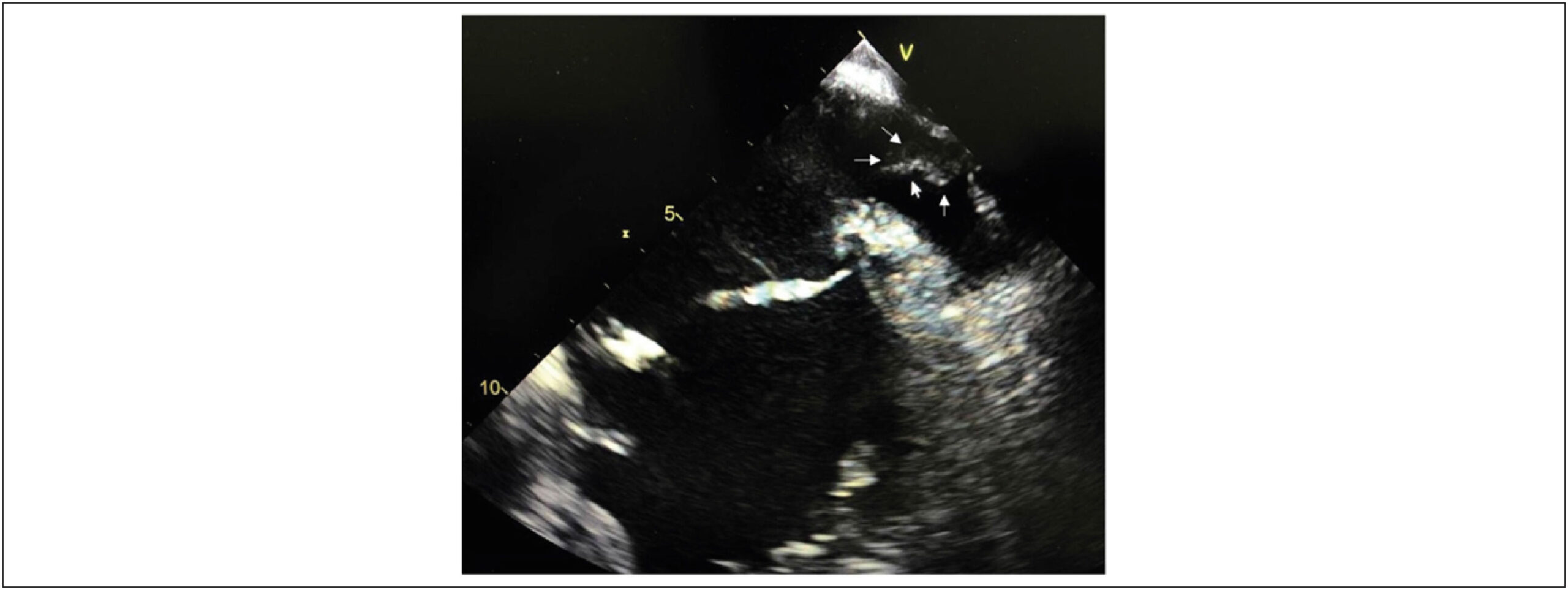
TEE is considered the gold standard test to detect LAA thrombi., With the incorporation of heart CT-angio into clinical practice, the diagnosis of a “false” thrombus, called pseudo-thrombus, in the LAA has become more frequent and is explained by contrast filling failure due to reduced flow velocity in the LAA trabeculae. The recent use of specific CT-angio protocols to evaluate thrombi and late imaging capture has significantly improved its sensitivity and specificity, which reaches near 100% in some series,, thus becoming a less invasive option than TEE. It has been widely used in preoperative catheter ablation to treat AF or LAA percutaneous occlusions.

In this case, it is noteworthy that the diagnosis of pseudo-thrombus by TEE corresponded to an extracardiac metallic structure (stent) artefact closely related to the LAA but only identified on the cardiac CT-angio and relevant in the patient’s treatment.

[…]

  • Atrial Appendage
  • Computed Tomography
  • Echocardiography
Read Full Article
Creative Commons Attribution (CC BY) Creative Commons license
11/06/2021 DOI: 10.47593/2675-312X/20213402eabc168 Views 2
Figure 1
ABCIC
Image

Spontaneous Pneumomediastinum

Luísa Gonçalves et al. Inês Pires João Santos Joana Correia Davide Moreira

Case

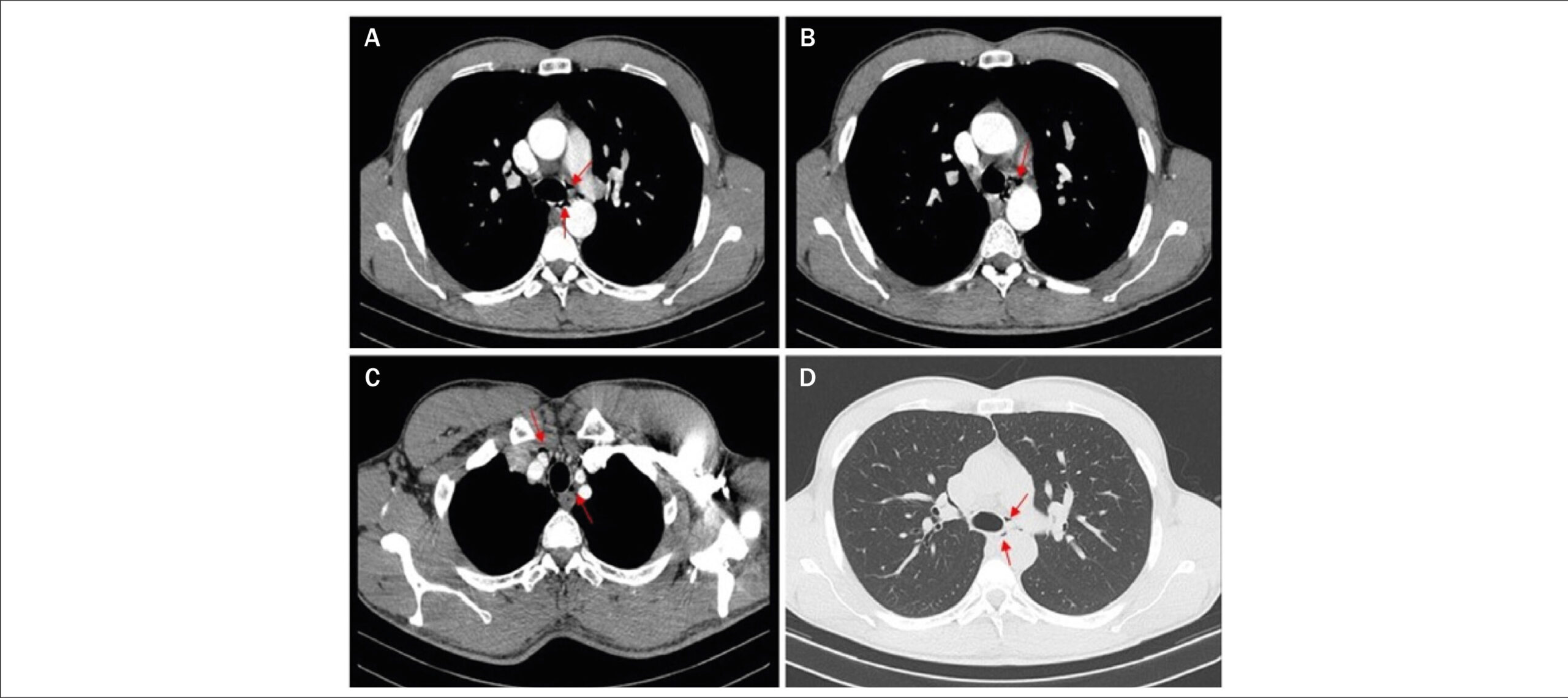
A 31-year-old male athlete presented with acute chest pain that was aggravated with swallowing after completing a marathon. Physical examination findings and his vital signs were normal. Electrocardiography revealed showed sinus rhythm, an incomplete right bundle block, and a negative T wave in V1-V2. A blood analysis showed normal D-dimer values and a slightly elevated troponin-I level (maximum at 72 hours: 0.52 ng/mL; normal, <0.07 ng/mL) with fluctuations. Chest X-ray, echocardiography, and coronary angiography findings were normal. Cardiac computed tomography (CT) revealed small gas collections on the superior and middle mediastinum compatible with spontaneous pneumomediastinum (SP). ( and ) SP is rare, usually benign, and often underdiagnosed. Intense physical exercise is a recognized cause. Cardiac CT assesses the extension, causative factors, and pathologies and discloses the diagnosis of SP when chest X-ray findings are normal.

[…]

  • Chest Pain
  • Computed Tomography
  • Pneumomediastinum
Read Full Article
Creative Commons Attribution (CC BY) Creative Commons license
11/06/2021 DOI: 10.47593/2675-312X/20213402eabc170 Views 2
ABC
Original Article

Safety, Efficacy, and Dose Protocol of Metoprolol for Heart Rate Reduction in Pediatric Outpatients Undergoing Cardiac CT Angiography

Mariana de Oliveira Nunes et al. Dawn R. Witt Susan A. Casey Larissa I. Stanberry … David J. Caye Bradford J.Chu B. Jana Lindberg John R. Lesser B. Kelly Han

Abstract

Background

Image quality and radiation dose are optimized with a slow, steady heart rate (HR) when imaging the coronary arteries during cardiac computed tomography angiography (CCTA). The safety, efficacy, and protocol for HR reduction with beta blocker medication is not well described in a pediatric patient population.

Objective

Provide a safe and efficient metoprolol dose protocol to be used in pediatric outpatients undergoing CCTA.

Methods

We conducted a retrospective review of all pediatric outpatients who received metoprolol during CCTA. Demographic and clinical characteristics were summarized and the average reduction in HR was estimated using a multivariate linear regression model. Images were evaluated on a 1-4 scale (1= optimal).

Results

Seventy-eight pediatric outpatients underwent a CCTA scan with the use of metoprolol. The median age was 13 years, median weight of 46 kg, and 36 (46%) were male. The median doses of metoprolol were 1.5 (IQR 1.1, 1.8) mg/kg and 0.4 (IQR 0.2, 0.7) mg/kg for oral and intravenous administrations, respectively. Procedural dose-length product was 57 (IQR 30, 119) mGy*cm. The average reduction in HR was 19 (IQR 12, 26) beats per minute, or 23%. No complications or adverse events were reported.

Conclusion

Use of metoprolol in a pediatric outpatient setting for HR reduction prior to CCTA is safe and effective. A metoprolol dose protocol can be reproduced when a slower HR is needed, ensuring faster acquisition times, clear images, and associated reduction in radiation exposure in this population. (Arq Bras Cardiol. 2021; 116(1):100-105)

  • Adrenergic, Antagonists
  • Computed Tomography
  • Coronary Vessels
  • Heart Defects, Congenital
  • Heart Rate
  • metoprolol
Read Full Article
Creative Commons Attribution (CC BY) Creative Commons license
03/02/2021 DOI: 10.36660/abc.20190892 Views 1
ABC
Letter to the Editor

Cardiovascular Imaging in Patients with COVID-19

Gabriel Blacher Grossman et al. Ronaldo de Souza Leão Lima

Dear Editor,

In the latest issue of the Arquivos Brasileiros de Cardiologia, Costa et al. reviewed the role of cardiovascular imaging and interventional procedures in patients with novel coronavirus infection.

[…]

  • Computed Tomography
  • Coronavirus, Betacoronavirus
  • Image Cardiovascular
  • Nuclear Medicine
  • Pneumonia
Read Full Article
Creative Commons Attribution (CC BY) Creative Commons license
07/12/2020 DOI: 10.36660/abc.20200881 Views 1
IJCS
Editorial

Lung Ultrasound in COVID 19 Outbreak: Can we Reduce the Burden from the Overloaded CT Departments?

Filipe Penna de Carvalho et al. Alex dos Santos Felix

Since the first descriptions on the use of lung ultrasound (LUS) for bedside evaluation of critically ill patients, important publications have underscored the utility of LUS for the detection of a wide array of pulmonary and pleural diseases, as well as for the differential diagnosis of acute respiratory failure, proving to be an important tool for a point-of-care based examination and an extended resource for examining critical patients, cited by many as the “new stethoscope”.,

The coronavirus disease-2019 (COVID-19) outbreak has emerged as a global health burden since late 2019, challenging healthcare systems around the world due to the constant need for intensive care support and isolation of patients, many of whom evolve with severe hemodynamic involvement, leading to high morbidity and mortality. This critical scenario highlights the importance of appropriateness and best appliance of all diagnostic and therapeutic resources. In this regard, the use of point-of-care ultrasound (POCUS) is proving to be a cost-effective tool for the management of these potentially critical patients, from the initial presentation (triage) to in-hospital management and post-discharge follow-up.

[…]

  • Computed Tomography
  • COVID-19/complications, Pandemics, Acute Failure Respiratory
  • Critic care
  • Morbidity
  • Mortality
  • Pleural Diseases/Diagnosis
  • Ultrasonography/methods
Read Full Article
Creative Commons Attribution (CC BY) Creative Commons license
25/11/2020 DOI: 10.36660/ijcs.20200283
Figure 1
ABC
Case Report

A Case of Acute Myocardial Infarction and Pericarditis Unmasking Metastatic Involvement of the Heart

Sofia Torres et al. Mariana Vasconcelos Carla Sousa Antonio J. Madureira … Alzira Nunes Maria Júlia Maciel

Introduction

Metastases to the heart and pericardium are much more common than primary cardiac tumors and are generally associated with a poor prognosis. , While they are most commonly asymptomatic, cardiac metastases can mimic primary cardiac diseases such as acute coronary syndromes, congestive heart failure and pericarditis. , Lung cancer is the most frequent source of metastatic cardiac disease, either from direct extension or by a combination of lymphatic, hematogenous, and transvenous dissemination. ,

[…]

  • Cardiac Magnetic Ressonance
  • Cardiac metastases
  • Computed Tomography
  • Lung Neoplasms
  • Multimodality imaging
  • Myocardial Infarction
  • Pericarditis
Read Full Article
Creative Commons Attribution (CC BY) Creative Commons license
14/09/2020 DOI: 10.36660/abc.20190534
Figure 1
ABC
Case Report

Myocardial Bridge and Angiotomography of the Coronary Arteries: Perfusion under Pharmacological Stress

Wilter dos Santos Ker et al. Daniel Gama Neves Alair Sarmet A. A. Damas Cláudio Tinoco Mesquita Marcelo Souto Nacif

Introduction

The myocardial bridge is one of the most prevalent congenital anomalies that involve the coronary circulation, and its incidence in the general population is high, affecting from 23 to 55% in necropsy studies.The impairment of the anterior descending artery is more frequent, on its proximal 2/3.In most patients, the myocardial bridges do not cause symptoms, because in order to have ischemia, there must be an imbalance between supply and consumption of oxygen. The superficial bridges, with small or slender muscle band, are the most common ones, and they may account for 75% of the cases, with average length of 1.5 cm and usually without causing symptoms. In approximately 24% of the cases, we observed deep myocardial bridges, with thicker muscle band.,

Atherosclerosis is the most common cause of ischemic heart disease. However, other causes for ischemia are frequent, and among them, we highlight the myocardial bridge, which may provoke typical or atypicalchest angina,acute myocardial infarction and sudden death.–

[…]

  • Computed Tomography
  • Coronary Artery Disease
  • Myocardial Bridge
  • Myocardial Ischemia
  • Perfusion
  • Radionuclide Imaging
Read Full Article
Creative Commons Attribution (CC BY) Creative Commons license
01/06/2017 DOI: 10.5935/abc.20170021
Figure 1
ABC
Artigo Original

Subcutaneous Tissue Thickness is an Independent Predictor of Image Noise in Cardiac CT

Henrique Lane Staniak et al. Rodolfo Sharovsky Alexandre Costa Pereira Cláudio Campi de Castro … Isabela M. Benseñor Paulo A. Lotufo Márcio Sommer Bittencourt

Background:

Few data on the definition of simple robust parameters to predict image noise in cardiac computed tomography (CT) exist.

Objectives:

To evaluate the value of a simple measure of subcutaneous tissue as a predictor of image noise in cardiac CT.

Methods:

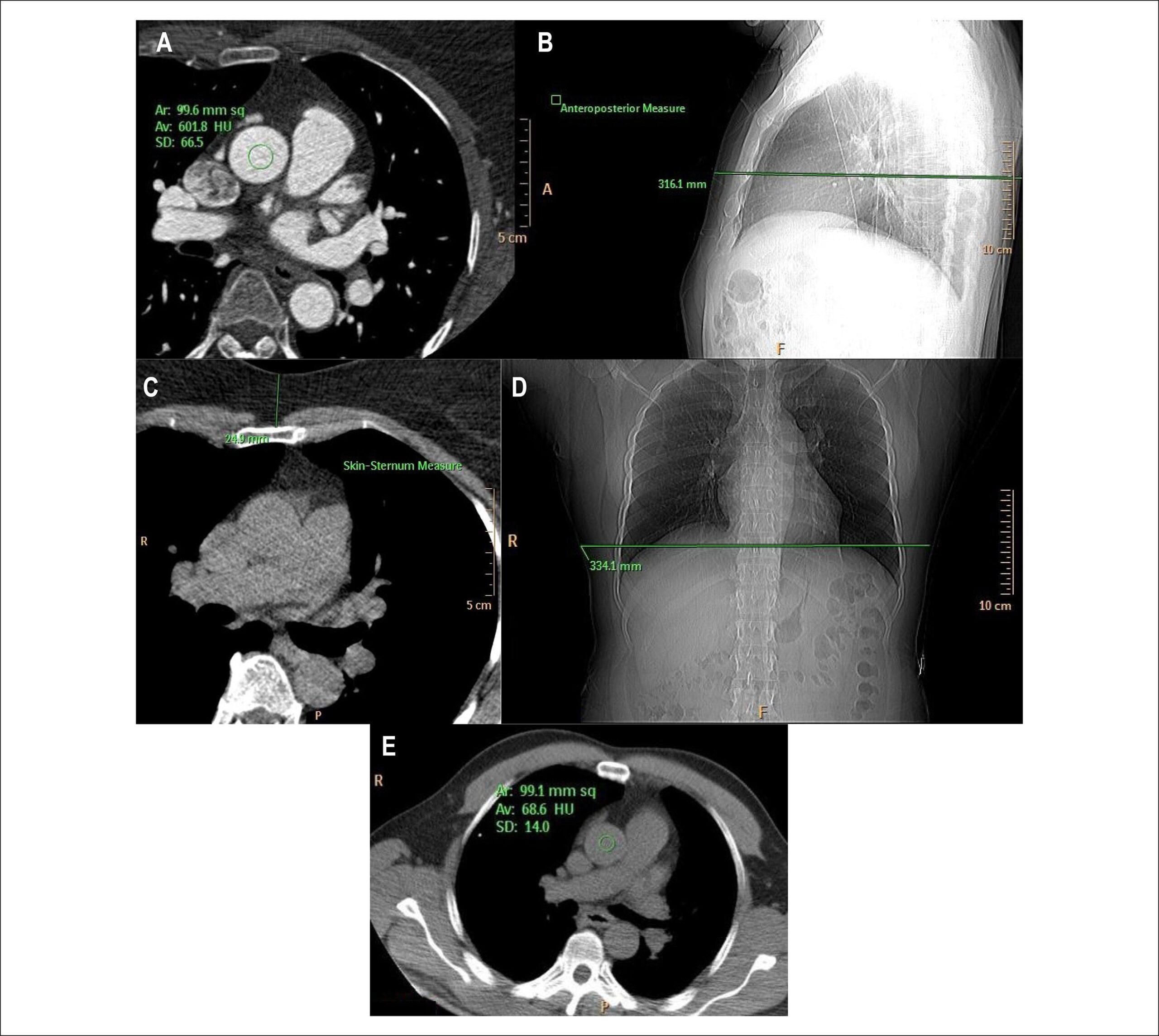
86 patients underwent prospective ECG-gated coronary computed tomographic angiography (CTA) and coronary calcium scoring (CAC) with 120 kV and 150 mA. The image quality was objectively measured by the image noise in the aorta in the cardiac CTA, and low noise was defined as noise < 30HU. The chest anteroposterior diameter and lateral width, the image noise in the aorta and the skin-sternum (SS) thickness were measured as predictors of cardiac CTA noise. The association of the predictors and image noise was performed by using Pearson correlation.

Results:

The mean radiation dose was 3.5 ± 1.5 mSv. The mean image noise in CT was 36.3 ± 8.5 HU, and the mean image noise in non-contrast scan was 17.7 ± 4.4 HU. All predictors were independently associated with cardiac CTA noise. The best predictors were SS thickness, with a correlation of 0.70 (p < 0.001), and noise in the non-contrast images, with a correlation of 0.73 (p < 0.001). When evaluating the ability to predict low image noise, the areas under the ROC curve for the non-contrast noise and for the SS thickness were 0.837 and 0.864, respectively.

Conclusion:

Both SS thickness and CAC noise are simple accurate predictors of cardiac CTA image noise. Those parameters can be incorporated in standard CT protocols to adequately adjust radiation exposure.

  • Artifacts
  • Computed Tomography
  • Radiation injuries
  • Sternum / radiation effects
Read Full Article
Creative Commons Attribution (CC BY) Creative Commons license
01/11/2013 DOI: 10.5935/abc.20130215 Views 1
Epicardial fat: definition, measurements and systematic review of main outcomes
ABC
Atualização

Epicardial fat: definition, measurements and systematic review of main outcomes

Angela Gallina Bertaso et al. Daniela Bertol Bruce Bartholow Duncan Murilo Foppa

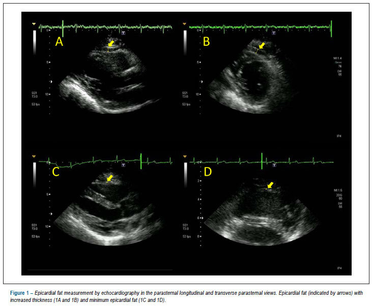
Epicardial fat (EF) is a visceral fat deposit, located between the heart and the pericardium, which shares many of the pathophysiological properties of other visceral fat deposits, It also potentially causes local inflammation and likely has direct effects on coronary atherosclerosis. Echocardiography, computed tomography and magnetic resonance imaging have been used to evaluate EF, but variations between methodologies limit the comparability between these modalities. We performed a systematic review of the literature finding associations of EF with metabolic syndrome and coronary artery disease. The summarization of these associations is limited by the heterogeneity of the methods used and the populations studied, where most of the subjects were at high cardiovascular disease risk. EF is also associated with other known factors, such as obesity, diabetes mellitus, age and hypertension, which makes the interpretation of its role as an independent risk marker intricate. Based on these data, we conclude that EF is a visceral fat deposit with potential implications in coronary artery disease. We describe the reference values of EF for the different imaging modalities, even though these have not yet been validated for clinical use. It is still necessary to better define normal reference values and the risk associated with EF to further evaluate its role in cardiovascular and metabolic risk assessment in relation to other criteria currently used.

  • Abdominal Fat
  • Computed Tomography
  • Echocardiography
  • Magnetic Resonance Imaging
  • Metabolic Syndrome
Read Full Article
Creative Commons Attribution (CC BY) Creative Commons license
31/07/2013 DOI: 10.5935/abc.20130138 Views 1
Correlation between myocardial scintigraphy and ct angiography in the evaluation of coronary disease
ABC
Artigo Original

Correlation between myocardial scintigraphy and ct angiography in the evaluation of coronary disease

Jader Cunha de Azevedo et al. Diógenes de Souza Ferreira Junior Felipe Carvalhinho Vieira Laís Santos Prezotti … Luciana Silveira Simões Marcelo Souto Nacif Carlos Eduardo Rochitte Amarino de Carvalho Oliveira Junior Evandro Tinoco Mesquita Cláudio Tinoco Mesquita

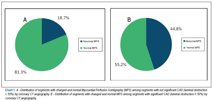
BACKGROUND: Coronary multidetector CT angiography (CTA) has shown good accuracy for detection of coronary stenosis. Although this is a promising technique for the evaluation of CAD, its correlation with the functional expression of the disease is not yet well established. OBJECTIVE: To evaluate whether the presence of CAD and the degree of coronary stenosis assessed by CT angiography are associated with changes in the Myocardial Perfusion Scintigraphy (MPS). METHODS: This is a retrospective observational study, which included 99 consecutive patients with known or suspected CAD. MPS and CTA were conducted. We compared the presence of perfusion defects by MPS with the presence of CAD and the degree of luminal obstruction by CTA. For statistical analysis, Student’s t-test, ANOVA, and Chi-square (or Fisher’s exact test for n <5) tests were used. Multivariate analysis was performed using logistic regression: the level of significance was 5%. RESULTS: Mean age was 62 ± 11.4 years, with 46 (71.7%) men. The variable analysis was performed per patient (n = 99) and per coronary irrigation territory (n = 297). Of the 67 territories that had significant CAD by CTA, 44.8% had abnormal MPS. Considering the degree of stenosis, abnormal MPS was present in 18.7% of the territories with no significant stenosis, 45.28% of the territories with moderate stenosis, and 42.8% of the territories with severe injuries. CONCLUSIONS: CTA is a good method for exclusion of CAD. However, its use to evaluate the severity of stenosis and its functional impact has not shown good correlation.

  • Computed Tomography
  • Coronary Disease
  • Heart
Read Full Article
Creative Commons Attribution (CC BY) Creative Commons license
15/04/2013 DOI: 10.5935/abc.20130042 Views 1
Calcium score to evaluate chest pain in the emergency room
ABC
Brief Communication

Calcium score to evaluate chest pain in the emergency room

Henrique Lane Staniak et al. Márcio Sommer Bittencourt Rodolfo Sharovsky Isabela Benseñor … Rodrigo Diaz Olmos Paulo A. Lotufo

Some authors have suggested that a zero calcium score (CAC) can be used to rule out the diagnosis of acute coronary syndrome. Objective this study is to evaluate the diagnostic accuracy of a zero CAC when compared to the coronary computed tomography angiography (CCTA) at the emergency department. 135 symptomatic patients with no previous coronary heart disease (CHD) who presented to the emergency department were submitted to CAC and CCTA to rule out CHD. All patients had normal electrocardiogram and cardiac biomarkers and were TIMI risk score 0 to 2. The CCTA was considered positive if any obstructive lesion (> 50%) was identified. The mean age was 51.7 ± 13.6 years with 50.6% of men. Seventy-three (54.1%) patients had a calcium score of zero. Of them, 3 (4.1%) had an obstruction > 50 % and underwent invasive coronary angiography. Calcium score showed a sensitivity of 92.9%, specificity of 75.3%, positive and negative predictive values of, respectively, 62.9% and 95.9%. Positive and negative likelihood ratios were respectively of 3.7 and 0.09 to detect lesions greater than 50% in the CCTA. A negative likelihood ratio of 0.09 is very good to rule out most cases of significant coronary obstruction in epidemiologic studies. However, it is important to understand that in a clinical scenario, all evidence including history, clinical examination, data from eletrocardiogram and myocardial biomarkers have to be interpreted together. In our study, three cases with a zero CAC score had coronary obstruction higher than 50% at the CCTA.

  • Calcium
  • Chest Pain
  • Computed Tomography
  • Coronary Disease
Read Full Article
Creative Commons Attribution (CC BY) Creative Commons license
29/01/2013 DOI: 10.1590/S0066-782X2013000100014 Views 1
Coronary tomography for predicting adverse events in patients with suspected coronary disease
ABC
Artigo Original

Coronary tomography for predicting adverse events in patients with suspected coronary disease

Márcio Vinicius Lins Barros et al. Daniel Rocha Rabelo Maria do Carmo Pereira Nunes Maria Helena Albernaz Siqueira

BACKGROUND: Although studies have demonstrated a high diagnostic accuracy of multi-detector Coronary CT (MDCT) in the detection of Coronary Artery Disease (CAD), data on the prognostic value of this method are limited. OBJECTIVE: Determine the value of MDCT in predicting adverse clinical outcomes in patients with suspected CAD. METHODS: We prospectively evaluated 355 consecutive patients (mean age 58 ± 12 years, 252 males) with suspected CAD between January 2008 and June 2010. CAD at MDCT was defined as the presence of atheromatous plaque observed in any coronary artery. The adverse clinical outcomes were defined as death, myocardial infarction, unstable angina or myocardial revascularization. RESULTS: During a mean follow-up of 15 months, there were 55 cardiac events. In the multivariate analysis using the Cox regression model, NYHA functional class, diabetes, smoking and atherosclerosis at the MDCT were predictors of adverse clinical outcome, and the presence of plaque at the MDCT was strongly associated with adverse clinical outcomes, regardless of established risk factors for CAD (hazard ratio 5.29; 95% confidence interval: 2.4 -11.8, p <0.001). CONCLUSION: The presence of atherosclerosis demonstrated by MDCT in patients with suspected CAD showed independent and incremental value when compared to conventional risk factors in the prediction of adverse clinical outcomes, and may prove useful in risk stratification of these patients.

  • Computed Tomography
  • Coronary Disease
  • Plaque, Atherosclerotic
  • Prognosis
Read Full Article
Creative Commons Attribution (CC BY) Creative Commons license
30/11/2012 DOI: 10.1590/S0066-782X2012005000113 Views 1
Clinical outcomes after investigation for pulmonary embolism using CT angiography and venography
ABC
Artigo Original

Clinical outcomes after investigation for pulmonary embolism using CT angiography and venography

Eduardo S. Darze et al. João F.M. Braghiroli Ricardo V. Almeida Ênio P. Araújo … Sergio M. Toscano César Augusto Araújo-Neto

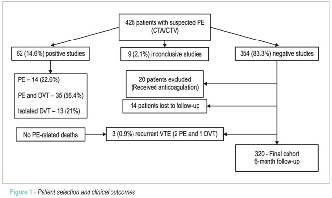
BACKGROUND: The diagnosis of pulmonary embolism (PE) still requires long work-up periods and multiple tests. OBJECTIVE: We aim to assess clinical outcomes after a negative investigation using a combined protocol of CT pulmonary angiography and CT venography (CTA/CTV) as a sole diagnostic test in unselected patients with suspected PE. METHODS: This retrospective cohort study enrolled consecutive patients with suspected PE who were investigated with a combined CTA/CTV protocol. Patients who had an initially negative investigation and were not anticoagulated were followed for 6 months for the occurrence of recurrent venous thromboembolic events. RESULTS: Out of 425 patients with suspected PE, 62 (14.6%) had venous thromboembolism diagnosed on the initial CTA/CTV. The mean age was 56 ± 19 years and 61% of the population fell into the low clinical probability category. Isolated deep vein thrombosis represented 21% of all venous thromboembolic events, and when considering the whole population, CTV was associated with an increment in diagnostic yield of 3.1%. Our cohort was composed of 320 patients with initially negative CTA/CTVs and who were not anticoagulated. After 6 months of follow up, only three patients presented with recurrent thromboembolic events (0.9%; 95% CI -0.1% – 2.0%) and none was fatal. There were no PE-related deaths. CONCLUSION: Our study suggests that a diagnostic strategy that utilizes CTA/CTV as a sole diagnostic test can safely rule out PE in a low to moderate risk population and is associated with favorable outcomes with a negative predictive value of 99.1%. (Arq Bras Cardiol. 2012; [online].ahead print, PP.0-0)

  • Computed Tomography
  • Phlebography
  • Pulmonary embolism
  • thorax
Read Full Article
Creative Commons Attribution (CC BY) Creative Commons license
26/06/2012 DOI: 10.1590/S0066-782X2012005000058
Dynamic myocardial perfusion imaging by dual-source computed tomography
ABC
Case Report

Dynamic myocardial perfusion imaging by dual-source computed tomography

José Rodrigues Parga Filho et al. Cintia Souza Lima Moraes Lima Felipe Gallego Lima Tiago da Silveira Jaques … Luiz Francisco Rodrigues de Ávila Roberto Kalil Filho

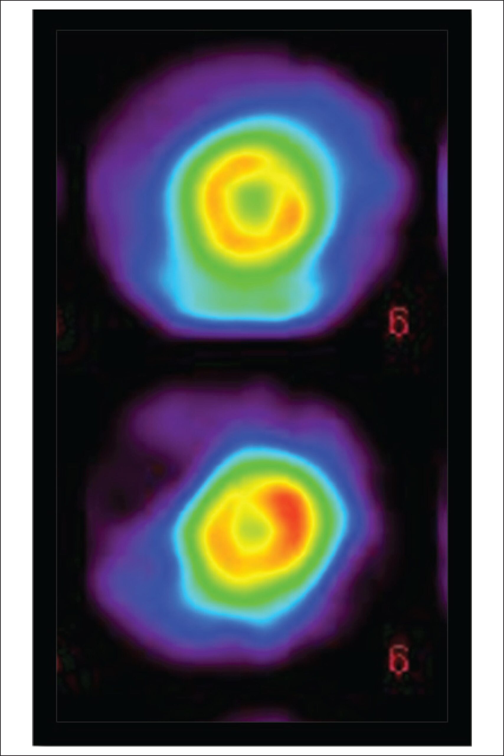
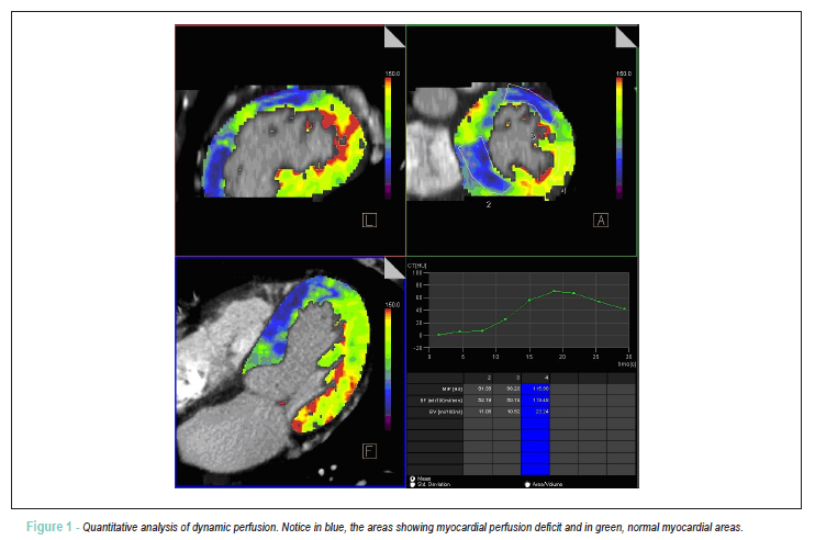
We report a dual-source computed tomography study of dynamic and quantitative myocardial perfusion in a 44-year-old patient with previous documented coronary artery disease. Quantitatively, the tomography showed myocardial perfusion deficit in the territories with significant coronary stenosis, confirmed by computed tomography angiography and conventional angiography. Dual-source computed tomography allowed dynamic perfusion and anatomic evaluation in a single study during the follow-up of this patient.

  • Computed Tomography
  • Coronary Disease
  • Evaluation
  • Myocardial Reperfusion
Read Full Article
Creative Commons Attribution (CC BY) Creative Commons license
23/04/2012 DOI: 10.1590/S0066-782X2012000300014
Imaging assessment of visceral adipose tissue area and its correlations with metabolic alterations
ABC
Artigo Original

Imaging assessment of visceral adipose tissue area and its correlations with metabolic alterations

Anna Karla Carneiro Roriz et al. Adriana Lima Mello Juliana Fontes Guimarães Fabiana Cajuhy dos Santos … Jairza Maria Barreto Medeiros Lílian Ramos Sampaio

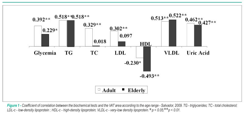
BACKGROUND: Androgenic obesity is associated with a higher risk of metabolic disorders, thus favoring the occurrence of cardiovascular diseases and other morbidities. OBJECTIVE: To verify the influence of the visceral adipose tissue (VAT) area, measured by computed tomography (CT), on the metabolic alterations in adult and elderly individuals. METHODS: CT results and lipoprotein levels, total cholesterol and fractions, triglycerides, glycemia and uric acid levels, were obtained from 194 individuals stratified by sex, age group and body mass and analyzed using the tests of correlation and means. RESULTS: The elderly individuals presented higher VAT area, glycemia, uric acid and total cholesterol levels. The most important correlations were observed between VAT area, triglycerides (TG) and VLDL-c (r > 0.5; p < 0.01), in both age groups. The mean VAT area was always higher when TG and glycemia levels were altered, in both age groups. CONCLUSION: Most tests showed a strong correlation with VAT area, which was considered as risk for metabolic alterations. In elderly individuals, the risk VAT area seems to be higher than that of adult individuals.

  • Abdominal Fat
  • Blood Glucose
  • Computed Tomography
  • Lipoproteins
  • uric acid
Read Full Article
Creative Commons Attribution (CC BY) Creative Commons license
10/12/2010 DOI: 10.1590/S0066-782X2010005000159 Views 1
Late outcome of sirolimus-eluting stents: comparison of multidetector computed tomography with quantitative coronary angiography and intravascular ultrasound
ABC
Artigo Original

Late outcome of sirolimus-eluting stents: comparison of multidetector computed tomography with quantitative coronary angiography and intravascular ultrasound

Ibraim Masciarelli F. Pinto et al. Amanda G. M. R. Sousa Walther Ishikama Luiz Alberto Mattos … Alexandre Abizaid Fausto Feres Luiz Fernando L. Tanajura Luiz C. B. Sousa J. Eduardo M. R. Sousa Adib Jatene

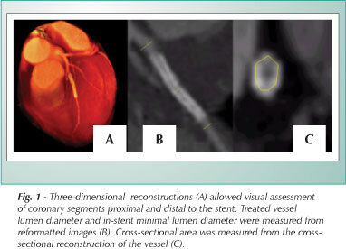
OBJECTIVE: To assess the performance of multidetector computed tomography in determining late clinical outcomes of patients undergoing sirolimus-eluting stent implantation. METHODS: Thirty patients, successfully submitted to sirolimus-eluting stent implantation for more than six months, were selected to participate in the study. All underwent invasive angiography and intravascular ultrasound following CT angiography using iodinated contrast medium at a dose of 1.5 ml/kg. RESULTS: Mean proximal reference diameter was 3.01 ± 0.31 mm by tomography and 3.14 ± 0.31 mm by angiography (p = 0.04). When the left circumflex artery was excluded from the analysis, the difference between both examinations was no longer significant (tomography = 3.01 ± 0.32 mm; angiography = 3.10 ± 0.30 mm, p = 0.65). Mean distal reference diameter was 2.86 ± 0.30 mm by tomography and 2.92 ± 0.32 by angiography (p = 0.25). Mean in-stent minimal lumen diameter was 2.85 ± 0.25 mm by tomography and 2.85 ± 0.29 mm by angiography (p = 0.27). Mean minimal in-stent cross-sectional area was 7.19 ± 1.47 mm² by tomography and 6.90 ± 1.52 mm² by intravascular ultrasound (p = 0.36), but there was only a weak correlation between these measurements (r = 0.33). CONCLUSION: Computed tomography allows the qualitative assessment of sirolimus-eluting stents, accurate estimate of proximal and distal reference diameters of the target vessel, and in-stent minimal lumen diameter. Its correlation with measurements performed using intravascular ultrasound, however, is less strong.

  • Computed Tomography
  • Coronary failure
  • intravascular ultrasound
  • quantitative angiography
Read Full Article
Creative Commons Attribution (CC BY) Creative Commons license
06/03/2007 DOI: 10.1590/S0066-782X2006001800005
Detection of coronary artery disease based on the calcification index obtained by helical computed tomography
ABC
Original Article

Detection of coronary artery disease based on the calcification index obtained by helical computed tomography

Carlos Jader Feldman et al. Domingos Vitola Nádia Schiavo

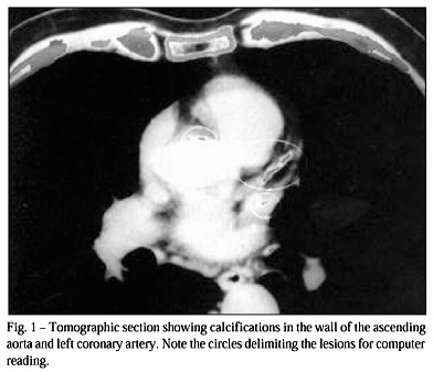
OBJECTIVE: To assess the relation between coronary artery disease and the calcification index on helical computed tomography. METHOD: We studied 22 patients (ages ranging from 40 to 70 years) who underwent coronary angiography because of chest pain suggestive of angina pectoris. Findings on coronary angiography were classified as follows: significant obstructive disease (stenosis > or = 50%), nonobstructive disease (stenosis <50%), and no disease. With no previous knowledge of the results of the coronary angiography and within 7 days, helical computed tomography of the chest was performed. Then, data of the coronary angiography were correlated with the calcification index obtained by helical computed tomography. RESULTS: The sensitivity of helical computed tomography to the presence of significant obstructive lesions on coronary angiography was 87.5%, specificity was 100%, and negative and positive predictive values were 75% and 100%, respectively. The mean calcification index was greater in patients with severe coronary lesions, mainly when involvement of 2 or 3 vessels occurred, than that in patients with no coronary artery disease or with nonobstructive coronary artery lesions (p<0.05). CONCLUSION: Helical computed tomography is an effective method for detecting and quantifying coronary artery calcification, and it has proved to be sensitive to and specific for the noninvasive diagnosis of coronary artery stenosis.

  • calcification
  • Computed Tomography
  • coronary arteries
  • Coronary Artery Disease
Read Full Article
Creative Commons Attribution (CC BY) Creative Commons license
08/01/2002 DOI: 10.1590/S0066-782X2000001200002 Views 1
ABC
Artigo Original

Late evaluation of bovine pericardium grafts as aortic substitutes by spiral computed tomography

Renato A. K. Kalil et al. Carlos Jader Feldman Fernando W. B. Ludwig Alessandro D'Avila da Silva … Paulo Roberto Prates João R. M. Sant'Anna Ivo A. Nesralla

PURPOSE: To evaluate morphologically by spiral computed tomography (SCT) bovine pericardial grafts as aortic substitutes. METHODS: Ten patients were submitted to aortic graft evaluation with SCT. There were 8 ascending and 2 descending grafts. Examination was done after 2 to 7 years post operative. Ages ranged from 49 to 67 years, with 6 male and 4 female patients. All had implanted smooth surface naked pericardial grafts. RESULTS: Surgical result was good in all. Distal dissection persisted in some cases. One had a peri-graft hematoma, 5 had no structural changes and 4 presented mild graft dilatation. There were no graft calcification or pseudoaneurysm. CONCLUSION: Glutaraldehyde preserved naked smooth surface bovine pericardial aortic grafts present satisfactory structural aspect, as seen by SCT, at medium term follow-up. Mild dilatation may be seen in some cases.

  • aortic grafts
  • bovine pericardium
  • Computed Tomography
Read Full Article
Creative Commons Attribution (CC BY) Creative Commons license
14/02/2001 DOI: 10.1590/S0066-782X1997000800006 Views 2

How to cite this article

EndNote RIS

Electronic Document Format (Vancouver)

Ferreira C. The “Value of 0”: Is There an Added Value in Using the Calcium Score in the Stratification of Symptomatic Individuals with a Zero Score?. Arquivos Brasileiros de Cardiologia 2023;120(3):e20230105.

Electronic Document Format (ABNT)

Ferreira, Catarina. The “Value of 0”: Is There an Added Value in Using the Calcium Score in the Stratification of Symptomatic Individuals with a Zero Score?. Arquivos Brasileiros de Cardiologia, v. 120, n. 3, e20230105, Apr. 2023.

Electronic Document Format (APA)

Ferreira, C. (2023). The “Value of 0”: Is There an Added Value in Using the Calcium Score in the Stratification of Symptomatic Individuals with a Zero Score?. Arquivos Brasileiros de Cardiologia, 120(3), e20230105.

Electronic Document Format (ISO)

Ferreira, Catarina. The “Value of 0”: Is There an Added Value in Using the Calcium Score in the Stratification of Symptomatic Individuals with a Zero Score?. Arquivos Brasileiros de Cardiologia [online]. 2023, vol. 120, n. 3, [cited 2026-10-14], e20230105. Available from: <https://journals.cardiol.br/en/article/the-value-of-0-is-there-an-added-value-in-using-the-calcium-score-in-the-stratification-of-symptomatic-individuals-with-a-zero-score/>. ISSN 0066-782X

EndNote RIS

Electronic Document Format (Vancouver)

Mesquita CT, Melo MDT, Leal ABP, Almeida ALC. Critical Analysis and Applicability of Imaging Methods in Monitoring and Diagnosing Ventricular Dysfunction in Patients with Cancer. ABC: Heart Failure & Cardiomyopathy 2022;2(4):386-94.

Electronic Document Format (ABNT)

Mesquita, Claudio Tinoco; Melo, Marcelo Dantas Tavares de; Leal, Ariane Binoti Pacheco; Almeida, André Luiz Cerqueira de. Critical Analysis and Applicability of Imaging Methods in Monitoring and Diagnosing Ventricular Dysfunction in Patients with Cancer. ABC: Heart Failure & Cardiomyopathy, v. 2, n. 4, p. 386-394, Dec. 2022.

Electronic Document Format (APA)

Mesquita, C. T., Melo, M. D. T. , Leal, A. B. P., & Almeida, A. L. C. (2022). Critical Analysis and Applicability of Imaging Methods in Monitoring and Diagnosing Ventricular Dysfunction in Patients with Cancer. ABC: Heart Failure & Cardiomyopathy, 2(4), 386-394.

Electronic Document Format (ISO)

Mesquita, Claudio Tinoco and Melo, Marcelo Dantas Tavares de and Leal, Ariane Binoti Pacheco and Almeida, André Luiz Cerqueira de. Critical Analysis and Applicability of Imaging Methods in Monitoring and Diagnosing Ventricular Dysfunction in Patients with Cancer. ABC: Heart Failure & Cardiomyopathy [online]. 2022, vol. 2, n. 4, [cited 2026-10-14], pp. 386-394. Available from: <https://journals.cardiol.br/en/article/critical-analysis-and-applicability-of-imaging-methods-in-monitoring-and-diagnosing-ventricular-dysfunction-in-patients-with-cancer/>

EndNote RIS

Electronic Document Format (Vancouver)

Morais TC, Assunção-Jr AN, Dantas Júnior RN, Silva CFG, Paula CB, Torres RA, Magalhães TA, et al. Diagnostic Performance of a Machine Learning-Based CT-Derived FFR in Detecting Flow-Limiting Stenosis. Arquivos Brasileiros de Cardiologia 2021;116(6):1091-8.

Electronic Document Format (ABNT)

Morais, Thamara Carvalho; Assunção-Jr, Antonildes Nascimento; Dantas Júnior, Roberto Nery; Silva, Carla Franco Grego da; Paula, Caroline Bastida de; Torres, Roberto Almeida; Magalhães, Tiago Augusto; Nomura, César Higa; Ávila, Luiz Francisco Rodrigues de; Parga Filho, José Rodrigues. Diagnostic Performance of a Machine Learning-Based CT-Derived FFR in Detecting Flow-Limiting Stenosis. Arquivos Brasileiros de Cardiologia, v. 116, n. 6, p. 1091-1098, Jun. 2021.

Electronic Document Format (APA)

Morais, T. C., Assunção-Jr, A. N., Dantas Júnior, R. N., Silva, C. F. G. , Paula, C. B. , Torres, R. A., Magalhães, T. A., Nomura, C. H., Ávila, L. F. R. , & Parga Filho, J. R. (2021). Diagnostic Performance of a Machine Learning-Based CT-Derived FFR in Detecting Flow-Limiting Stenosis. Arquivos Brasileiros de Cardiologia, 116(6), 1091-1098.

Electronic Document Format (ISO)

Morais, Thamara Carvalho and Assunção-Jr, Antonildes Nascimento and Dantas Júnior, Roberto Nery and Silva, Carla Franco Grego da and Paula, Caroline Bastida de and Torres, Roberto Almeida and Magalhães, Tiago Augusto and Nomura, César Higa and Ávila, Luiz Francisco Rodrigues de and Parga Filho, José Rodrigues. Diagnostic Performance of a Machine Learning-Based CT-Derived FFR in Detecting Flow-Limiting Stenosis. Arquivos Brasileiros de Cardiologia [online]. 2021, vol. 116, n. 6, [cited 2026-10-14], pp. 1091-1098. Available from: <https://journals.cardiol.br/en/article/diagnostic-performance-of-a-machine-learning-based-ct-derived-ffr-in-detecting-flow-limiting-stenosis/>. ISSN 0066-782X

EndNote RIS

Electronic Document Format (Vancouver)

Silva MA, Pazolini M, Cazelli JG, Peixoto DEB, Rua RS, Farinazzo RJM. Left Atrial Appendage Pseudo-Thrombus. ABC Imagem Cardiovascular 2021;34(2):eabc168.

Electronic Document Format (ABNT)

Silva, Marcio Augusto; Pazolini, Marcelo; Cazelli, José Guilherme; Peixoto, Daniel Escobar Bueno; Rua, Rafael Soares; Farinazzo, Rodolpho Jacques de Melo. Left Atrial Appendage Pseudo-Thrombus. ABC Imagem Cardiovascular, v. 34, n. 2, eabc168, Jun. 2021.

Electronic Document Format (APA)

Silva, M. A., Pazolini, M., Cazelli, J. G., Peixoto, D. E. B., Rua, R. S., & Farinazzo, R. J. M. (2021). Left Atrial Appendage Pseudo-Thrombus. ABC Imagem Cardiovascular, 34(2), eabc168.

Electronic Document Format (ISO)

Silva, Marcio Augusto and Pazolini, Marcelo and Cazelli, José Guilherme and Peixoto, Daniel Escobar Bueno and Rua, Rafael Soares and Farinazzo, Rodolpho Jacques de Melo. Left Atrial Appendage Pseudo-Thrombus. ABC Imagem Cardiovascular [online]. 2021, vol. 34, n. 2, [cited 2026-10-14], eabc168. Available from: <https://journals.cardiol.br/en/article/left-atrial-appendage-pseudo-thrombus/>. ISSN 2318-8219

EndNote RIS

Electronic Document Format (Vancouver)

Gonçalves L, Pires I, Santos J, Correia J, Moreira D. Spontaneous Pneumomediastinum. ABC Imagem Cardiovascular 2021;34(2):eabc170.

Electronic Document Format (ABNT)

Gonçalves, Luísa; Pires, Inês; Santos, João; Correia, Joana; Moreira, Davide. Spontaneous Pneumomediastinum. ABC Imagem Cardiovascular, v. 34, n. 2, eabc170, Jun. 2021.

Electronic Document Format (APA)

Gonçalves, L., Pires, I., Santos, J., Correia, J., & Moreira, D. (2021). Spontaneous Pneumomediastinum. ABC Imagem Cardiovascular, 34(2), eabc170.

Electronic Document Format (ISO)

Gonçalves, Luísa and Pires, Inês and Santos, João and Correia, Joana and Moreira, Davide. Spontaneous Pneumomediastinum. ABC Imagem Cardiovascular [online]. 2021, vol. 34, n. 2, [cited 2026-10-14], eabc170. Available from: <https://journals.cardiol.br/en/article/spontaneous-pneumomediastinum/>. ISSN 2318-8219

EndNote RIS

Electronic Document Format (Vancouver)

Nunes MO, Witt DR, Casey SA, Stanberry LI, Caye DJ, J.Chu B, Lindberg BJ, et al. Safety, Efficacy, and Dose Protocol of Metoprolol for Heart Rate Reduction in Pediatric Outpatients Undergoing Cardiac CT Angiography. Arquivos Brasileiros de Cardiologia 2021;116(1):100-5.

Electronic Document Format (ABNT)

Nunes, Mariana de Oliveira; Witt, Dawn R.; Casey, Susan A.; Stanberry, Larissa I.; Caye, David J.; J.Chu, Bradford; Lindberg, B. Jana; Lesser, John R.; Han, B. Kelly. Safety, Efficacy, and Dose Protocol of Metoprolol for Heart Rate Reduction in Pediatric Outpatients Undergoing Cardiac CT Angiography. Arquivos Brasileiros de Cardiologia, v. 116, n. 1, p. 100-105, Feb. 2021.

Electronic Document Format (APA)

Nunes, M. O., Witt, D. R., Casey, S. A., Stanberry, L. I., Caye, D. J., J.Chu, B., Lindberg, B. J., Lesser, J. R., & Han, B. K. (2021). Safety, Efficacy, and Dose Protocol of Metoprolol for Heart Rate Reduction in Pediatric Outpatients Undergoing Cardiac CT Angiography. Arquivos Brasileiros de Cardiologia, 116(1), 100-105.

Electronic Document Format (ISO)

Nunes, Mariana de Oliveira and Witt, Dawn R. and Casey, Susan A. and Stanberry, Larissa I. and Caye, David J. and J.Chu, Bradford and Lindberg, B. Jana and Lesser, John R. and Han, B. Kelly. Safety, Efficacy, and Dose Protocol of Metoprolol for Heart Rate Reduction in Pediatric Outpatients Undergoing Cardiac CT Angiography. Arquivos Brasileiros de Cardiologia [online]. 2021, vol. 116, n. 1, [cited 2026-10-14], pp. 100-105. Available from: <https://journals.cardiol.br/en/article/safety-efficacy-and-dose-protocol-of-metoprolol-for-heart-rate-reduction-in-pediatric-outpatients-undergoing-cardiac-ct-angiography/>. ISSN 0066-782X

EndNote RIS

Electronic Document Format (Vancouver)

Grossman GB, Lima RSL. Cardiovascular Imaging in Patients with COVID-19. Arquivos Brasileiros de Cardiologia 2020;115(5):973-4.

Electronic Document Format (ABNT)

Grossman, Gabriel Blacher; Lima, Ronaldo de Souza Leão. Cardiovascular Imaging in Patients with COVID-19. Arquivos Brasileiros de Cardiologia, v. 115, n. 5, p. 973-974, Dec. 2020.

Electronic Document Format (APA)

Grossman, G. B., & Lima, R. S. L. (2020). Cardiovascular Imaging in Patients with COVID-19. Arquivos Brasileiros de Cardiologia, 115(5), 973-974.

Electronic Document Format (ISO)

Grossman, Gabriel Blacher and Lima, Ronaldo de Souza Leão. Cardiovascular Imaging in Patients with COVID-19. Arquivos Brasileiros de Cardiologia [online]. 2020, vol. 115, n. 5, [cited 2026-10-14], pp. 973-974. Available from: <https://journals.cardiol.br/en/article/cardiovascular-imaging-in-patients-with-covid-19/>. ISSN 0066-782X

EndNote RIS

Electronic Document Format (Vancouver)

Carvalho FP, Felix AS. Lung Ultrasound in COVID 19 Outbreak: Can we Reduce the Burden from the Overloaded CT Departments?. International Journal of Cardiovascular Sciences 2020;33(6):604-5.

Electronic Document Format (ABNT)

Carvalho, Filipe Penna de; Felix, Alex dos Santos. Lung Ultrasound in COVID 19 Outbreak: Can we Reduce the Burden from the Overloaded CT Departments?. International Journal of Cardiovascular Sciences, v. 33, n. 6, p. 604-605, Nov. 2020.

Electronic Document Format (APA)

Carvalho, F. P. , & Felix, A. S. (2020). Lung Ultrasound in COVID 19 Outbreak: Can we Reduce the Burden from the Overloaded CT Departments?. International Journal of Cardiovascular Sciences, 33(6), 604-605.

Electronic Document Format (ISO)

Carvalho, Filipe Penna de and Felix, Alex dos Santos. Lung Ultrasound in COVID 19 Outbreak: Can we Reduce the Burden from the Overloaded CT Departments?. International Journal of Cardiovascular Sciences [online]. 2020, vol. 33, n. 6, [cited 2026-10-14], pp. 604-605. Available from: <https://journals.cardiol.br/en/article/lung-ultrasound-in-covid-19-outbreak-can-we-reduce-the-burden-from-the-overloaded-ct-departments/>. ISSN 2359-4802

EndNote RIS

Electronic Document Format (Vancouver)

Torres S, Vasconcelos M, Sousa C, Madureira AJ, Nunes A, Maciel MJ. A Case of Acute Myocardial Infarction and Pericarditis Unmasking Metastatic Involvement of the Heart. Arquivos Brasileiros de Cardiologia 2020;115(1 suppl 1):22-4.

Electronic Document Format (ABNT)

Torres, Sofia; Vasconcelos, Mariana; Sousa, Carla; Madureira, Antonio J.; Nunes, Alzira; Maciel, Maria Júlia. A Case of Acute Myocardial Infarction and Pericarditis Unmasking Metastatic Involvement of the Heart. Arquivos Brasileiros de Cardiologia, v. 115, n. 1 suppl 1, p. 22-24, Sep. 2020.

Electronic Document Format (APA)

Torres, S., Vasconcelos, M., Sousa, C., Madureira, A. J., Nunes, A., & Maciel, M. J. (2020). A Case of Acute Myocardial Infarction and Pericarditis Unmasking Metastatic Involvement of the Heart. Arquivos Brasileiros de Cardiologia, 115(1 suppl 1), 22-24.

Electronic Document Format (ISO)

Torres, Sofia and Vasconcelos, Mariana and Sousa, Carla and Madureira, Antonio J. and Nunes, Alzira and Maciel, Maria Júlia. A Case of Acute Myocardial Infarction and Pericarditis Unmasking Metastatic Involvement of the Heart. Arquivos Brasileiros de Cardiologia [online]. 2020, vol. 115, n. 1 suppl 1, [cited 2026-10-14], pp. 22-24. Available from: <https://journals.cardiol.br/en/article/a-case-of-acute-myocardial-infarction-and-pericarditis-unmasking-metastatic-involvement-of-the-heart/>. ISSN 0066-782X

EndNote RIS

Electronic Document Format (Vancouver)

Ker WS, Neves DG, Damas ASAA, Mesquita CT, Nacif MS. Myocardial Bridge and Angiotomography of the Coronary Arteries: Perfusion under Pharmacological Stress. Arquivos Brasileiros de Cardiologia 2017;108(6):572-5.

Electronic Document Format (ABNT)

Ker, Wilter dos Santos; Neves, Daniel Gama; Damas, Alair Sarmet A. A.; Mesquita, Cláudio Tinoco; Nacif, Marcelo Souto. Myocardial Bridge and Angiotomography of the Coronary Arteries: Perfusion under Pharmacological Stress. Arquivos Brasileiros de Cardiologia, v. 108, n. 6, p. 572-575, May. 2017.

Electronic Document Format (APA)

Ker, W. S., Neves, D. G., Damas, A. S. A. A., Mesquita, C. T., & Nacif, M. S. (2017). Myocardial Bridge and Angiotomography of the Coronary Arteries: Perfusion under Pharmacological Stress. Arquivos Brasileiros de Cardiologia, 108(6), 572-575.

Electronic Document Format (ISO)

Ker, Wilter dos Santos and Neves, Daniel Gama and Damas, Alair Sarmet A. A. and Mesquita, Cláudio Tinoco and Nacif, Marcelo Souto. Myocardial Bridge and Angiotomography of the Coronary Arteries: Perfusion under Pharmacological Stress. Arquivos Brasileiros de Cardiologia [online]. 2017, vol. 108, n. 6, [cited 2026-10-14], pp. 572-575. Available from: <https://journals.cardiol.br/en/article/myocardial-bridge-and-angiotomography-of-the-coronary-arteries-perfusion-under-pharmacological-stress/>. ISSN 0066-782X

EndNote RIS

Electronic Document Format (Vancouver)

Staniak HL, Sharovsky R, Pereira AC, Castro CC, Benseñor IM, Lotufo PA, Bittencourt MS, et al. Subcutaneous Tissue Thickness is an Independent Predictor of Image Noise in Cardiac CT. Arquivos Brasileiros de Cardiologia 2013;102(1):86-92.

Electronic Document Format (ABNT)

Staniak, Henrique Lane; Sharovsky, Rodolfo; Pereira, Alexandre Costa; Castro, Cláudio Campi de; Benseñor, Isabela M.; Lotufo, Paulo A.; Bittencourt, Márcio Sommer. Subcutaneous Tissue Thickness is an Independent Predictor of Image Noise in Cardiac CT. Arquivos Brasileiros de Cardiologia, v. 102, n. 1, p. 86-92, Oct. 2013.

Electronic Document Format (APA)

Staniak, H. L., Sharovsky, R., Pereira, A. C., Castro, C. C. , Benseñor, I. M., Lotufo, P. A., & Bittencourt, M. S. (2013). Subcutaneous Tissue Thickness is an Independent Predictor of Image Noise in Cardiac CT. Arquivos Brasileiros de Cardiologia, 102(1), 86-92.

Electronic Document Format (ISO)

Staniak, Henrique Lane and Sharovsky, Rodolfo and Pereira, Alexandre Costa and Castro, Cláudio Campi de and Benseñor, Isabela M. and Lotufo, Paulo A. and Bittencourt, Márcio Sommer. Subcutaneous Tissue Thickness is an Independent Predictor of Image Noise in Cardiac CT. Arquivos Brasileiros de Cardiologia [online]. 2013, vol. 102, n. 1, [cited 2026-10-14], pp. 86-92. Available from: <https://journals.cardiol.br/en/article/subcutaneous-tissue-thickness-is-an-independent-predictor-of-image-noise-in-cardiac-ct/>. ISSN 0066-782X

EndNote RIS

Electronic Document Format (Vancouver)

Bertaso AG, Bertol D, Duncan BB, Foppa M. Epicardial fat: definition, measurements and systematic review of main outcomes. Arquivos Brasileiros de Cardiologia 2013;101(1):e18-2.

Electronic Document Format (ABNT)

Bertaso, Angela Gallina; Bertol, Daniela; Duncan, Bruce Bartholow; Foppa, Murilo. Epicardial fat: definition, measurements and systematic review of main outcomes. Arquivos Brasileiros de Cardiologia, v. 101, n. 1, p. e18-e28, Jul. 2013.

Electronic Document Format (APA)

Bertaso, A. G., Bertol, D., Duncan, B. B., & Foppa, M. (2013). Epicardial fat: definition, measurements and systematic review of main outcomes. Arquivos Brasileiros de Cardiologia, 101(1), e18-e28.

Electronic Document Format (ISO)

Bertaso, Angela Gallina and Bertol, Daniela and Duncan, Bruce Bartholow and Foppa, Murilo. Epicardial fat: definition, measurements and systematic review of main outcomes. Arquivos Brasileiros de Cardiologia [online]. 2013, vol. 101, n. 1, [cited 2026-10-14], pp. e18-e28. Available from: <https://journals.cardiol.br/en/article/epicardial-fat-definition-measurements-and-systematic-review-of-main-outcomes/>. ISSN 0066-782X

EndNote RIS

Electronic Document Format (Vancouver)

Azevedo JC, Ferreira Junior DS, Vieira FC, Prezotti LS, Simões LS, Nacif MS, Rochitte CE, et al. Correlation between myocardial scintigraphy and ct angiography in the evaluation of coronary disease. Arquivos Brasileiros de Cardiologia 2013;100(3):238-45.

Electronic Document Format (ABNT)

Azevedo, Jader Cunha de; Ferreira Junior, Diógenes de Souza; Vieira, Felipe Carvalhinho; Prezotti, Laís Santos; Simões, Luciana Silveira; Nacif, Marcelo Souto; Rochitte, Carlos Eduardo; Oliveira Junior, Amarino de Carvalho; Mesquita, Evandro Tinoco; Mesquita, Cláudio Tinoco. Correlation between myocardial scintigraphy and ct angiography in the evaluation of coronary disease. Arquivos Brasileiros de Cardiologia, v. 100, n. 3, p. 238-245, Apr. 2013.

Electronic Document Format (APA)

Azevedo, J. C. , Ferreira Junior, D. S., Vieira, F. C., Prezotti, L. S., Simões, L. S., Nacif, M. S., Rochitte, C. E., Oliveira Junior, A. C., Mesquita, E. T., & Mesquita, C. T. (2013). Correlation between myocardial scintigraphy and ct angiography in the evaluation of coronary disease. Arquivos Brasileiros de Cardiologia, 100(3), 238-245.

Electronic Document Format (ISO)

Azevedo, Jader Cunha de and Ferreira Junior, Diógenes de Souza and Vieira, Felipe Carvalhinho and Prezotti, Laís Santos and Simões, Luciana Silveira and Nacif, Marcelo Souto and Rochitte, Carlos Eduardo and Oliveira Junior, Amarino de Carvalho and Mesquita, Evandro Tinoco and Mesquita, Cláudio Tinoco. Correlation between myocardial scintigraphy and ct angiography in the evaluation of coronary disease. Arquivos Brasileiros de Cardiologia [online]. 2013, vol. 100, n. 3, [cited 2026-10-14], pp. 238-245. Available from: <https://journals.cardiol.br/en/article/correlation-between-myocardial-scintigraphy-and-ct-angiography-in-the-evaluation-of-coronary-disease/>. ISSN 0066-782X

EndNote RIS

Electronic Document Format (Vancouver)

Staniak HL, Bittencourt MS, Sharovsky R, Benseñor I, Olmos RD, Lotufo PA. Calcium score to evaluate chest pain in the emergency room. Arquivos Brasileiros de Cardiologia 2013;100(1):90-3.

Electronic Document Format (ABNT)

Staniak, Henrique Lane; Bittencourt, Márcio Sommer; Sharovsky, Rodolfo; Benseñor, Isabela; Olmos, Rodrigo Diaz; Lotufo, Paulo A.. Calcium score to evaluate chest pain in the emergency room. Arquivos Brasileiros de Cardiologia, v. 100, n. 1, p. 90-93, Jan. 2013.

Electronic Document Format (APA)

Staniak, H. L., Bittencourt, M. S., Sharovsky, R., Benseñor, I., Olmos, R. D., & Lotufo, P. A. (2013). Calcium score to evaluate chest pain in the emergency room. Arquivos Brasileiros de Cardiologia, 100(1), 90-93.

Electronic Document Format (ISO)

Staniak, Henrique Lane and Bittencourt, Márcio Sommer and Sharovsky, Rodolfo and Benseñor, Isabela and Olmos, Rodrigo Diaz and Lotufo, Paulo A.. Calcium score to evaluate chest pain in the emergency room. Arquivos Brasileiros de Cardiologia [online]. 2013, vol. 100, n. 1, [cited 2026-10-14], pp. 90-93. Available from: <https://journals.cardiol.br/en/article/calcium-score-to-evaluate-chest-pain-in-the-emergency-room/>. ISSN 0066-782X

EndNote RIS

Electronic Document Format (Vancouver)

Barros MVL, Rabelo DR, Nunes MCP, Siqueira MHA. Coronary tomography for predicting adverse events in patients with suspected coronary disease. Arquivos Brasileiros de Cardiologia 2012;99(6):1142-8.

Electronic Document Format (ABNT)

Barros, Márcio Vinicius Lins; Rabelo, Daniel Rocha; Nunes, Maria do Carmo Pereira; Siqueira, Maria Helena Albernaz. Coronary tomography for predicting adverse events in patients with suspected coronary disease. Arquivos Brasileiros de Cardiologia, v. 99, n. 6, p. 1142-1148, Nov. 2012.

Electronic Document Format (APA)

Barros, M. V. L., Rabelo, D. R., Nunes, M. C. P., & Siqueira, M. H. A. (2012). Coronary tomography for predicting adverse events in patients with suspected coronary disease. Arquivos Brasileiros de Cardiologia, 99(6), 1142-1148.

Electronic Document Format (ISO)

Barros, Márcio Vinicius Lins and Rabelo, Daniel Rocha and Nunes, Maria do Carmo Pereira and Siqueira, Maria Helena Albernaz. Coronary tomography for predicting adverse events in patients with suspected coronary disease. Arquivos Brasileiros de Cardiologia [online]. 2012, vol. 99, n. 6, [cited 2026-10-14], pp. 1142-1148. Available from: <https://journals.cardiol.br/en/article/coronary-tomography-for-predicting-adverse-events-in-patients-with-suspected-coronary-disease/>. ISSN 0066-782X

EndNote RIS

Electronic Document Format (Vancouver)

Darze ES, Braghiroli JF, Almeida RV, Araújo P, Toscano SM, Araújo-Neto CA. Clinical outcomes after investigation for pulmonary embolism using CT angiography and venography. Arquivos Brasileiros de Cardiologia 2012;99(2):740-6.

Electronic Document Format (ABNT)

Darze, Eduardo S.; Braghiroli, João F.M.; Almeida, Ricardo V.; Araújo, Ênio P.; Toscano, Sergio M.; Araújo-Neto, César Augusto. Clinical outcomes after investigation for pulmonary embolism using CT angiography and venography. Arquivos Brasileiros de Cardiologia, v. 99, n. 2, p. 740-746, Jun. 2012.

Electronic Document Format (APA)

Darze, E. S., Braghiroli, J. F., Almeida, R. V., Araújo, P., Toscano, S. M., & Araújo-Neto, C. A. (2012). Clinical outcomes after investigation for pulmonary embolism using CT angiography and venography. Arquivos Brasileiros de Cardiologia, 99(2), 740-746.

Electronic Document Format (ISO)

Darze, Eduardo S. and Braghiroli, João F.M. and Almeida, Ricardo V. and Araújo, Ênio P. and Toscano, Sergio M. and Araújo-Neto, César Augusto. Clinical outcomes after investigation for pulmonary embolism using CT angiography and venography. Arquivos Brasileiros de Cardiologia [online]. 2012, vol. 99, n. 2, [cited 2026-10-14], pp. 740-746. Available from: <https://journals.cardiol.br/en/article/clinical-outcomes-after-investigation-for-pulmonary-embolism-using-ct-angiography-and-venography/>. ISSN 0066-782X

EndNote RIS

Electronic Document Format (Vancouver)

Parga Filho JR, Lima CSM, Lima FG, Jaques TS, Ávila LFR, Kalil Filho R. Dynamic myocardial perfusion imaging by dual-source computed tomography. Arquivos Brasileiros de Cardiologia 2012;98(3):54-8.

Electronic Document Format (ABNT)

Parga Filho, José Rodrigues; Lima, Cintia Souza Moraes; Lima, Felipe Gallego; Jaques, Tiago da Silveira; Ávila, Luiz Francisco Rodrigues de; Kalil Filho, Roberto. Dynamic myocardial perfusion imaging by dual-source computed tomography. Arquivos Brasileiros de Cardiologia, v. 98, n. 3, p. 54-58, Apr. 2012.

Electronic Document Format (APA)

Parga Filho, J. R., Lima, C. S. M., Lima, F. G., Jaques, T. S., Ávila, L. F. R. , & Kalil Filho, R. (2012). Dynamic myocardial perfusion imaging by dual-source computed tomography. Arquivos Brasileiros de Cardiologia, 98(3), 54-58.

Electronic Document Format (ISO)

Parga Filho, José Rodrigues and Lima, Cintia Souza Moraes and Lima, Felipe Gallego and Jaques, Tiago da Silveira and Ávila, Luiz Francisco Rodrigues de and Kalil Filho, Roberto. Dynamic myocardial perfusion imaging by dual-source computed tomography. Arquivos Brasileiros de Cardiologia [online]. 2012, vol. 98, n. 3, [cited 2026-10-14], pp. 54-58. Available from: <https://journals.cardiol.br/en/article/dynamic-myocardial-perfusion-imaging-by-dual-source-computed-tomography/>. ISSN 0066-782X

EndNote RIS

Electronic Document Format (Vancouver)

Roriz AKC, Mello AL, Guimarães JF, Santos FC, Medeiros JMB, Sampaio LR. Imaging assessment of visceral adipose tissue area and its correlations with metabolic alterations. Arquivos Brasileiros de Cardiologia 2010;95(6):698-704.

Electronic Document Format (ABNT)

Roriz, Anna Karla Carneiro; Mello, Adriana Lima; Guimarães, Juliana Fontes; Santos, Fabiana Cajuhy dos; Medeiros, Jairza Maria Barreto; Sampaio, Lílian Ramos. Imaging assessment of visceral adipose tissue area and its correlations with metabolic alterations. Arquivos Brasileiros de Cardiologia, v. 95, n. 6, p. 698-704, Dec. 2010.

Electronic Document Format (APA)

Roriz, A. K. C., Mello, A. L., Guimarães, J. F., Santos, F. C. , Medeiros, J. M. B., & Sampaio, L. R. (2010). Imaging assessment of visceral adipose tissue area and its correlations with metabolic alterations. Arquivos Brasileiros de Cardiologia, 95(6), 698-704.

Electronic Document Format (ISO)

Roriz, Anna Karla Carneiro and Mello, Adriana Lima and Guimarães, Juliana Fontes and Santos, Fabiana Cajuhy dos and Medeiros, Jairza Maria Barreto and Sampaio, Lílian Ramos. Imaging assessment of visceral adipose tissue area and its correlations with metabolic alterations. Arquivos Brasileiros de Cardiologia [online]. 2010, vol. 95, n. 6, [cited 2026-10-14], pp. 698-704. Available from: <https://journals.cardiol.br/en/article/imaging-assessment-of-visceral-adipose-tissue-area-and-its-correlations-with-metabolic-alterations/>. ISSN 0066-782X

EndNote RIS

Electronic Document Format (Vancouver)

Pinto IMF, Sousa AGMR, Ishikama W, Mattos LA, Abizaid A, Feres F, Tanajura LFL, et al. Late outcome of sirolimus-eluting stents: comparison of multidetector computed tomography with quantitative coronary angiography and intravascular ultrasound. Arquivos Brasileiros de Cardiologia 2007;87(5):575-82.

Electronic Document Format (ABNT)

Pinto, Ibraim Masciarelli F.; Sousa, Amanda G. M. R.; Ishikama, Walther; Mattos, Luiz Alberto; Abizaid, Alexandre; Feres, Fausto; Tanajura, Luiz Fernando L.; Sousa, Luiz C. B.; Sousa, J. Eduardo M. R.; Jatene, Adib. Late outcome of sirolimus-eluting stents: comparison of multidetector computed tomography with quantitative coronary angiography and intravascular ultrasound. Arquivos Brasileiros de Cardiologia, v. 87, n. 5, p. 575-582, Mar. 2007.

Electronic Document Format (APA)

Pinto, I. M. F., Sousa, A. G. M. R., Ishikama, W., Mattos, L. A., Abizaid, A., Feres, F., Tanajura, L. F. L., Sousa, L. C. B., Sousa, J. E. M. R., & Jatene, A. (2007). Late outcome of sirolimus-eluting stents: comparison of multidetector computed tomography with quantitative coronary angiography and intravascular ultrasound. Arquivos Brasileiros de Cardiologia, 87(5), 575-582.

Electronic Document Format (ISO)

Pinto, Ibraim Masciarelli F. and Sousa, Amanda G. M. R. and Ishikama, Walther and Mattos, Luiz Alberto and Abizaid, Alexandre and Feres, Fausto and Tanajura, Luiz Fernando L. and Sousa, Luiz C. B. and Sousa, J. Eduardo M. R. and Jatene, Adib. Late outcome of sirolimus-eluting stents: comparison of multidetector computed tomography with quantitative coronary angiography and intravascular ultrasound. Arquivos Brasileiros de Cardiologia [online]. 2007, vol. 87, n. 5, [cited 2026-10-14], pp. 575-582. Available from: <https://journals.cardiol.br/en/article/late-outcome-of-sirolimus-eluting-stents-comparison-of-multidetector-computed-tomography-with-quantitative-coronary-angiography-and-intravascular-ultrasound/>. ISSN 0066-782X

EndNote RIS

Electronic Document Format (Vancouver)

Feldman CJ, Vitola D, Schiavo N. Detection of coronary artery disease based on the calcification index obtained by helical computed tomography. Arquivos Brasileiros de Cardiologia 2002;75(6):476-80.

Electronic Document Format (ABNT)

Feldman, Carlos Jader; Vitola, Domingos; Schiavo, Nádia. Detection of coronary artery disease based on the calcification index obtained by helical computed tomography. Arquivos Brasileiros de Cardiologia, v. 75, n. 6, p. 476-480, Jan. 2002.

Electronic Document Format (APA)

Feldman, C. J., Vitola, D., & Schiavo, N. (2002). Detection of coronary artery disease based on the calcification index obtained by helical computed tomography. Arquivos Brasileiros de Cardiologia, 75(6), 476-480.

Electronic Document Format (ISO)

Feldman, Carlos Jader and Vitola, Domingos and Schiavo, Nádia. Detection of coronary artery disease based on the calcification index obtained by helical computed tomography. Arquivos Brasileiros de Cardiologia [online]. 2002, vol. 75, n. 6, [cited 2026-10-14], pp. 476-480. Available from: <https://journals.cardiol.br/en/article/detection-of-coronary-artery-disease-based-on-the-calcification-index-obtained-by-helical-computed-tomography/>. ISSN 0066-782X

EndNote RIS

Electronic Document Format (Vancouver)

Kalil RAK, Feldman CJ, Ludwig FWB, Silva AD, Prates PR, Sant’Anna JRM, Nesralla IA, et al. Late evaluation of bovine pericardium grafts as aortic substitutes by spiral computed tomography. Arquivos Brasileiros de Cardiologia 2001;69(2):111-5.

Electronic Document Format (ABNT)

Kalil, Renato A. K.; Feldman, Carlos Jader; Ludwig, Fernando W. B.; Silva, Alessandro D’Avila da; Prates, Paulo Roberto; Sant’Anna, João R. M.; Nesralla, Ivo A.. Late evaluation of bovine pericardium grafts as aortic substitutes by spiral computed tomography. Arquivos Brasileiros de Cardiologia, v. 69, n. 2, p. 111-115, Feb. 2001.

Electronic Document Format (APA)

Kalil, R. A. K., Feldman, C. J., Ludwig, F. W. B., Silva, A. D. , Prates, P. R., Sant’Anna, J. R. M., & Nesralla, I. A. (2001). Late evaluation of bovine pericardium grafts as aortic substitutes by spiral computed tomography. Arquivos Brasileiros de Cardiologia, 69(2), 111-115.

Electronic Document Format (ISO)

Kalil, Renato A. K. and Feldman, Carlos Jader and Ludwig, Fernando W. B. and Silva, Alessandro D’Avila da and Prates, Paulo Roberto and Sant’Anna, João R. M. and Nesralla, Ivo A.. Late evaluation of bovine pericardium grafts as aortic substitutes by spiral computed tomography. Arquivos Brasileiros de Cardiologia [online]. 2001, vol. 69, n. 2, [cited 2026-10-14], pp. 111-115. Available from: <https://journals.cardiol.br/en/article/late-evaluation-of-bovine-pericardium-grafts-as-aortic-substitutes-by-spiral-computed-tomography/>. ISSN 0066-782X

  • About ABC Family
  • Internal Regulation
  • Editorial Board
  • Brazilian Society of Cardiology (SBC)

Issues

SBC Guidelines

  • Videos
  • Podcasts
  • News