Cynthia Aparecida Castro et al. Tereza Cristina Buzinari Rafael Luis Bressani Lino Heloisa Sobreiro Selistre de Araújo … Fernanda de Freitas Aníbal Roberto Mario Machado Verzola Vanderlei Salvador Bagnato Natalia Mayumi Inada Gerson Jhonatan Rodrigues
Abstract
Background
The formation of foam cells occurs due to the increase in low-density plasma lipoprotein (LDL) and dysregulation of inflammation, which is important for the development of atherosclerosis.
Objective
To evaluate the profile of tumor necrosis factor-alpha (TNF-α) and Interleukin-6 (IL-6) in the existing foam cell formation method, optimizing this protocol.
Methods
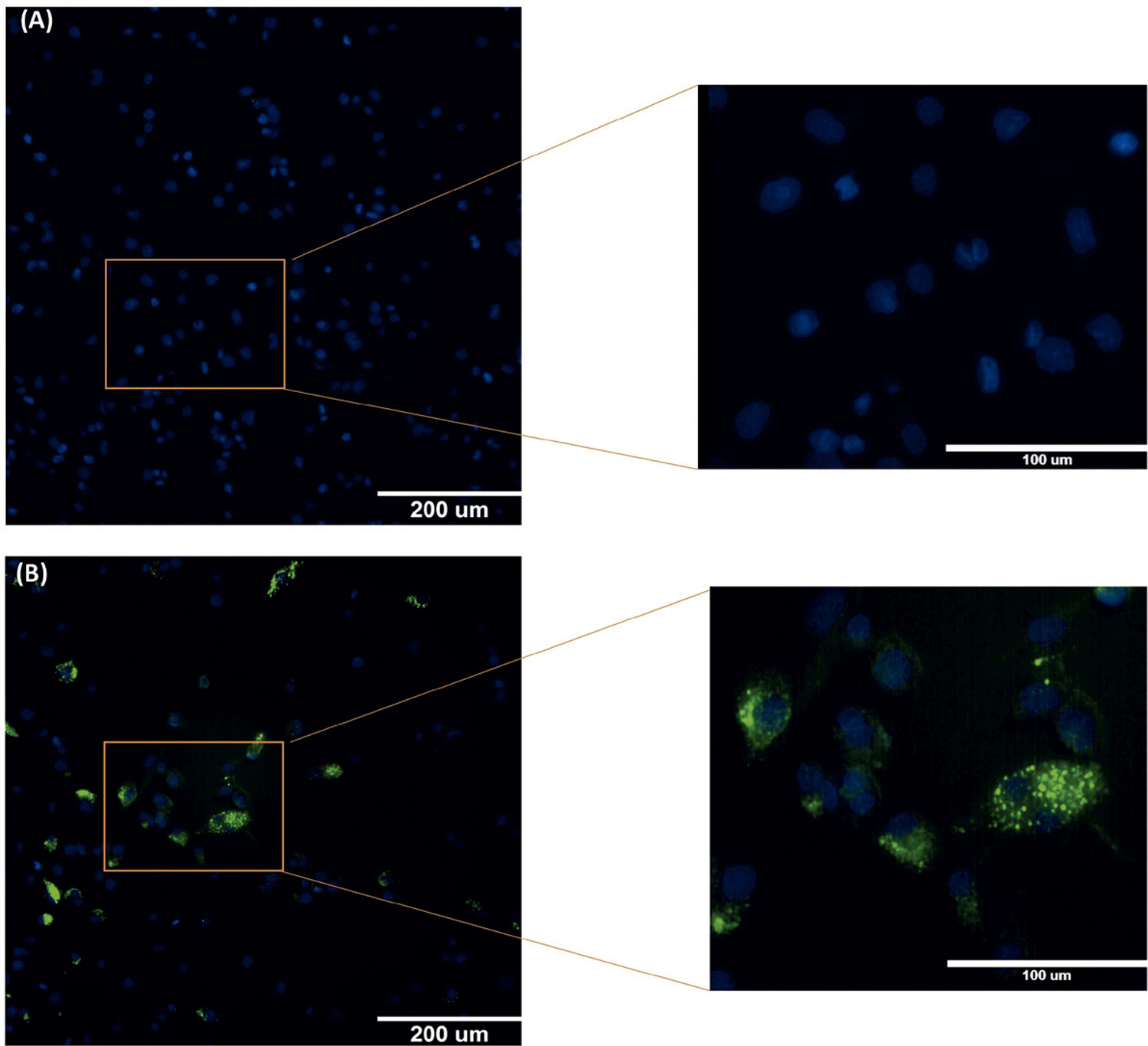
The LDL was isolated, oxidized, and labeled with a Fluorescein isothiocyanate (FITC) probe. Foam cells were generated from THP-1 human monocyte-derived cells and incubated in the absence (control) or presence of FITC-ox-LDL (10, 50, 100, 150, or 200 μg/mL), for 12, 24, 48, or 72 hours. The accumulated FITC-ox-LDL in the cell was quantified by microscopy. The enzyme-linked immunosorbent assay was evaluated to quantify the IL-6 and TNF-α, with p < 0.05 considered significant.
Results
All the FITC-ox-LDL concentrations tested showed a higher fluorescence when compared to the control, showing a greater accumulation of lipoprotein in cells. The higher the concentration of FITC-ox-LDL, the greater the production of TNF-α and IL-6. The production of IL-6 by foam cells was detected up to the value of 150 µg/mL of the maximum stimulus for LDL. Concentrations above 50 μg/mL LDL stimulated greater release of TNF-α compared to control.
Conclusions
Our model contributes to the understanding of the release of IL-6 and TNF-α in response to different concentrations of ox-LDL, using an optimized method for the formation of foam cells.