ABC Family Arquivos Brasileiros de Cardiologia International Journal of Cardiovascular Sciences ABC Heart Failure & Cardiomyopathy ABC Imagem Cardiovascular
Português English
ABC Family
Português English
  • About
    • About ABC Family
    • Internal Regulation
    • Editorial Board
    • Brazilian Society of Cardiology (SBC)
  • Issues
  • SBC Guidelines
  • Content
    • Videos
    • Podcasts
    • News
  • About
    • About ABC Family
    • Internal Regulation
    • Editorial Board
    • Brazilian Society of Cardiology (SBC)
  • Issues
  • SBC Guidelines
  • Content
    • Videos
    • Podcasts
    • News

ABC Family

  • ABC Family
  • Arquivos Brasileiros de Cardiologia
  • International Journal of Cardiovascular Sciences
  • ABC Heart Failure & Cardiomyopathy
  • ABC Imagem Cardiovascular
Search in

No content found.

Journals
Ordering
Article types
Others

  1. Home
  2. Keyword: body surface potential mapping
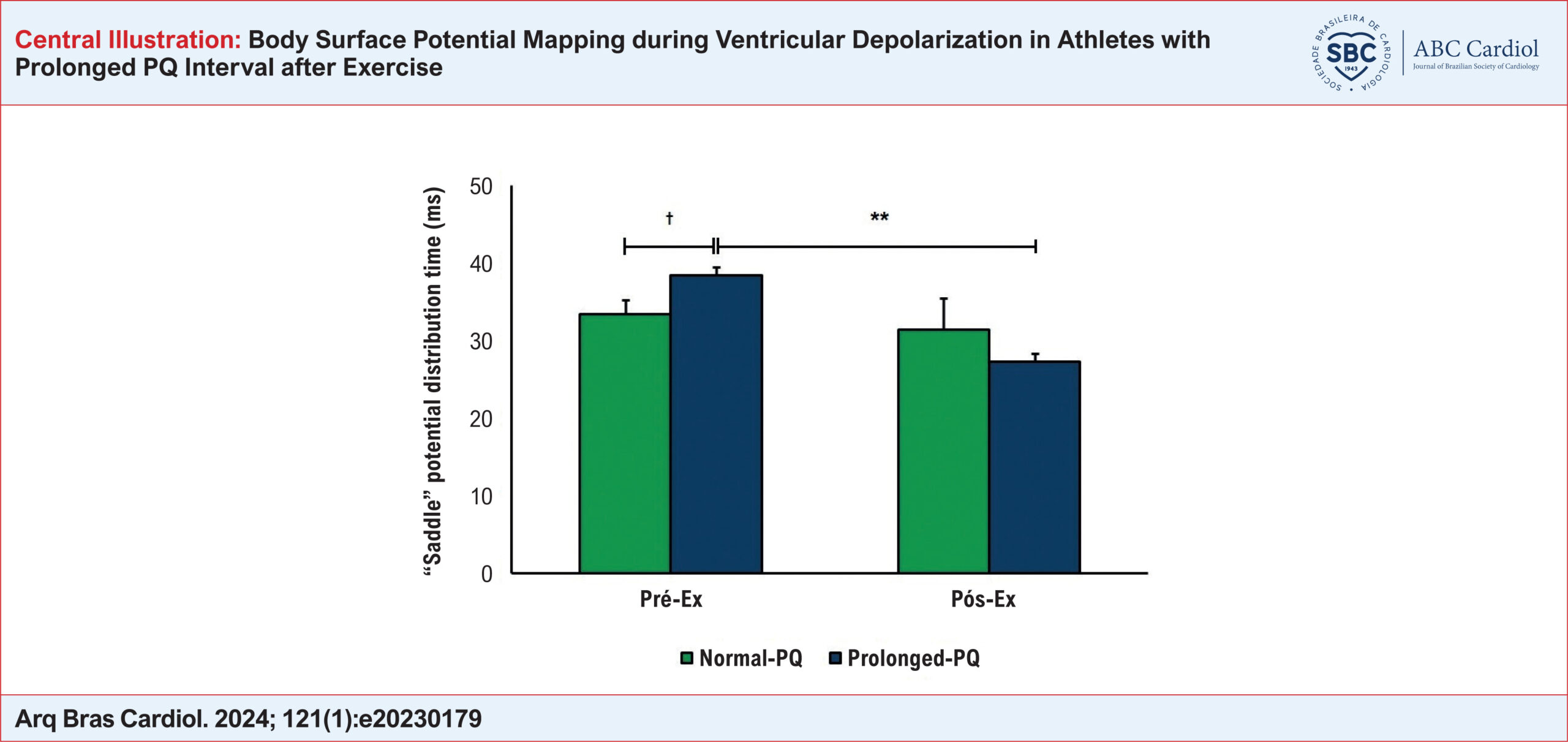
Body Surface Potential Mapping during Ventricular Depolarization in Athletes with Prolonged PQ Interval after Exercise
ABC
Original Article

Body Surface Potential Mapping during Ventricular Depolarization in Athletes with Prolonged PQ Interval after Exercise

Natalya I. Ivonina et al. Alexey G. Ivonin Irina M. Roshchevskaya

Abstract

Background:

Prolongation of the PQ interval, generally associated with an atrioventricular conduction delay, may be related to changes in intraventricular impulse spreading.

Objective:

To assess, using body surface potential mapping (BSPM), the process of ventricular depolarization in athletes with prolonged PQ intervals at rest and after exercise.

Methods:

The study included 7 cross-country skiers with a PQ interval of more than 200 ms (Prolonged-PQ group) and 7 with a PQ interval of less than 200 ms (Normal-PQ group). The BSPM from 64 unipolar torso leads was performed before (Pre-Ex) and after the bicycle exercise test (Post-Ex). Body surface equipotential maps were analyzed during ventricular depolarization. The significance level was 5%.

Results:

Compared to Normal-PQ athletes, the first and second periods of the stable position of cardiac potentials on the torso surface were longer, and the formation of the “saddle” potential distribution occurred later, at Pre-Ex, in Prolonged-PQ athletes. At Post-Ex, the Prolonged-PQ group showed a shortening of the first and second periods of stable potential distributions and a decrease in appearance time of the “saddle” phenomenon relative to Pre-Ex (to the values near to those of the Normal-PQ group). Additionally, at Post-Ex, the first inversion of potential distributions and the total duration of ventricular depolarization in Prolonged-PQ athletes decreased compared to Pre-Ex and with similar values in Normal-PQ athletes. Compared to Normal-PQ athletes, the second inversion was longer at Pre-Ex and Post-Ex in Prolonged-PQ athletes.

Conclusion:

Prolonged-PQ athletes had significant differences in the temporal characteristics of BSPM during ventricular depolarization both at rest and after exercise as compared to Normal-PQ athletes.
  • Athletes
  • Atrioventricular block
  • body surface potential mapping
  • Exercise Test
  • Heart Ventricles
Read Full Article
Creative Commons Attribution (CC BY) Creative Commons license
04/03/2024 DOI: 10.36660/abc.20230179 Views 1
ABC
Editorial

Applicability of Body Surface Potential Mapping Through Exercise in Small Animals

Carlos Alberto Pastore

The electrical potentials of the heart have been recorded for over a hundred years to represent the distribution of these internal electrical events on the torso surface. Such recordings that started with Waller tried to establish a resultant vector from a bipolar source, the “heart vector,” assuming that this distribution of potentials would act as if an electrical bipole was inserted inside the thorax and applied to the body surface, with a third electrode adding the sagittal component of the vector. The three would be enough to supply all the electrocardiogram (ECG) information extracted from measurements from the body surface. It was not until the 1930s and 1940s that the number of electrodes on the chest surface was tentatively increased to detect events occurring in cardiac areas near the precordial leads. Finally, after 1950 some studies demonstrated that the complexity of the electrical information generated inside the heart was far greater than that generated by a sole bipole, with multiple wavefronts in the ventricles creating currents that flow into and from the heart in several places; the potentials, therefore, would exhibit maximum and minimum distributions varying with time, usually located in areas unexplored by the conventional electrocardiogram.

New methodologies complementing the ECG and vectorcardiogram (ECG) added new electrodes (dorsal and right precordial leads), and, since Wilson, the 12-lead ECG started to have three bipolar and three unipolar modified leads in the frontal plane, in addition to six unipolar (precordial) leads on the anterior chest, which can record most of the information about electrical events in the heart. However, the myocardial electrical activity global expression cannot be captured without a larger number of simultaneously recorded leads.

[…]

  • Animal
  • body surface potential mapping
  • Exercise
  • Heart Ventricle
  • Myocardial
  • Physical Conditionning
  • Rats
Read Full Article
Creative Commons Attribution (CC BY) Creative Commons license
25/11/2022 DOI: 10.36660/abc.20220646 Views 1
Figure 1
ABC
Original Article

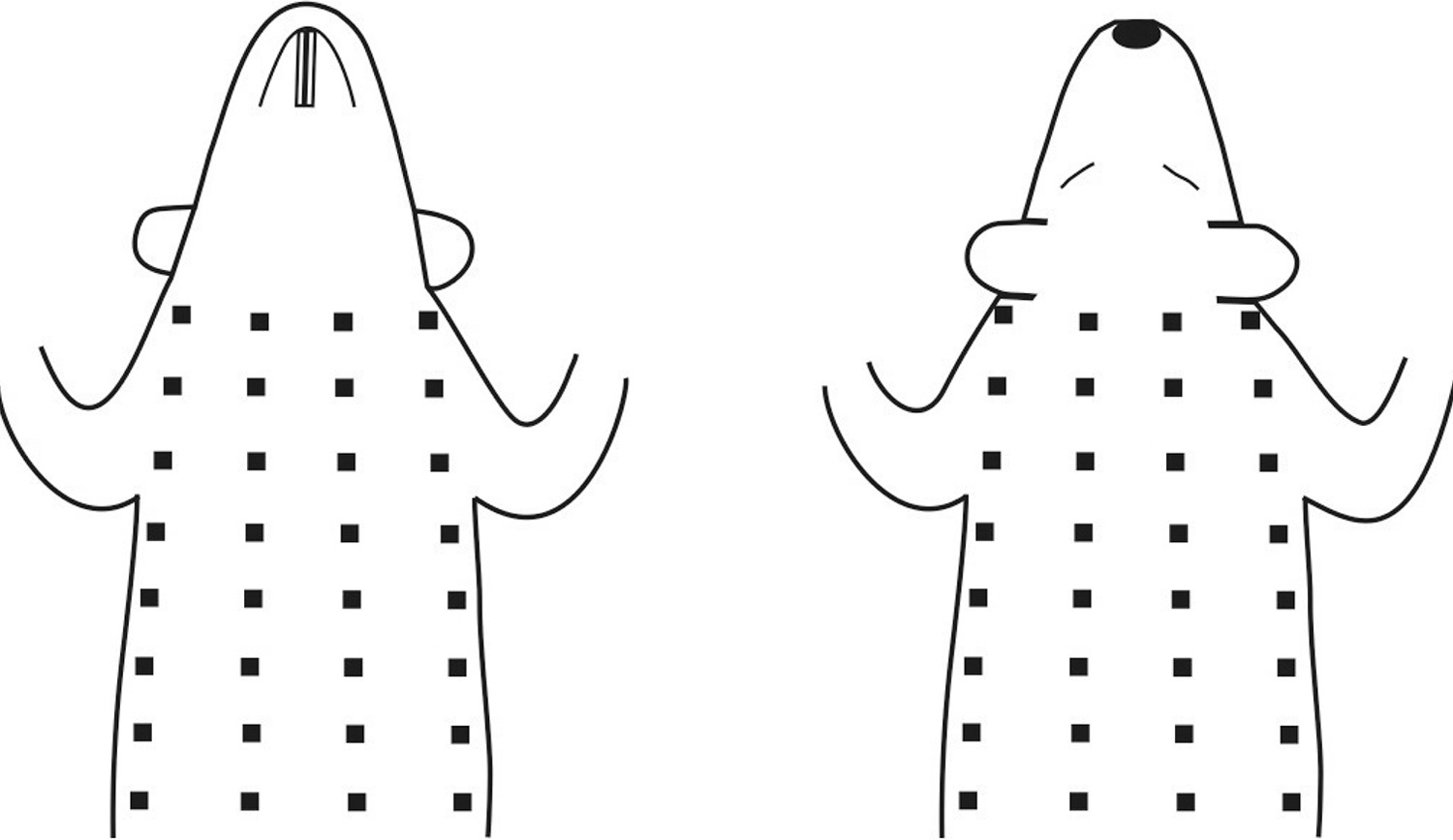
Body Surface Potential Mapping during Ventricular Depolarization in Rats after Acute Exhaustive Exercise

Alexey G. Ivonin et al. Svetlana L. Smirnova Irina M. Roshchevskaya

Abstract

Background

Exhaustive physical exercise can cause substantial changes in the electrical properties of the myocardium.

Objective

To evaluate, using body surface potential mapping, the electrical activity of the heart in rats during ventricular depolarization after acute exhaustive exercise.

Methods

Twelve-week-old male rats were submitted to acute treadmill exercise at 36 m/min until exhaustion. Unipolar electrocardiograms (ECGs) from the torso surface were recorded in zoletil-anesthetized rats three to five days before (Pre-Ex), 5 and 10 minutes after exhaustive exercise (Post-Ex 5 and Post-Ex 10, respectively) simultaneously with ECGs in limb leads. The instantaneous body surface potential maps (BSPMs) were analyzed during ventricular depolarization. P values <0.05 were considered statistically significant.

Results

Compared with Pre-Ex, an early completion of the second inversion of potential distributions, an early completion of ventricular depolarization, as well as a decrease in the duration of the middle phase and the total duration of ventricular depolarization on BSPMs were revealed at Post-Ex 5. Also, compared with Pre-Ex, an increase in the amplitude of negative BSPM extremum at the R-wave peak on the ECG in lead II (RII-peak) and a decrease in the amplitude of negative BSPM extremum at 3 and 4 ms after RII-peak were showed at Post-Ex 5. At Post-Ex 10, parameters of BSPMs did not differ from those at Pre-Ex.

Conclusion

In rats, acute exhaustive exercise causes reversible changes in the temporal and amplitude characteristics of BSPMs during ventricular depolarization, most likely related to alterations in the excitation of the main mass of the ventricular myocardium.

  • acute
  • body surface potential mapping
  • Exercises
  • Heart Ventricles
  • Myocardium
  • Rats
Read Full Article
Creative Commons Attribution (CC BY) Creative Commons license
06/09/2022 DOI: 10.36660/abc.20211058
Ventricular electrical activation in cardiac resynchronization as characterized by body surface potential mapping
ABC
Artigo Original

Ventricular electrical activation in cardiac resynchronization as characterized by body surface potential mapping

Carlos Alberto Pastore et al. Nancy Tobias Nelson Samesima Martino Martinelli Filho … Anisio Pedrosa Silvana Nishioka Roberto A. Douglas Luiz Felipe Pinho Moreira José Franchini Ramires

OBJECTIVES: To assess cardiac electrical activation by using body surface potential mapping (BSPM), in patients with congestive heart failure (CHF) and left bundle branch block (LBBB) undergoing cardiac resynchronization therapy (CRT) with biventricular pacemaker (BIV-PM) implantation. METHODS: Mean cardiac electrical activation times were analyzed in the right ventricle (RV) (mean RV activation time = mRV), anteroseptal area (mAS), and left ventricle (mLV) of 28 patients (mean age 61.2 ± 9.5 years; NYHA class III-IV CHF; ejection fraction <40%; LBBB of mean QRS 181.2±19.4ms, SÂQRS -8.5º±68.6º), as shown in their BSPM isochronous maps, before and after implantation of atriobiventricular pacemaker, comparing those with values obtained from a control group of normal individuals [CG], in three situations: (1) native LBBB; (2) RV pacing; and (3) atriobiventricular pacing. RESULTS: Situation (1): mRV and mAS values were similar (41.0±11.8ms x 43.6±13.4ms), with delayed mLV (81.0±12.5ms, p<0.01) and asynchronous with RV and AS areas; situation (2): mRV was greater than in CG (86.8±22.9ms, p<0.001), with greater difference between mAS and mLV (63.4±20.7ms vs. 102.7±20.3ms; p<0,001); situation (3): mLV and mRV were similar (72.0±32.0ms vs. 71.6±32.3ms), mRV was greater than in CG and native LBBB (71.6±32.3ms vs. 35.1±10.9ms and 41.0±11.8ms; p<0.001), and mAS was close to CG and native LBBB values (51.3±32.8ms vs. 50.1±11.4ms and 43.6±13.4ms). CONCLUSION: The body surface potential mapping showed that RV and LV activation times which are similar, and are close to those of the AS area, suggest patterns of synchronized ventricular activation in patients with CHF and LBBB during atriobiventricular pacing.

  • body surface potential mapping
  • Bundle Branch Block
  • Cardiac Pacing, Artificial
  • heart failure, congestive
Read Full Article
Creative Commons Attribution (CC BY) Creative Commons license
21/05/2007 DOI: 10.1590/S0066-782X2007000300001 Views 1

How to cite this article

EndNote RIS

Electronic Document Format (Vancouver)

Ivonina NI, Ivonin AG, Roshchevskaya IM. Body Surface Potential Mapping during Ventricular Depolarization in Athletes with Prolonged PQ Interval after Exercise. Arquivos Brasileiros de Cardiologia 2024;121(1):e20230179.

Electronic Document Format (ABNT)

Ivonina, Natalya I.; Ivonin, Alexey G.; Roshchevskaya, Irina M.. Body Surface Potential Mapping during Ventricular Depolarization in Athletes with Prolonged PQ Interval after Exercise. Arquivos Brasileiros de Cardiologia, v. 121, n. 1, e20230179, Mar. 2024.

Electronic Document Format (APA)

Ivonina, N. I., Ivonin, A. G., & Roshchevskaya, I. M. (2024). Body Surface Potential Mapping during Ventricular Depolarization in Athletes with Prolonged PQ Interval after Exercise. Arquivos Brasileiros de Cardiologia, 121(1), e20230179.

Electronic Document Format (ISO)

Ivonina, Natalya I. and Ivonin, Alexey G. and Roshchevskaya, Irina M.. Body Surface Potential Mapping during Ventricular Depolarization in Athletes with Prolonged PQ Interval after Exercise. Arquivos Brasileiros de Cardiologia [online]. 2024, vol. 121, n. 1, [cited 2026-10-14], e20230179. Available from: <https://journals.cardiol.br/en/article/body-surface-potential-mapping-during-ventricular-depolarization-in-athletes-with-prolonged-pq-interval-after-exercise/>. ISSN 0066-782X

EndNote RIS

Electronic Document Format (Vancouver)

Pastore CA. Applicability of Body Surface Potential Mapping Through Exercise in Small Animals. Arquivos Brasileiros de Cardiologia 2022;119(5):776-7.

Electronic Document Format (ABNT)

Pastore, Carlos Alberto. Applicability of Body Surface Potential Mapping Through Exercise in Small Animals. Arquivos Brasileiros de Cardiologia, v. 119, n. 5, p. 776-777, Nov. 2022.

Electronic Document Format (APA)

Pastore, C. A. (2022). Applicability of Body Surface Potential Mapping Through Exercise in Small Animals. Arquivos Brasileiros de Cardiologia, 119(5), 776-777.

Electronic Document Format (ISO)

Pastore, Carlos Alberto. Applicability of Body Surface Potential Mapping Through Exercise in Small Animals. Arquivos Brasileiros de Cardiologia [online]. 2022, vol. 119, n. 5, [cited 2026-10-14], pp. 776-777. Available from: <https://journals.cardiol.br/en/article/applicability-of-body-surface-potential-mapping-through-exercise-in-small-animals/>. ISSN 0066-782X

EndNote RIS

Electronic Document Format (Vancouver)

Ivonin AG, Smirnova SL, Roshchevskaya IM. Body Surface Potential Mapping during Ventricular Depolarization in Rats after Acute Exhaustive Exercise. Arquivos Brasileiros de Cardiologia 2022;119(5):766-75.

Electronic Document Format (ABNT)

Ivonin, Alexey G.; Smirnova, Svetlana L.; Roshchevskaya, Irina M.. Body Surface Potential Mapping during Ventricular Depolarization in Rats after Acute Exhaustive Exercise. Arquivos Brasileiros de Cardiologia, v. 119, n. 5, p. 766-775, Sep. 2022.

Electronic Document Format (APA)

Ivonin, A. G., Smirnova, S. L., & Roshchevskaya, I. M. (2022). Body Surface Potential Mapping during Ventricular Depolarization in Rats after Acute Exhaustive Exercise. Arquivos Brasileiros de Cardiologia, 119(5), 766-775.

Electronic Document Format (ISO)

Ivonin, Alexey G. and Smirnova, Svetlana L. and Roshchevskaya, Irina M.. Body Surface Potential Mapping during Ventricular Depolarization in Rats after Acute Exhaustive Exercise. Arquivos Brasileiros de Cardiologia [online]. 2022, vol. 119, n. 5, [cited 2026-10-14], pp. 766-775. Available from: <https://journals.cardiol.br/en/article/body-surface-potential-mapping-during-ventricular-depolarization-in-rats-after-acute-exhaustive-exercise/>. ISSN 0066-782X

EndNote RIS

Electronic Document Format (Vancouver)

Pastore CA, Tobias N, Samesima N, Martinelli Filho M, Pedrosa A, Nishioka S, Douglas RA, et al. Ventricular electrical activation in cardiac resynchronization as characterized by body surface potential mapping. Arquivos Brasileiros de Cardiologia 2007;88(3):251-7.

Electronic Document Format (ABNT)

Pastore, Carlos Alberto; Tobias, Nancy; Samesima, Nelson; Martinelli Filho, Martino; Pedrosa, Anisio; Nishioka, Silvana; Douglas, Roberto A.; Moreira, Luiz Felipe Pinho; Ramires, José Franchini. Ventricular electrical activation in cardiac resynchronization as characterized by body surface potential mapping. Arquivos Brasileiros de Cardiologia, v. 88, n. 3, p. 251-257, May. 2007.

Electronic Document Format (APA)

Pastore, C. A., Tobias, N., Samesima, N., Martinelli Filho, M., Pedrosa, A., Nishioka, S., Douglas, R. A., Moreira, L. F. P., & Ramires, J. F. (2007). Ventricular electrical activation in cardiac resynchronization as characterized by body surface potential mapping. Arquivos Brasileiros de Cardiologia, 88(3), 251-257.

Electronic Document Format (ISO)

Pastore, Carlos Alberto and Tobias, Nancy and Samesima, Nelson and Martinelli Filho, Martino and Pedrosa, Anisio and Nishioka, Silvana and Douglas, Roberto A. and Moreira, Luiz Felipe Pinho and Ramires, José Franchini. Ventricular electrical activation in cardiac resynchronization as characterized by body surface potential mapping. Arquivos Brasileiros de Cardiologia [online]. 2007, vol. 88, n. 3, [cited 2026-10-14], pp. 251-257. Available from: <https://journals.cardiol.br/en/article/ventricular-electrical-activation-in-cardiac-resynchronization-as-characterized-by-body-surface-potential-mapping/>. ISSN 0066-782X

  • About ABC Family
  • Internal Regulation
  • Editorial Board
  • Brazilian Society of Cardiology (SBC)

Issues

SBC Guidelines

  • Videos
  • Podcasts
  • News