ABC Family Arquivos Brasileiros de Cardiologia International Journal of Cardiovascular Sciences ABC Heart Failure & Cardiomyopathy ABC Imagem Cardiovascular
Português English
ABC Family
Português English
  • About
    • About ABC Family
    • Internal Regulation
    • Editorial Board
    • Brazilian Society of Cardiology (SBC)
  • Issues
  • SBC Guidelines
  • Content
    • Videos
    • Podcasts
    • News
  • About
    • About ABC Family
    • Internal Regulation
    • Editorial Board
    • Brazilian Society of Cardiology (SBC)
  • Issues
  • SBC Guidelines
  • Content
    • Videos
    • Podcasts
    • News

ABC Family

  • ABC Family
  • Arquivos Brasileiros de Cardiologia
  • International Journal of Cardiovascular Sciences
  • ABC Heart Failure & Cardiomyopathy
  • ABC Imagem Cardiovascular
Search in

No content found.

Journals
Ordering
Article types
Others

  1. Home
  2. Keyword: Coronary sinus
Assessment of Coronary Sinus Flow and Dimensions by Transthoracic Echocardiography in Patients with Atrioventricular Nodal Reentrant Tachycardia
ABC
Original Article

Assessment of Coronary Sinus Flow and Dimensions by Transthoracic Echocardiography in Patients with Atrioventricular Nodal Reentrant Tachycardia

Ali Duygu et al. Alkame Akgümüş Ahmet Balun Fahri Er

Abstract

Background:

The coronary sinus (CS) plays a pivotal role in cardiac venous drainage and is anatomically adjacent to the atrioventricular nodal region. Although structural enlargement of the CS has been described in patients with atrioventricular nodal reentrant tachycardia (AVNRT), coronary sinus flow (CSF) assessed by transthoracic echocardiography (TTE) has not previously been investigated in this population.

Objective:

To evaluate the relationship between AVNRT and CSF.

Methods:

This retrospective single-center study included 35 patients with AVNRT who underwent successful slow-pathway ablation and 34 age- and sex-matched healthy controls. All participants underwent comprehensive transthoracic echocardiography. CS ostial diameter, CS diameter, and CSF were measured using pulsed-wave Doppler echocardiography. CSF was calculated using standard geometric and Doppler-derived parameters. A p-value <0.05 was considered statistically significant.

Results:

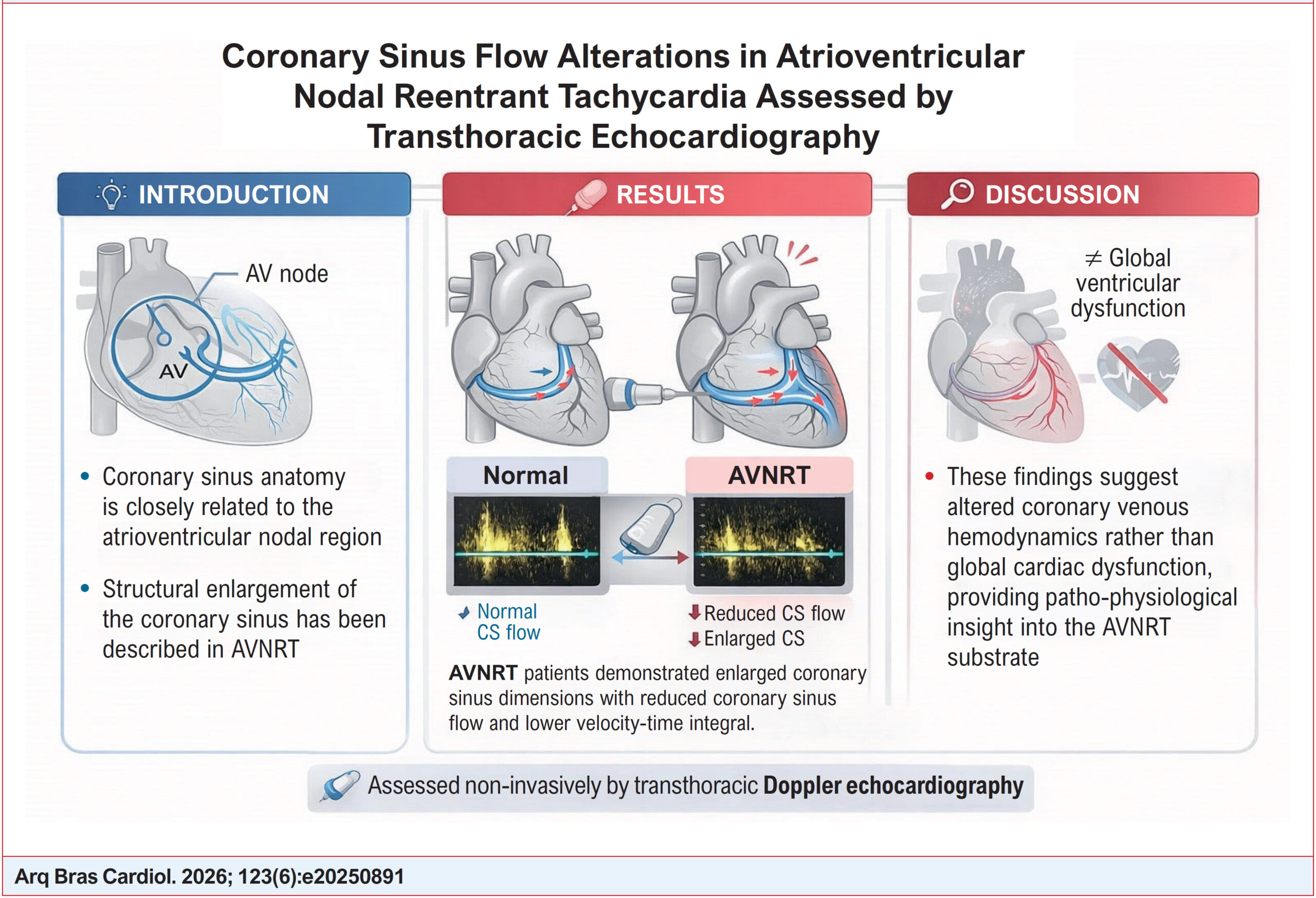
Global left and right ventricular systolic and diastolic function parameters were similar between groups. Patients with AVNRT exhibited significantly larger CS ostial and CS diameters compared with controls (both p<0.001). Despite this enlargement, CSF was significantly reduced in the AVNRT group (460.8 ± 83.3 vs. 523.8 ± 90.1 mL/min, p=0.004), accompanied by a lower CS velocity–time integral. Notably, CSF indexed to left ventricular mass also remained significantly lower in AVNRT patients, indicating that reduced flow was independent of myocardial mass.

Conclusion:

Patients with AVNRT demonstrate a distinct coronary sinus hemodynamic profile characterized by CS enlargement and reduced CSF as assessed by transthoracic echocardiography. These findings suggest altered coronary venous hemodynamics in AVNRT and provide novel pathophysiological insights using a non-invasive imaging modality.
  • Atrioventricular Nodal Reentry Tachycardia
  • Coronary sinus
  • Doppler Echocardiography
Read Full Article
Creative Commons Attribution (CC BY) Creative Commons license
24/07/2026 DOI: 10.36660/abc.20250891i Views 1
Figure 1
ABC
Image

Silent Giant: Right Coronary Artery Ectasia with a Hidden Fistula to the Coronary Sinus in an Asymptomatic Patient

Inês Ferreira Neves et al. André Ferreira Inês Almeida Tânia Branco Mano Lídia de Sousa

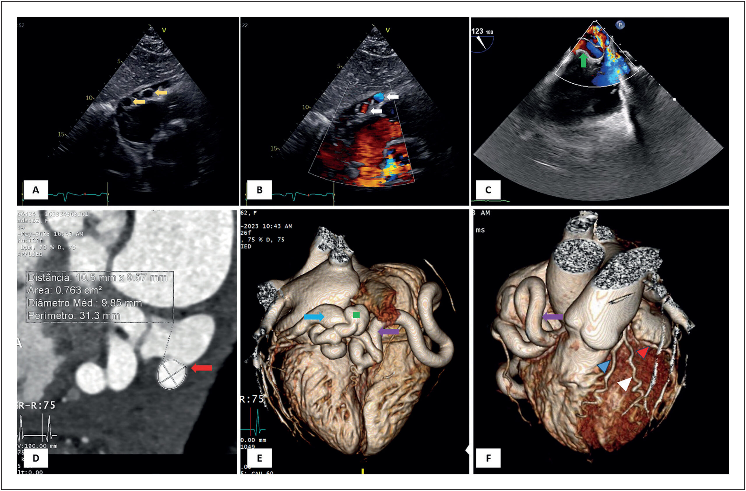
A 62-year-old woman, asymptomatic and without significant medical history, was referred to a routine transthoracic echocardiogram (TTE). The TTE had no abnormal findings but showed, in subcostal view, three round images adjacent to the lateral wall of the right atrium. Color Doppler showed that these were apparently vascularized (, panels A and B, supplemental and ). A transesophageal echocardiogram showed a giant right coronary artery (RCA), apparently originating from the RCA ostium, with turbulent flow inside (, panel C, supplemental ). It was not possible to observe shunts or fistulae in this imaging modality. Given the suspicion of a fistula to the RCA, computed tomography coronary angiography was performed, showing an ectatic RCA with a normal origin, a diameter of 10,5 mm, and a tortuous trajectory, with fistulation to the coronary sinus in its final segment ( panels D to F). Right heart catheterization demonstrated a non-significant left-to-right shunt (Qp: Qs ratio of 1.60). Cardiac magnetic resonance imaging (MRI) had no evidence of perfusion defects during hyperemia, thus excluding the coronary “steal phenomenon”. After a multidisciplinary team discussion, since the patient was asymptomatic and the fistula had no hemodynamic significance, no invasive treatment was performed at this time.

Coronary arteriovenous fistulas are rare, with an incidence of 0.002%., The majority of these are congenital and asymptomatic at presentation. Coronary steal can lead to several complications, including ischemia., The treatment approach remains controversial. The presence of symptoms, significant left-right shunt, and large fistula are the most frequent indications for percutaneous or surgical approach.,

[…]

  • Arteriovenous Fistula
  • Congenital Heart Defects
  • Coronary sinus
  • Coronary Vessels
Read Full Article
Creative Commons Attribution (CC BY) Creative Commons license
28/07/2025 DOI: 10.36660/abc.20240574i
Figure 1
ABCIC
Case Report

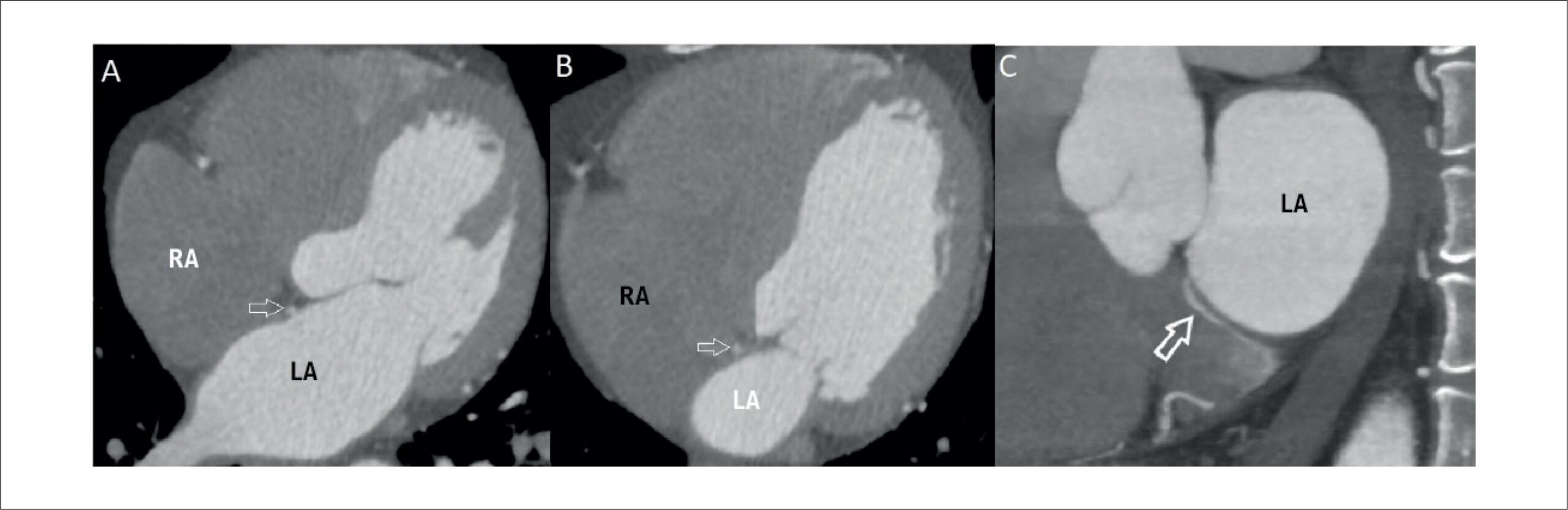
A Bridging Vein Connecting Left Atrium and Coronary Sinus: Coronary Computed Tomography Angiography Findings

Sercan Şahin et al. Nilgün Işıksalan Özbülbül

Introduction

Until recent years, coronary venous system imaging was overshadowed by coronary artery imaging. However, in recent times, the significance of coronary venous system imaging has risen markedly, particularly due to its pivotal role in diverse interventional cardiology procedures. These interventions encompass a spectrum of tasks, including left ventricular pacing, mapping and ablation of arrhythmias, and retrograde cardioplegia. These procedures are facilitated by utilizing the potential of the coronary sinus (CS). As a result, comprehending the anatomy and anomalies of the CS holds paramount importance, and coronary computed tomography (CT) angiography has proven invaluable for achieving this objective.

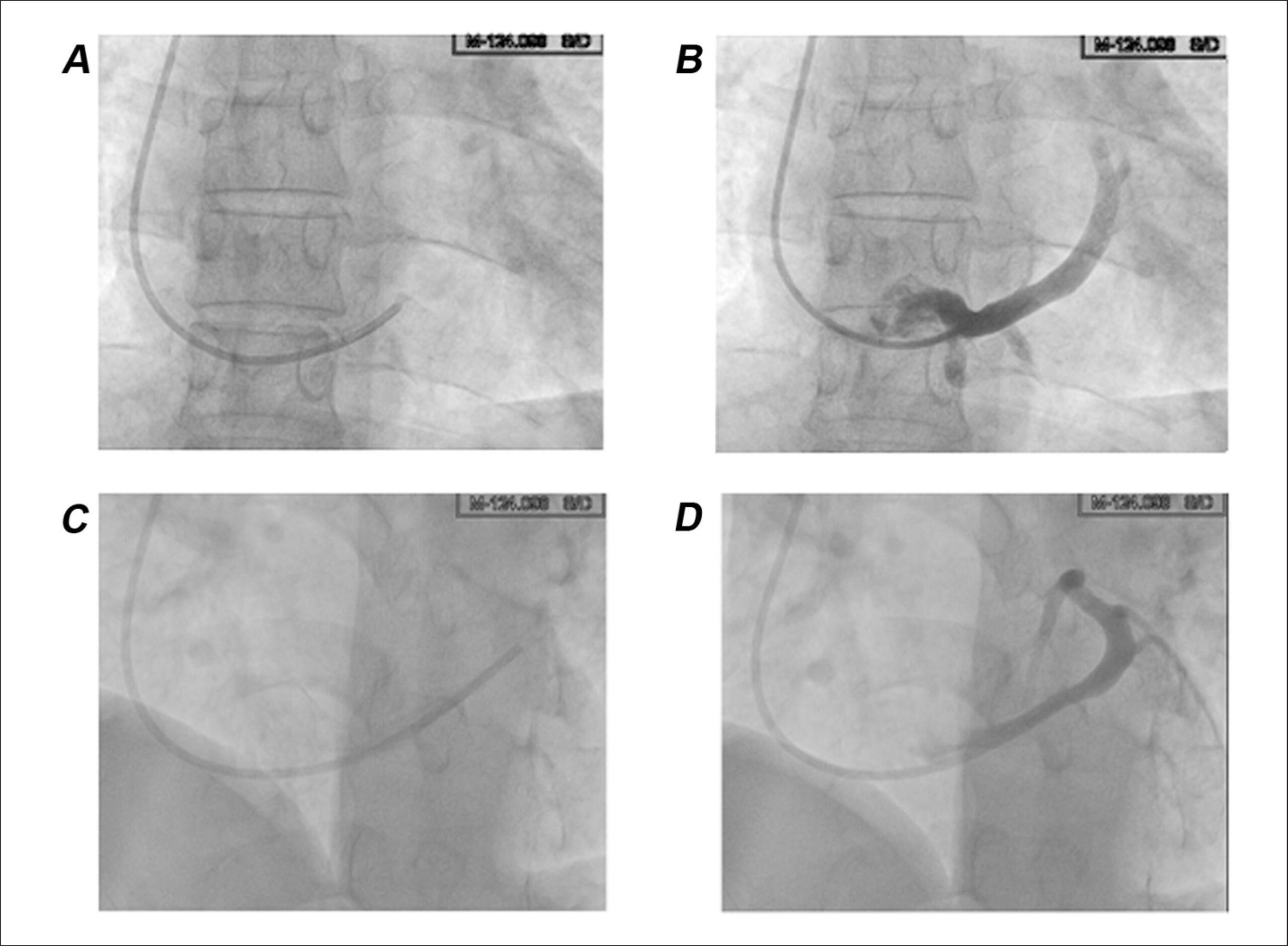
In this case report, we present an extraordinary cardiac venous anomaly: a bridging vein connecting the left atrium to the CS with non-specific chest pain. We believe that this case serves as an illustrative example for understanding rarely observed cardiac venous vascular anomalies, while also making a valuable contribution to the existing literature through the utilization of clear and direct coronary CT angiography images.

[…]

  • Computed Tomography Angiography
  • Coronary sinus
  • Heart Atria
Read Full Article
Creative Commons Attribution (CC BY) Creative Commons license
12/03/2024 DOI: 10.36660/abcimg.20230111i Views 1
ABCIC
Case Report

Ruptured Right Sinus of Valsalva Aneurysm with Right Ventricular Fistula

Carolina Santana dos Reis Gonçalves et al. Rafael Rocha Silva Marta Fernandes Lima

[…]

  • Aneurysm, Ruptured
  • Coronary sinus
  • Fistula
  • Heart Defects, Congenital
  • Heart Failure
  • Sinus of Valsalva
  • Ventricular Dysfunction Right
Read Full Article
Creative Commons Attribution (CC BY) Creative Commons license
04/02/2022 DOI: 10.5935/2318-8219.20180038 Views 2
ABCIC
Case Report

A Rare Cause of Hypoxemia

Lisete Lopes et al. Patrícia Vaz Silva Dina Rodrigues António Pires

Abstract

An unroofed coronary sinus is a very rare cardiac anomaly that is commonly associated with a persistent left superior vena cava. Its diagnosis is often difficult, but clinical signs and echocardiographic findings of an interatrial shunt should raise its suspicion. The authors describe the case of a child with chronic hypoxemia whose diagnostic work-up revealed an unroofed coronary sinus and persistent superior left vena cava.

  • Contrast Echocardiography
  • Coronary sinus
  • Hypoxemia
Read Full Article
Creative Commons Attribution (CC BY) Creative Commons license
14/01/2022 DOI: 10.5935/2318-8219.20200038
Figure 1
ABC
Original Article

Correlation between C-Reactive Protein in Peripheral Vein and Coronary Sinus in Stable and Unstable Angina

Weverton Ferreira Leite et al. José Antonio Franchini Ramires Luiz Felipe Pinho Moreira Célia Maria Cassaro Strunz José Armando Mangione

Background:

High sensitivity C-reactive protein (hs-CRP) is commonly used in clinical practice to assess cardiovascular risk. However, a correlation has not yet been established between the absolute levels of peripheral and central hs-CRP.

Objective:

To assess the correlation between serum hs-CRP levels (mg/L) in a peripheral vein in the left forearm (LFPV) with those in the coronary sinus (CS) of patients with coronary artery disease (CAD) and a diagnosis of stable angina (SA) or unstable angina (UA).

Methods:

This observational, descriptive, and cross-sectional study was conducted at the Instituto do Coração, Hospital das Clinicas, Faculdade de Medicina, Universidade de São Paulo, and at the Hospital Beneficência Portuguesa de Sao Paulo, where CAD patients referred to the hospital for coronary angiography were evaluated.

Results:

Forty patients with CAD (20 with SA and 20 with UA) were included in the study. Blood samples from LFPV and CS were collected before coronary angiography. Furthermore, analysis of the correlation between serum levels of hs-CRP in LFPV versus CS showed a strong linear correlation for both SA (r = 0.993, p < 0.001) and UA (r = 0.976, p < 0.001) and for the entire sample (r = 0.985, p < 0.001).

Conclusion:

Our data suggest a strong linear correlation between hs-CRP levels in LFPV versus CS in patients with SA and UA.

  • Angina, Stable
  • Angina, Unstable
  • C-Reactive Protein
  • Coronary Artery Disease
  • Coronary sinus
Read Full Article
Creative Commons Attribution (CC BY) Creative Commons license
09/12/2014 DOI: 10.5935/abc.20140188 Views 2
Study of Coronary Sinus Flow Reserve Through Transesophageal Doppler Echocardiography in Normal Subjects
ABC
Original Article

Study of Coronary Sinus Flow Reserve Through Transesophageal Doppler Echocardiography in Normal Subjects

José Ramos Filho et al. José A. F. Ramires Marko Turina Caio J. Medeiros … Mário Lachat Jeane Tsutsui

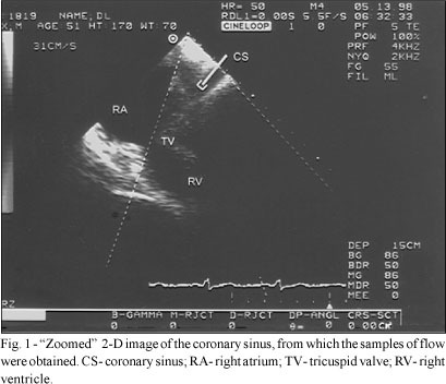
OBJECTIVE – To evaluate the Coronary Flow Reserve in the Coronary Sinus through transesophageal Doppler echocardiography in normal subjects. METHODS – We obtained technically adequate flow samples for analysis in 10 healthy volunteers (37±8 years, 5 men) with no history of heart or systemic disease and with mean left ventricular mass index by transthoracic echocardiography of 87±18 g/m². Coronary sinus flow velocity was recorded within the coronary sinus with the patient in a resting condition and during intravenous adenosine infusion at a dose of 140 µg/kg/min for 4 minutes. Recording of coronary sinus blood flow was possible in all cases with measurement of peak systolic, diastolic, and retrograde velocities (PSV, PDV, and PRV, cm/sec), mean systolic and diastolic velocities (MSV and MDV, cm/sec), and systolic and diastolic velocity time integral (VTI S and VTI D, cm/sec). RESULTS – The coronary flow reserve was calculated as the ratio between the blood flow in the basal state and the maximum measured hyperemic blood flow with adenosine infusion. Results are shown as mean and standard deviations. (CFR = PSV + PDV — PRV/basal PSV): 1st min = 2.2±0.21; 2nd min = 3±0.3; 3rd min = 3.4±0.37; 4th min = 3.6 ± 0.33. CONCLUSION – Although coronary sinus flow had significantly increased in the first minute, higher velocities were seen at third and fourth minutes, indicating that these should be the best times to study coronary sinus flow with intravenous adenosine in continuous infusion.

  • coronary reserve
  • Coronary sinus
  • transesophageal Doppler echocardiography
Read Full Article
Creative Commons Attribution (CC BY) Creative Commons license
10/09/2002 DOI: 10.1590/S0066-782X2002001100001

How to cite this article

EndNote RIS

Electronic Document Format (Vancouver)

Duygu A, Akgümüş A, Balun A, Er F. Assessment of Coronary Sinus Flow and Dimensions by Transthoracic Echocardiography in Patients with Atrioventricular Nodal Reentrant Tachycardia. Arquivos Brasileiros de Cardiologia 2026;123(6):e20250891.

Electronic Document Format (ABNT)

Duygu, Ali; Akgümüş, Alkame; Balun, Ahmet; Er, Fahri. Assessment of Coronary Sinus Flow and Dimensions by Transthoracic Echocardiography in Patients with Atrioventricular Nodal Reentrant Tachycardia. Arquivos Brasileiros de Cardiologia, v. 123, n. 6, e20250891, Jul. 2026.

Electronic Document Format (APA)

Duygu, A., Akgümüş, A., Balun, A., & Er, F. (2026). Assessment of Coronary Sinus Flow and Dimensions by Transthoracic Echocardiography in Patients with Atrioventricular Nodal Reentrant Tachycardia. Arquivos Brasileiros de Cardiologia, 123(6), e20250891.

Electronic Document Format (ISO)

Duygu, Ali and Akgümüş, Alkame and Balun, Ahmet and Er, Fahri. Assessment of Coronary Sinus Flow and Dimensions by Transthoracic Echocardiography in Patients with Atrioventricular Nodal Reentrant Tachycardia. Arquivos Brasileiros de Cardiologia [online]. 2026, vol. 123, n. 6, [cited 2026-10-14], e20250891. Available from: <https://journals.cardiol.br/en/article/assessment-of-coronary-sinus-flow-and-dimensions-by-transthoracic-echocardiography-in-patients-with-atrioventricular-nodal-reentrant-tachycardia/>. ISSN 0066-782X

EndNote RIS

Electronic Document Format (Vancouver)

Neves IF, Ferreira A, Almeida I, Mano TB, Sousa L. Silent Giant: Right Coronary Artery Ectasia with a Hidden Fistula to the Coronary Sinus in an Asymptomatic Patient. Arquivos Brasileiros de Cardiologia 2025;122(6):e20240574.

Electronic Document Format (ABNT)

Neves, Inês Ferreira; Ferreira, André; Almeida, Inês; Mano, Tânia Branco; Sousa, Lídia de. Silent Giant: Right Coronary Artery Ectasia with a Hidden Fistula to the Coronary Sinus in an Asymptomatic Patient. Arquivos Brasileiros de Cardiologia, v. 122, n. 6, e20240574, Jul. 2025.

Electronic Document Format (APA)

Neves, I. F., Ferreira, A., Almeida, I., Mano, T. B., & Sousa, L. (2025). Silent Giant: Right Coronary Artery Ectasia with a Hidden Fistula to the Coronary Sinus in an Asymptomatic Patient. Arquivos Brasileiros de Cardiologia, 122(6), e20240574.

Electronic Document Format (ISO)

Neves, Inês Ferreira and Ferreira, André and Almeida, Inês and Mano, Tânia Branco and Sousa, Lídia de. Silent Giant: Right Coronary Artery Ectasia with a Hidden Fistula to the Coronary Sinus in an Asymptomatic Patient. Arquivos Brasileiros de Cardiologia [online]. 2025, vol. 122, n. 6, [cited 2026-10-14], e20240574. Available from: <https://journals.cardiol.br/en/article/silent-giant-right-coronary-artery-ectasia-with-a-hidden-fistula-to-the-coronary-sinus-in-an-asymptomatic-patient/>. ISSN 0066-782X

EndNote RIS

Electronic Document Format (Vancouver)

Şahin S, Özbülbül NI. A Bridging Vein Connecting Left Atrium and Coronary Sinus: Coronary Computed Tomography Angiography Findings. ABC Imagem Cardiovascular 2024;37(1):e20230111.

Electronic Document Format (ABNT)

Şahin, Sercan; Özbülbül, Nilgün Işıksalan. A Bridging Vein Connecting Left Atrium and Coronary Sinus: Coronary Computed Tomography Angiography Findings. ABC Imagem Cardiovascular, v. 37, n. 1, e20230111, Mar. 2024.

Electronic Document Format (APA)

Şahin, S., & Özbülbül, N. I. (2024). A Bridging Vein Connecting Left Atrium and Coronary Sinus: Coronary Computed Tomography Angiography Findings. ABC Imagem Cardiovascular, 37(1), e20230111.

Electronic Document Format (ISO)

Şahin, Sercan and Özbülbül, Nilgün Işıksalan. A Bridging Vein Connecting Left Atrium and Coronary Sinus: Coronary Computed Tomography Angiography Findings. ABC Imagem Cardiovascular [online]. 2024, vol. 37, n. 1, [cited 2026-10-14], e20230111. Available from: <https://journals.cardiol.br/en/article/a-bridging-vein-connecting-left-atrium-and-coronary-sinus-coronary-computed-tomography-angiography-findings/>. ISSN 2318-8219

EndNote RIS

Electronic Document Format (Vancouver)

Gonçalves CSR, Silva RR, Lima MF. Ruptured Right Sinus of Valsalva Aneurysm with Right Ventricular Fistula. Arquivos Brasileiros de Cardiologia Imagem cardiovascular 2022;31(4):294-8.

Electronic Document Format (ABNT)

Gonçalves, Carolina Santana dos Reis; Silva, Rafael Rocha; Lima, Marta Fernandes. Ruptured Right Sinus of Valsalva Aneurysm with Right Ventricular Fistula. Arquivos Brasileiros de Cardiologia Imagem cardiovascular, v. 31, n. 4, p. 294-298, Feb. 2022.

Electronic Document Format (APA)

Gonçalves, C. S. R., Silva, R. R., & Lima, M. F. (2022). Ruptured Right Sinus of Valsalva Aneurysm with Right Ventricular Fistula. Arquivos Brasileiros de Cardiologia Imagem cardiovascular, 31(4), 294-298.

Electronic Document Format (ISO)

Gonçalves, Carolina Santana dos Reis and Silva, Rafael Rocha and Lima, Marta Fernandes. Ruptured Right Sinus of Valsalva Aneurysm with Right Ventricular Fistula. Arquivos Brasileiros de Cardiologia Imagem cardiovascular [online]. 2022, vol. 31, n. 4, [cited 2026-10-14], pp. 294-298. Available from: <https://journals.cardiol.br/en/article/ruptured-right-sinus-of-valsalva-aneurysm-with-right-ventricular-fistula/>. ISSN 2318-8219

EndNote RIS

Electronic Document Format (Vancouver)

Lopes L, Silva PV, Rodrigues D, Pires A. A Rare Cause of Hypoxemia. Arquivos Brasileiros de Cardiologia Imagem cardiovascular 2022;33(3):1-3.

Electronic Document Format (ABNT)

Lopes, Lisete; Silva, Patrícia Vaz; Rodrigues, Dina; Pires, António. A Rare Cause of Hypoxemia. Arquivos Brasileiros de Cardiologia Imagem cardiovascular, v. 33, n. 3, p. 1-3, Jan. 2022.

Electronic Document Format (APA)

Lopes, L., Silva, P. V., Rodrigues, D., & Pires, A. (2022). A Rare Cause of Hypoxemia. Arquivos Brasileiros de Cardiologia Imagem cardiovascular, 33(3), 1-3.

Electronic Document Format (ISO)

Lopes, Lisete and Silva, Patrícia Vaz and Rodrigues, Dina and Pires, António. A Rare Cause of Hypoxemia. Arquivos Brasileiros de Cardiologia Imagem cardiovascular [online]. 2022, vol. 33, n. 3, [cited 2026-10-14], pp. 1-3. Available from: <https://journals.cardiol.br/en/article/a-rare-cause-of-hypoxemia/>. ISSN 2318-8219

EndNote RIS

Electronic Document Format (Vancouver)

Leite WF, Ramires JAF, Moreira LFP, Strunz CMC, Mangione JA. Correlation between C-Reactive Protein in Peripheral Vein and Coronary Sinus in Stable and Unstable Angina. Arquivos Brasileiros de Cardiologia 2014;104(3):202-8.

Electronic Document Format (ABNT)

Leite, Weverton Ferreira; Ramires, José Antonio Franchini; Moreira, Luiz Felipe Pinho; Strunz, Célia Maria Cassaro; Mangione, José Armando. Correlation between C-Reactive Protein in Peripheral Vein and Coronary Sinus in Stable and Unstable Angina. Arquivos Brasileiros de Cardiologia, v. 104, n. 3, p. 202-208, Dec. 2014.

Electronic Document Format (APA)

Leite, W. F., Ramires, J. A. F., Moreira, L. F. P., Strunz, C. M. C., & Mangione, J. A. (2014). Correlation between C-Reactive Protein in Peripheral Vein and Coronary Sinus in Stable and Unstable Angina. Arquivos Brasileiros de Cardiologia, 104(3), 202-208.

Electronic Document Format (ISO)

Leite, Weverton Ferreira and Ramires, José Antonio Franchini and Moreira, Luiz Felipe Pinho and Strunz, Célia Maria Cassaro and Mangione, José Armando. Correlation between C-Reactive Protein in Peripheral Vein and Coronary Sinus in Stable and Unstable Angina. Arquivos Brasileiros de Cardiologia [online]. 2014, vol. 104, n. 3, [cited 2026-10-14], pp. 202-208. Available from: <https://journals.cardiol.br/en/article/correlation-between-c-reactive-protein-in-peripheral-vein-and-coronary-sinus-in-stable-and-unstable-angina/>. ISSN 0066-782X

EndNote RIS

Electronic Document Format (Vancouver)

Ramos Filho J, Ramires JAF, Turina M, Medeiros CJ, Lachat M, Tsutsui J. Study of Coronary Sinus Flow Reserve Through Transesophageal Doppler Echocardiography in Normal Subjects. Arquivos Brasileiros de Cardiologia 2002;79(2):102-6.

Electronic Document Format (ABNT)

Ramos Filho, José; Ramires, José A. F.; Turina, Marko; Medeiros, Caio J.; Lachat, Mário; Tsutsui, Jeane. Study of Coronary Sinus Flow Reserve Through Transesophageal Doppler Echocardiography in Normal Subjects. Arquivos Brasileiros de Cardiologia, v. 79, n. 2, p. 102-106, Sep. 2002.

Electronic Document Format (APA)

Ramos Filho, J., Ramires, J. A. F., Turina, M., Medeiros, C. J., Lachat, M., & Tsutsui, J. (2002). Study of Coronary Sinus Flow Reserve Through Transesophageal Doppler Echocardiography in Normal Subjects. Arquivos Brasileiros de Cardiologia, 79(2), 102-106.

Electronic Document Format (ISO)

Ramos Filho, José and Ramires, José A. F. and Turina, Marko and Medeiros, Caio J. and Lachat, Mário and Tsutsui, Jeane. Study of Coronary Sinus Flow Reserve Through Transesophageal Doppler Echocardiography in Normal Subjects. Arquivos Brasileiros de Cardiologia [online]. 2002, vol. 79, n. 2, [cited 2026-10-14], pp. 102-106. Available from: <https://journals.cardiol.br/en/article/study-of-coronary-sinus-flow-reserve-through-transesophageal-doppler-echocardiography-in-normal-subjects/>. ISSN 0066-782X

  • About ABC Family
  • Internal Regulation
  • Editorial Board
  • Brazilian Society of Cardiology (SBC)

Issues

SBC Guidelines

  • Videos
  • Podcasts
  • News