Silent Giant: Right Coronary Artery Ectasia with a Hidden Fistula to the Coronary Sinus in an Asymptomatic Patient
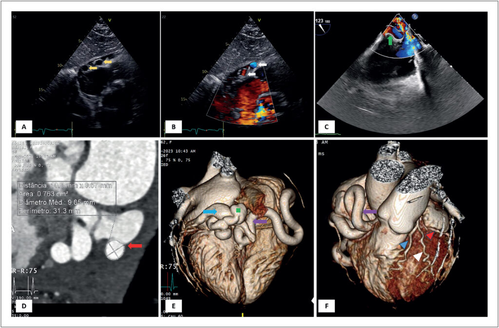
A 62-year-old woman, asymptomatic and without significant medical history, was referred to a routine transthoracic echocardiogram (TTE). The TTE had no abnormal findings but showed, in subcostal view, three round images adjacent to the lateral wall of the right atrium. Color Doppler showed that these were apparently vascularized (, panels A and B, supplemental and ). A transesophageal echocardiogram showed a giant right coronary artery (RCA), apparently originating from the RCA ostium, with turbulent flow inside (, panel C, supplemental ). It was not possible to observe shunts or fistulae in this imaging modality. Given the suspicion of a fistula to the RCA, computed tomography coronary angiography was performed, showing an ectatic RCA with a normal origin, a diameter of 10,5 mm, and a tortuous trajectory, with fistulation to the coronary sinus in its final segment ( panels D to F). Right heart catheterization demonstrated a non-significant left-to-right shunt (Qp: Qs ratio of 1.60). Cardiac magnetic resonance imaging (MRI) had no evidence of perfusion defects during hyperemia, thus excluding the coronary “steal phenomenon”. After a multidisciplinary team discussion, since the patient was asymptomatic and the fistula had no hemodynamic significance, no invasive treatment was performed at this time.
Coronary arteriovenous fistulas are rare, with an incidence of 0.002%., The majority of these are congenital and asymptomatic at presentation. Coronary steal can lead to several complications, including ischemia., The treatment approach remains controversial. The presence of symptoms, significant left-right shunt, and large fistula are the most frequent indications for percutaneous or surgical approach.,
[…]
Images
Image gallery fullscreen viewer
Comments are moderated by the editorial team before publication. Discussions should be evidence-based and use scientific language. Conflicts of interest should be declared.