| ABCIC
ABC Family Arquivos Brasileiros de Cardiologia International Journal of Cardiovascular Sciences ABC Heart Failure & Cardiomyopathy ABC Imagem Cardiovascular
Português English
ABC Family | ABCIC
Português English
  • About
    • About the Journal
    • Internal Regulation
    • Editors Section
    • Editorial Board
    • SBC Award
  • Instructions to Authors
    • Guidelines for Publication
    • Editorial Policy
    • Ethics Policy
    • Policy on the Use of Artificial Intelligence
    • Mandatory Publication Form
  • Issues
  • SBC Guidelines
  • About
    • About the Journal
    • Internal Regulation
    • Editors Section
    • Editorial Board
    • SBC Award
  • Instructions to Authors
    • Guidelines for Publication
    • Editorial Policy
    • Ethics Policy
    • Policy on the Use of Artificial Intelligence
    • Mandatory Publication Form
  • Issues
  • SBC Guidelines

ABC Family

  • ABC Family
  • Arquivos Brasileiros de Cardiologia
  • International Journal of Cardiovascular Sciences
  • ABC Heart Failure & Cardiomyopathy
  • ABC Imagem Cardiovascular
Search in

No content found.

Journals
Ordering
Article types
Others

  1. Home
  2. ABCIC
  3. Issues
  4. 2025, Volume 38, Number 2
Figure 1
Case Report

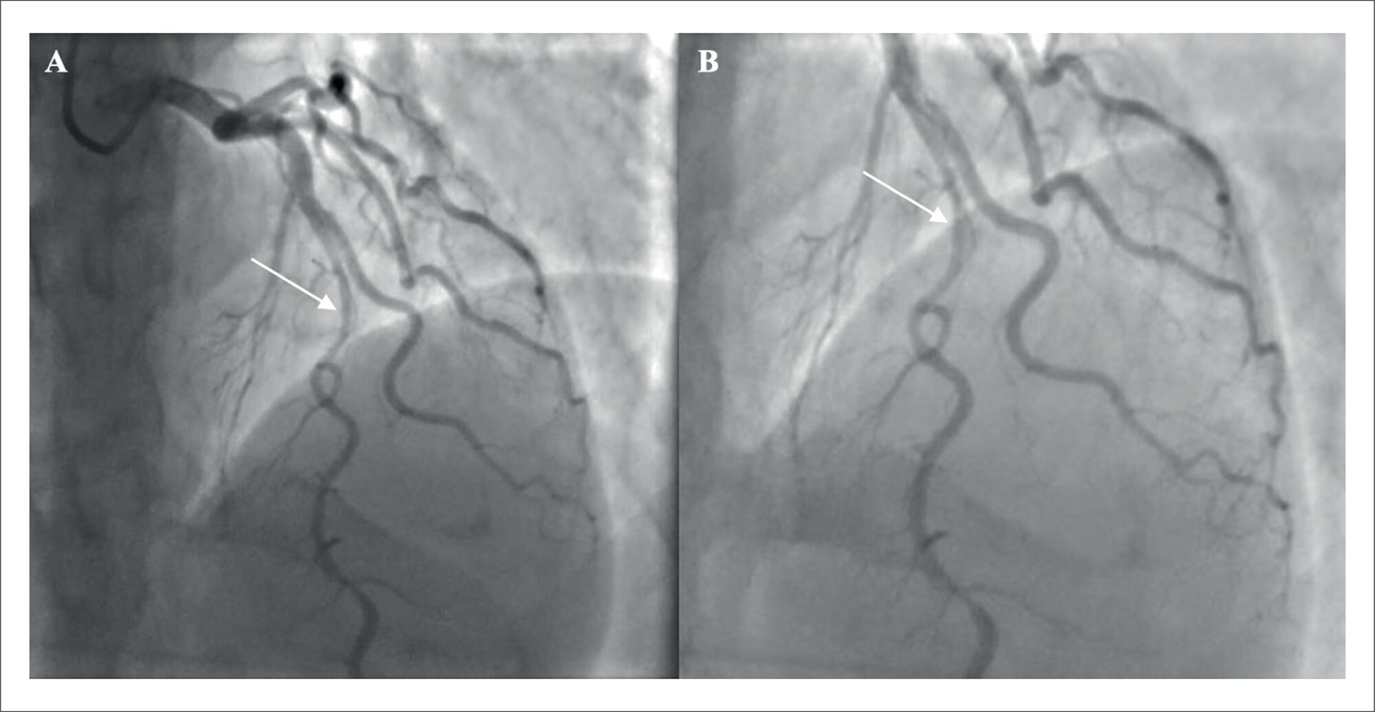
Coronary Artery Dissection Following Pharmacologic Stress Echocardiography and the Follow-Up of Clinical Management: A Case Report

Thiago Artioli et al. Bruno Querido Marcondes Santos Denis Cárdenas Monteiro Layane Bonfante Batista … Matheus Zavaris Lorenzoni Hsu Gwo Jen Kelvyn Melo Vital Kelvin Henrique Vilalva

Introduction

Spontaneous coronary artery dissection (SCAD) remains a diagnostic and therapeutic challenge in the evaluation of chest pain. Misinterpretation of symptom characteristics and failure to consider the patient’s epidemiological profile may lead to inappropriate diagnostic workups and treatment strategies. The case described herein represents only the second report with similar characteristics published in major medical databases. This case underscores the importance of considering uncommon causes of chest pain and acute coronary syndrome (ACS), while also providing a brief review of relevant literature. The report was prepared in accordance with the CAse REport (CARE) guidelines.

[…]

  • Blood Vessel Dissection
  • Case Reports
  • Coronary Disease
  • Stress echocardiography
Read Full Article
Creative Commons Attribution (CC BY) Creative Commons license
01/07/2025 DOI: 10.36660/abcimg.20250034i Views 2
Editorial

"Triple Rule-Out": Including the Abdominal Aorta With a Clear Conscience?

Tiago A. Magalhães

Less is More – Less Radiation Dose, More Patient Safety

The main strength of the study lies in its methodological approach. The authors selected patients who had previously undergone the TRO CTA protocol including the abdominal aorta with full electrocardiographic synchronization. After obtaining consent, they performed a new examination using the partial electrocardiographic synchronization protocol, enabling a robust comparison through a “matched case-control” model. This approach minimizes bias related to anatomical and technical variability among different individuals.

The results were interesting. The dose reduction was significant: the mean effective dose of the protocol with partial ECG synchronization was 47.9% lower than that of the protocol with full synchronization (17.0 mSv vs. 32.6 mSv, p<0.001). It is noteworthy that this reduction in radiation dose was not accompanied by a compromise in image quality. The quantitative assessment of attenuation values in critical vascular regions – such as the ascending aorta, descending aorta, abdominal aorta, and pulmonary trunk – showed no statistically significant differences. Similarly, the subjective qualitative evaluation demonstrated equivalence between the two protocols in terms of image quality of the assessed anatomical structures.

[…]

  • Angiography
  • Patient Safety
  • X-Ray Computed Tomography
Read Full Article
Creative Commons Attribution (CC BY) Creative Commons license
01/07/2025 DOI: 10.36660/abcimg.20250041i
Figure 1
Review Article

My Approach to Diagnose Fabry Disease

Sandra Marques e Silva

Abstract

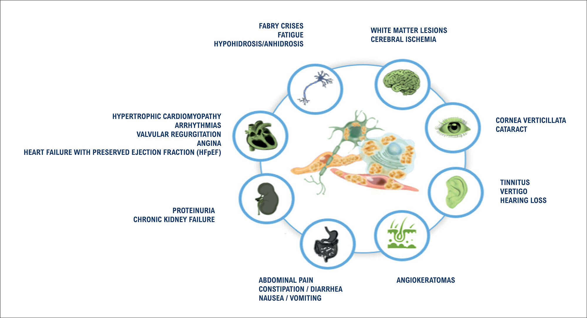
Fabry disease (FD) is an inherited lysosomal storage disorder caused by a deficiency of the enzyme alpha-galactosidase A (α-Gal A). This enzymatic defect leads to the cytoplasmic lysosomal accumulation of globotriaosylceramides (GB3 and LysoGB3), resulting in multisystemic clinical manifestations. Cardiovascular involvement, often mimicking hypertrophic cardiomyopathy, is the main determinant of morbidity and mortality, due to the development of arrhythmia, myocardial ischemia, and heart failure. Although FD is a rare condition in the general population, the availability of specific enzyme replacement therapy, which can alter the natural course of the disease, underscores the importance of including FD as a key differential diagnosis among storage cardiomyopathies. Diagnostic evaluation should encompass a thorough clinical assessment, with particular attention to patient history and physical examination, complemented by laboratory testing and imaging studies, such as electrocardiography and echocardiography. Cardiac magnetic resonance imaging, including late gadolinium enhancement and T1 and T2 parametric mapping, provides additional diagnostic and prognostic information and should ideally be performed at the time of initial diagnosis. Definitive diagnosis is established by genetic sequencing of the GLA gene, located on the long arm of the X chromosome, enabling the selection of the most appropriate therapeutic strategy for each patient.

  • Fabry Disease
  • Glycosphingolipids
  • Hypertrophic Cardiomyopathy
Read Full Article
Creative Commons Attribution (CC BY) Creative Commons license
01/07/2025 DOI: 10.36660/abcimg.20240091i
Figure 1
Case Report

Assessment of Coronary Flow Reserve by Myocardial Perfusion Scintigraphy using CZT Equipment

Wilter dos Santos Ker et al. Davi Shunji Yahiro Gabriel de Moraes Mangas Isabelle Mendes Rodrigues Salomão … José Augusto Antonio Alves Flor Hugo Matheus Silvestre Vianna Cláudio Tinoco Mesquita

Abstract

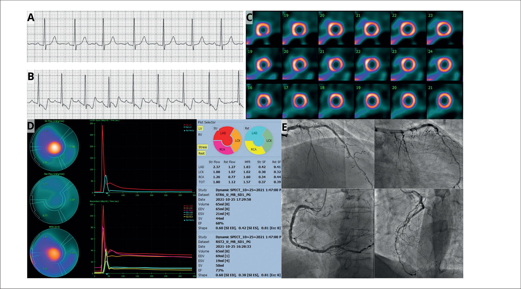
This case report presents a myocardial perfusion analysis in a 67-year-old asymptomatic male patient with known coronary artery disease (CAD) and multiple risk factors. While myocardial perfusion Imaging (MPI) with single-photon emission computed tomography (SPECT) revealed a 5% area of ischemia, the myocardial blood flow quantification assessment in a camera equipped with cadmium-zinc-telluride (CZT) detectors demonstrated a reduced global myocardial flow reserve (MFR) of 1.57. Coronary angiography confirmed extensive multivessel disease, including an 80% stenosis in the left anterior descending artery (LAD) and an occlusion of the circumflex artery (LCx), findings not fully identified by the semi-quantitative SPECT MPI. This case underscores the importance of MFR assessment, particularly using CZT technology, in detecting significant CAD that may be underestimated by traditional SPECT methods. It highlights the potential of dynamic CZT SPECT to provide more comprehensive functional information, enhancing risk stratification and potentially influencing patient management in complex cases of suspected or known CAD.

  • Coronary Artery Disease
  • Myocardial Ischemia
  • Single-Photon Emission-Computed Tomography
Read Full Article
Creative Commons Attribution (CC BY) Creative Commons license
01/07/2025 DOI: 10.36660/abcimg.20250020i Views 1
Editorial

The Use of POCUS in Daily Practice: Are We Really Ready to Use it Efficiently?

Angelo Antunes Salgado et al. Marcos Paulo Lacerda Bernardo

Research on congestion in patients with heart failure (HF) is crucial for clinical management and prognosis of the disease. Congestion (i.e., the accumulation of fluids in the body) is the main cause of hospital admission and readmission of patients with HF, and it is associated with increased mortality. Early detection and identification of the extent of congestion are essential for choosing the most appropriate therapy, preventing complications, and improving prognosis. Clinical signs of congestion may be insufficiently sensitive and specific for the diagnosis and management of patients with HF.

The point-of-care ultrasound (POCUS) is a diagnostic tool that allows immediate and dynamic visualization of ultrasound images. This tool can be used at the bedside to obtain additional clinical information for the physical examination of a specific disease or procedure. The practicality and speed with which images are obtained result in their high use at hospitals for critically ill patients. POCUS has been used in various medical specialties, especially in pulmonary and cardiac ultrasound.

[…]

  • Clinical Diagnosis
  • Heart Failure
  • Ultrasonography
Read Full Article
Creative Commons Attribution (CC BY) Creative Commons license
01/07/2025 DOI: 10.36660/abcimg.20250026i
Figure 1
Editorial

New Tools for Optimizing HF Management: The Role of POCUS in the Office

Luiz Claudio Danzmann et al. Elisa Kalil Soraya Abunader Kalil

A Reflection on Clinical and Subclinical Circulatory Congestion

The management of heart failure (HF) has improved over the past 20 years, mainly due to advances in diagnostic methods and circulatory decongestion strategies. Several management strategies have demonstrated efficacy in reducing symptoms, lowering rates of HF decompensation events, decreasing mortality indices, and improving surrogate markers of clinical events, such as left ventricular ejection fraction (LVEF). In HF management, the need for detecting and monitoring circulatory congestion is justified by its strong association with neurohormonal activation and systemic inflammation, which contribute to the progressive worsening of HF and, consequently, a higher incidence of clinical events.

But what is the evidence supporting decongestion strategies with diuretics in reducing hard outcomes? In the outpatient setting, the STRONG-HF clinical trial is a good example. Comparing intensive optimization strategy versus usual HF treatment after hospital discharge, the study demonstrated that the group with more frequent visits and more intensive management achieved an absolute risk reduction of 8.1% (15.2% vs. 23.3%, P=0.0021) in the combined outcome of all-cause mortality and/or HF hospitalization within 180 days of follow-up. The statistically significant reduction in clinical signs of congestion, along with a 27% decrease in natriuretic peptide (NP) levels after three months, provided proof of concept for the relationship between decongestion and clinical benefit. This evidence reinforces the need to detect subclinical congestion, especially during the vulnerable phase of HF, which occurs between one and three months after an HF hospitalization, a period in which most potentially preventable events take place.

[…]

  • Ambulatory Care
  • Heart Failure
  • Ultrasonography
Read Full Article
Creative Commons Attribution (CC BY) Creative Commons license
30/06/2025 DOI: 10.36660/abcimg.20250039i Views 2
Editorial

Do Patients with Homozygosity and Compound Heterozygosity for Variants in the Transthyretin Amyloidosis TTR Gene Have More Severe and Earlier Clinical Conditions?

Tonnison de Oliveira Silva et al. Samuel Ulisses Chaves Nogueira do Nascimento

The prevalence of homozygotes for the Val142Ile genetic variant in the transthyretin (TTR) gene is extremely low in the general population (0.72%). This prevalence is slightly higher in endomyocardial biopsy studies (6% to 10%) and in cohorts involving reference centers for TTR cardiac amyloidosis (4% to 14%).,

Homozygotes represent a distinct subpopulation within the spectrum of cardiac amyloidosis, with clinical characteristics that appear to differ substantially from those of heterozygotes. This genetic variation has been associated with an onset of clinical manifestations in younger patients compared with heterozygotes and also with a higher risk of serious cardiovascular events, such as heart failure and ventricular arrhythmias.³ Besides the earlier onset, observational studies indicate that the disease has a more aggressive phenotypic behavior in homozygous patients, characterized by greater ventricular thickening, worse diastolic function, and a higher load of amyloid fibrils in the heart.³ Thus, homozygotes usually have a higher risk of developing early and more severe heart failure than heterozygotes.–

[…]

  • Amyloidosis
  • Homozygote
  • Prealbumin
Read Full Article
Creative Commons Attribution (CC BY) Creative Commons license
30/06/2025 DOI: 10.36660/abcimg.20250027i Views 2
My Approach to Estimate Pulmonary Pressures: Practical Aspects
Review Article

My Approach to Estimate Pulmonary Pressures: Practical Aspects

Halsted Alarcão Gomes Pereira da Silva et al. Helder Moura Gomes

Abstract

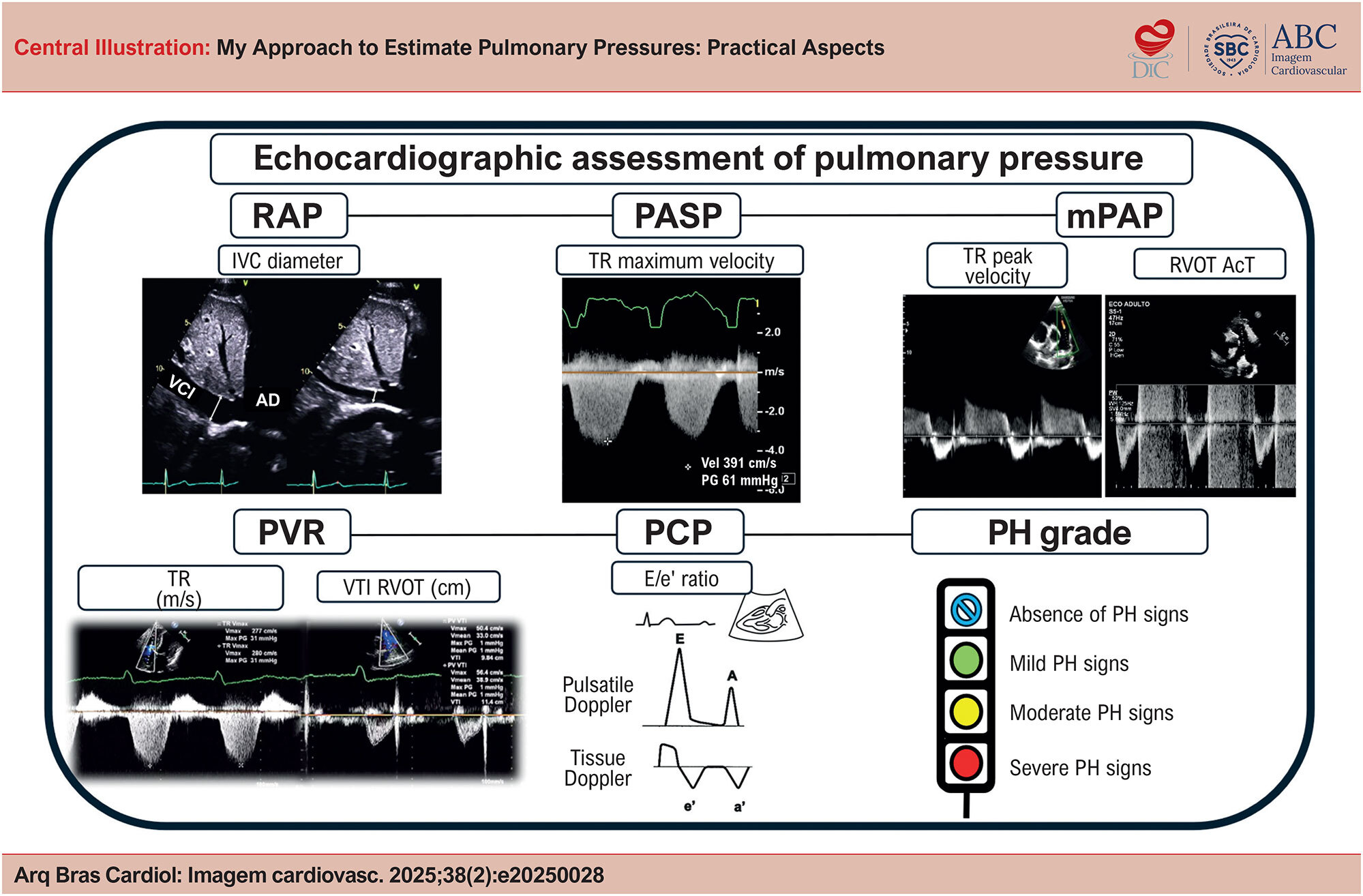
Pulmonary hypertension (PH) is a global disease and is increasingly present in echocardiography laboratories, given the progressive rise in its prevalence with population aging. Previously diagnosed only through invasive methods, echocardiographic assessment of pulmonary pressures has become fundamental in screening and monitoring patients with PH. This topic was revisited by the American Society of Echocardiography in a new guideline published in March 2025, reinforcing the importance of echocardiography in clinical stratification, considering recent updates in the definition of PH. Among the main assessment parameters, systolic pulmonary artery pressure, mean pulmonary artery pressure (mPAP), pulmonary vascular resistance (PVR), and pulmonary capillary pressure (PCP) are described, along with a series of precautions for the correct estimation and interpretation of these values. The classification of PH levels has also been revised in a new framework. As an accessible and reproducible method, the assessment of pulmonary pressures through echocardiography stands out as an essential tool in the evaluation and follow-up of PH.

  • Doppler ultrasonography
  • Echocardiography
  • Pulmonary Hypertension
  • Pulmonary Wedge Pressure
Read Full Article
Creative Commons Attribution (CC BY) Creative Commons license
30/06/2025 DOI: 10.36660/abcimg.20250028i Views 1
Editorial

Mental Traps That Impact Diagnostic Imaging

Maurício Rodrigues Jordão et al. Marcelo Tavares Fábio Fernandes

Daniel Kahneman reinforced and popularized the idea of two distinct modes of thinking. System 1 relies on pattern recognition, detecting simple relationships, correlating stereotypes, and associating ideas in a search for coherence. It is fast, intuitive, automatic, and requires little or no effort or energy. It cannot be turned off and continuously monitors the environment, driven by a survival need rooted in our ancestral past. System 2, on the other hand, demands attention, compares objects based on various attributes, and makes deliberate choices among options. It evaluates scenarios using logical and statistical reasoning. Because it requires attention, its activity is interrupted when that attention is diverted. Therefore, tasks that require effort interfere with one another (for instance, making a turn while driving and performing a complex calculation simultaneously is impossible). However, System 2 is slow, deliberate, effortful, and orderly. The two systems are interconnected: System 1 effortlessly generates impressions and feelings that serve as the main source for the explicit beliefs and deliberate choices made by System 2. In daily life, we often make decisions based on heuristics—mental shortcuts closely linked to System 1—used to save time and energy. While extremely efficient, this faster way of thinking opens the door to cognitive biases, which are systematic tendencies that distort logical reasoning and lead to flawed decisions.

The causes of error in diagnostic medicine can be categorized into two main groups: perceptual errors and interpretive errors. Perceptual errors occur when an abnormality is present on the image but goes undetected. These may be related to image acquisition, post-processing, or equipment quality. Interpretive errors arise when the abnormality is seen but its significance is misunderstood. In such cases, cognitive biases may be involved and are estimated to account for up to 74% of diagnostic errors.– Approximately 30 types of cognitive biases have been described in literature. In diagnostic imaging, the most relevant include anchoring, confirmation bias, availability bias, attribution bias, framing effect, and satisfaction of search.-,

[…]
  • Bias
  • Diagnostic Errors
  • Healthcare Near Miss
  • Heuristics
Read Full Article
Creative Commons Attribution (CC BY) Creative Commons license
30/06/2025 DOI: 10.36660/abcimg.20250040i Views 1
Figure 1
Editorial

Photon-Counting Computed Tomography in Cardiovascular Imaging: Where We Are and What Lies Ahead

Paulo Savoia et al. Rodrigo Bello

Computed Tomography Angiography (CTA) has been an important asset in cardiovascular diagnosis for decades. The continuous improvement of rapid data acquisition with often higher spatial resolution has established the method as indispensable for diagnosing and managing multiple cardiovascular entities such as aneurysms, aortic dissection, and pulmonary embolism., The use of CTA in heart and coronary imaging has also gained important territory and relevance., Computed Tomography (CT) techniques have been advancing significantly since its introduction in the medical field. Starting with single detectors, then helicoidal scanners, multidetector scanners with high pitch, improvement of reconstruction algorithms, the appearance of dual-energy technology, and lately the ability to acquire images of the whole heart in just one heartbeat with information from the entire cardiac cycle, are incredible milestones. Fractional Flow Reserve CT (FFR-CT) is also a very promising technique. Progression does not come without downsides: ionizing radiation is still concerning, and despite ways to reduce it such as dose modulation, some cardiovascular CTA exams still show relatively high levels of absorbed radiation, especially exams with multiple series, extremely thin slices, and retrospective reconstruction of larger parts of the cardiac cycle. Following this trend of improving image quality with less ionizing radiation as possible, a new complex technique emerged: Photon-counting CT (PCCT).

The main distinction between conventional CT and PCCT is in the detectors of X-rays. The conventional CT detector is based on receiving the X-ray photons that interact with the patient, converting them into light photons (it is an indirect system), and then converting this light into electrical signals that ultimately are going to be converted into digital signals. This system is called an Energy-Integrating Detector (EID). Basically, more light means more X-rays arriving and less interaction / more penetration in the patient’s tissues. It is not possible to differentiate exactly how many X-rays are arriving, nor the different energy levels of each X-ray photon. Moreover, the septa between each of the detectors also limit the reception of X-rays., On the other hand, PCCT detectors can detect separately each X-ray photon that is arriving and measure the energy of each one of the X-ray photons directly. This is possible because the PCCT detector has a high-voltage crystal layer that relocates electrons for each X-ray photon received, and the number of electrons relocated is proportional to the energy of each X-ray. The relocated electrons generate the electrical pulse that is going to be finally converted into the digital signal. And even smaller energy X-rays can be detected, improving the image contrast-to-noise ratio. It is a direct system, and there is no septa between the detectors. In the end, less X-rays are necessary (therefore less radiation) and it is possible to separate the X-rays according to the energy of each one, leading to applications of tissue recognition (different known energy X-rays are likely to have interacted with different known tissues) functioning as a dual energy CT.,

[…]

  • Cardiology
  • Cardiovascular Diagnostic Techniques
  • Computed Tomography Angiography
  • Radiology
Read Full Article
Creative Commons Attribution (CC BY) Creative Commons license
27/06/2025 DOI: 10.36660/abcimg.20250035i Views 1
Figure 1
Review Article

My Approach To Echocardiographic Evaluation in Pediatric Patients with Sickle Cell Disease

Viviane Thome Gonçalves Dias et al. Mirela Frederico de Almeida

Abstract

Sickle cell disease (SCD) is the most prevalent hereditary genetic disorder worldwide, and cardiovascular alterations are the main cause of death among patients with this disease.

Therefore, the early identification of markers of sickle cell cardiomyopathy using echocardiographic parameters (Central Illustration) is essential for the appropriate management of the cardiovascular condition in these patients.

This review provides a comprehensive overview of the use of echocardiography in pediatric patients with SCD, highlighting the main characteristics of the condition and the importance of regular follow-up.

  • Child
  • Echocardiography
  • Sickle Cell Anemia
Read Full Article
Creative Commons Attribution (CC BY) Creative Commons license
27/06/2025 DOI: 10.36660/abcimg.20240128i Views 1
Hypertrophic Cardiomyopathy: Analysis of Septal Thickness with Gradient Reduction in Patients Undergoing Radiofrequency Septal Ablation
Original Article

Hypertrophic Cardiomyopathy: Analysis of Septal Thickness with Gradient Reduction in Patients Undergoing Radiofrequency Septal Ablation

Andrea de Andrade Vilela et al. Mariane Higa Shinzato Bruno Pereira Valdigem Antonio Tito Paladino … Edileide de Barros Correia Jorge Eduardo Assef

Abstract

Background:

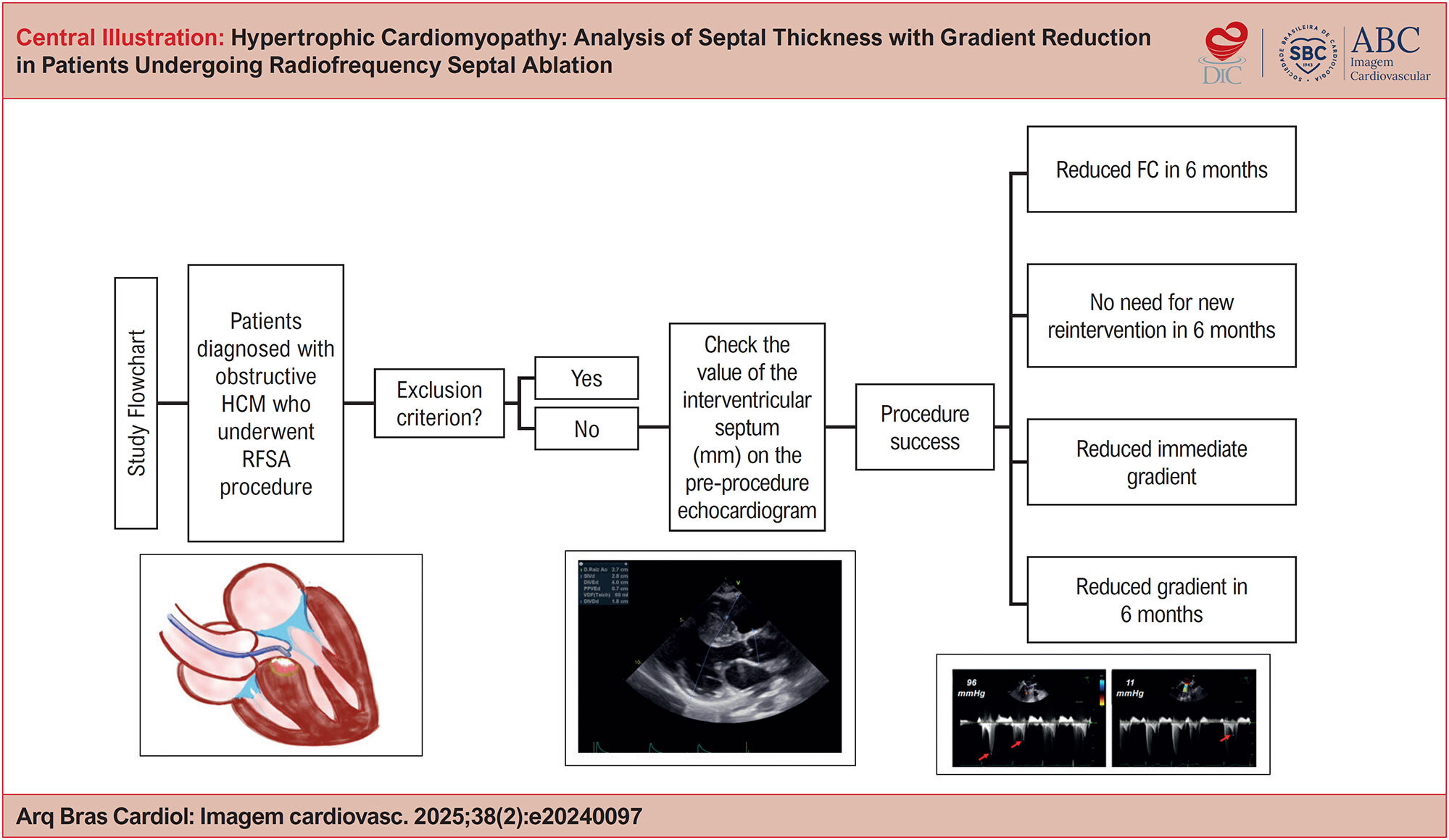
Hypertrophic obstructive cardiomyopathy (HOCM) is an autosomal dominant genetic disorder. Invasive treatment for intraventricular gradient reduction is indicated in patients (p) who are refractory to clinical treatment with gradient ≥ 50 mmHg. Radiofrequency ablation (RFA) is one of the current options for invasive management.

Objectives:

To evaluate the correlation between interventricular septal diameter and procedural success, defined as immediate post-procedural gradient reduction, improvement in functional class (FC), and sustained gradient reduction at 6-month follow-up.

Methods:

Twenty-two patients with HOCM were included. Transthoracic echocardiography was performed before and six months after RFA, while transesophageal echocardiography (TEE) was carried out intraprocedure.

Results:

The mean age was 56.64 years (± 12.23), with 68.18% of patients being female and 73% hypertensive. An immediate gradient reduction greater than 50% was observed in 72.7% of patients. At the 6-month follow-up, 60% of patients showed a ≥ 50% gradient reduction along with an improvement in FC. A median septal thickness of 18 mm was associated with greater procedural success compared to a median of 15 mm, and no significant change in septal thickness was observed over the 6-month follow-up period.

Conclusion:

Radiofrequency septal ablation (RFSA) is an effective and safe technique for the invasive management of HOCM. The procedure enables a significant reduction in the intraventricular gradient, accompanied by improved FC and sustained short-term results. Thicker septal were associated with greater intraprocedural gradient reductions; however, there was no reduction in thickness during the 6-month follow-up.

  • Hypertrophic Cardiomyopathy
  • Radiofrequency Ablation
Read Full Article
Creative Commons Attribution (CC BY) Creative Commons license
27/06/2025 DOI: 10.36660/abcimg.20240097i Views 3
Figure 1
Editorial

Radiofrequency in Obstructive Hypertrophic Cardiomyopathy: The Role of Imaging in the Assessment of Septal Thickness and Gradient Reduction

Alexandre Costa Souza et al. Marcus Vinicius Silva Freire de Carvalho

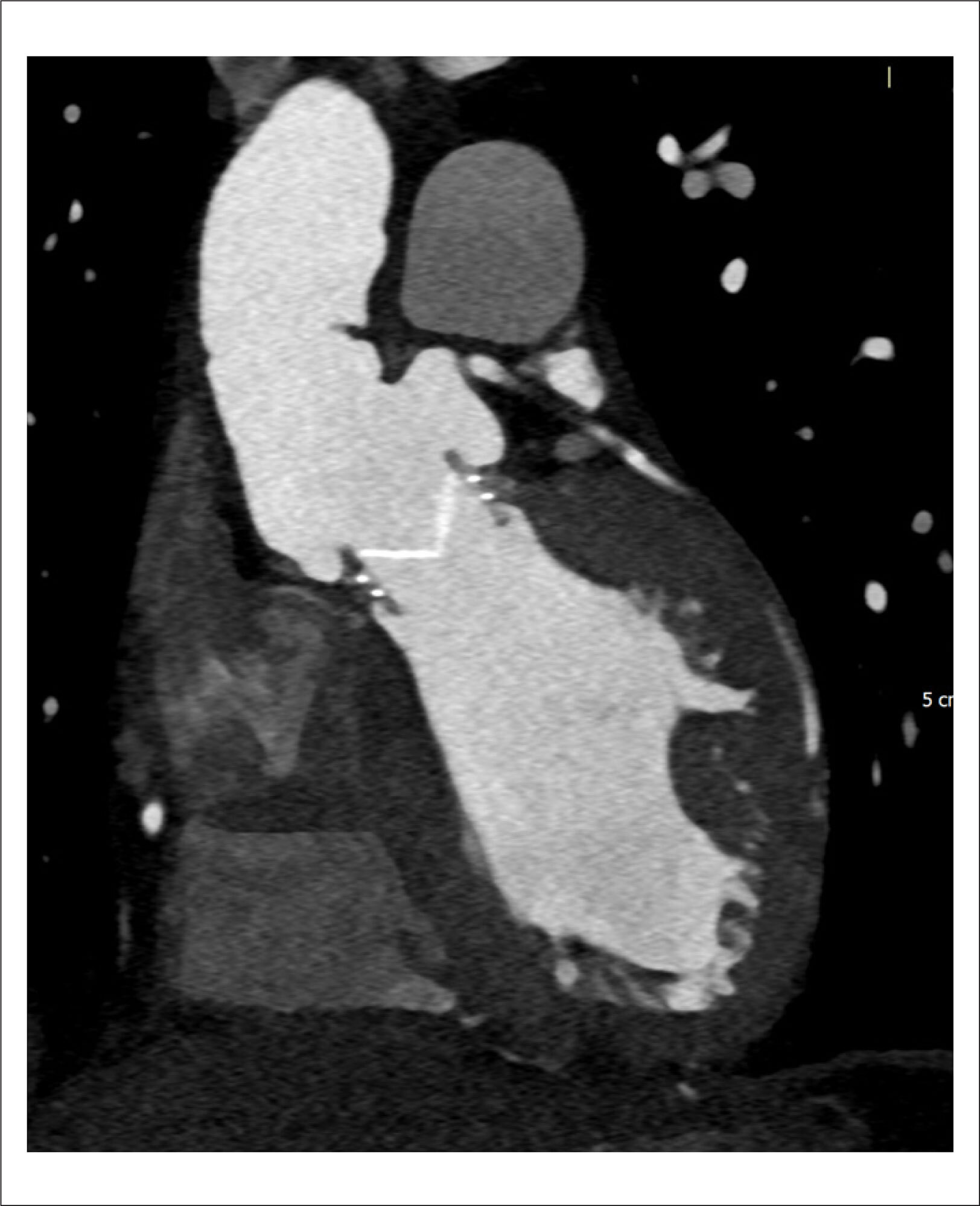
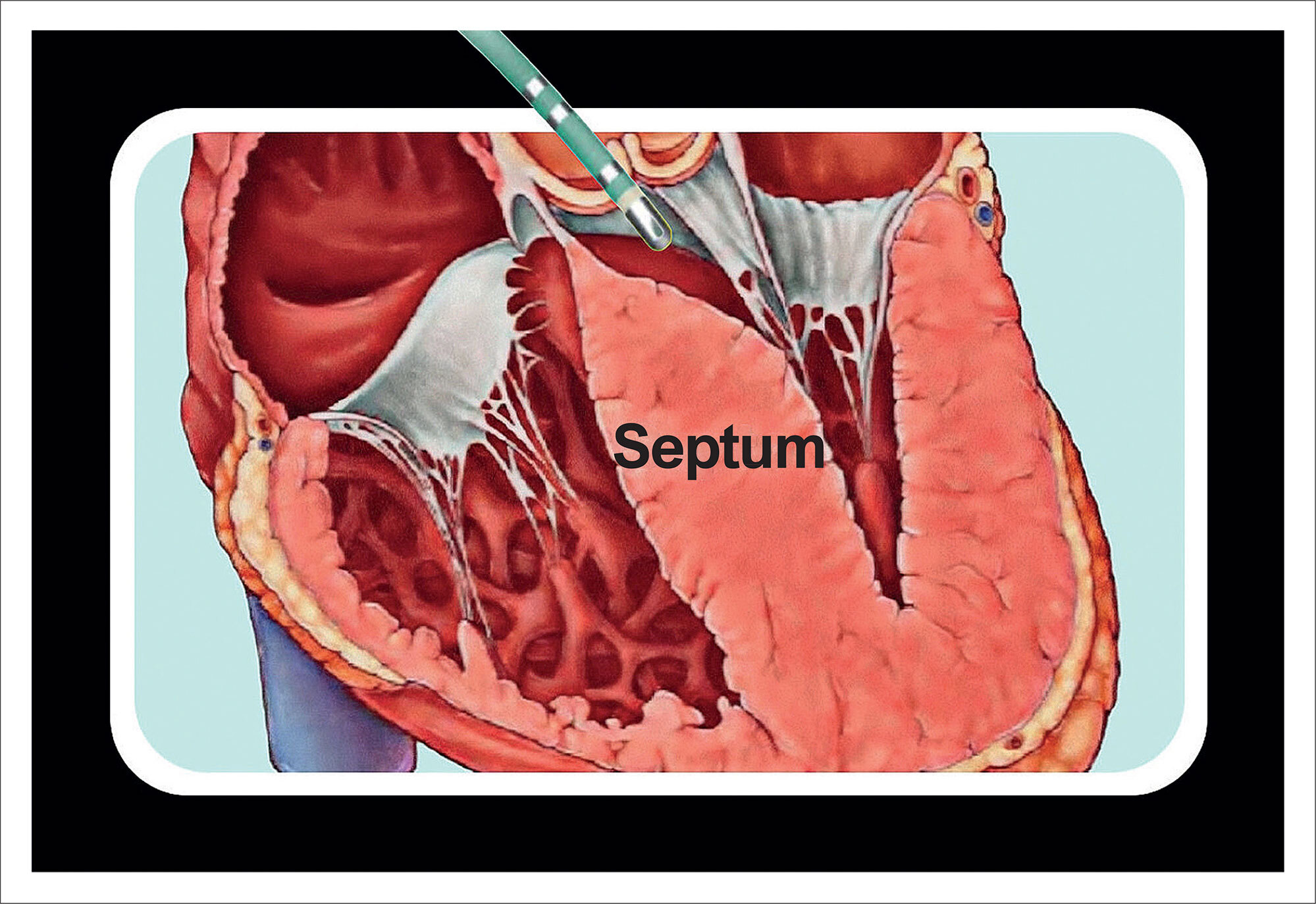
Obstructive hypertrophic cardiomyopathy (HCM) is a genetic disorder characterized by pronounced myocardial hypertrophy and varying degrees of fibrosis. The obstruction of the left ventricular outflow tract (LVOT) is an anatomical condition with dynamic behavior that can lead to symptoms such as exercise intolerance and reduced functional class, and is also associated with cases of sudden cardiac death.

The indication for invasive treatment aimed at reducing the intraventricular gradient is reserved for symptomatic patients —those who are refractory to medical therapy — with a gradient ≥ 50 mmHg. Common interventional therapies for this condition include surgical myectomy or, in a less invasive approach, alcohol septal ablation of coronary septal branches. However, these interventions have shown highly variable outcomes in the literature and remain infrequently used in clinical practice.,

[…]

  • Cardiomyopathies
  • Left Ventricular Outflow Obstruction
  • Radiofrequency Ablation
Read Full Article
Creative Commons Attribution (CC BY) Creative Commons license
27/06/2025 DOI: 10.36660/abcimg.20250024i Views 1
Figure 1
Case Report

Important Mitral Regurgitation and Ventricular Dysfunction in Hypereosinophilic Syndrome: A Case Report

Marcel Pina Ciuffo et al. Lucas Mori Tais Araújo Ingrid Kowatsch Ana Clara Tude Rodrigues

Introduction

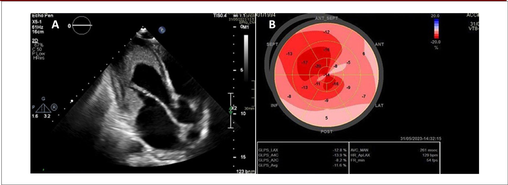
Hypereosinophilic syndrome (HES) represents a group of rare hematologic diseases characterized by an increased number of eosinophils in the peripheral blood. Once activated, these eosinophils can cause damage to host tissues and organs. Patients with HES generally present with dermatological, respiratory, and gastrointestinal signs and symptoms. Involvement of the cardiovascular system is uncommon and can lead to serious consequences such as heart failure, arrhythmias, and thromboembolic phenomena.

This article describes a case of eosinophilic endomyocardial disease, also known as Loeffler syndrome, a manifestation of HES characterized by eosinophil-mediated cardiac damage. Despite adequate treatment and control of the hematologic disease, the patient developed biventricular systolic dysfunction and important mitral regurgitation (MR).

[…]

  • Heart Failure
  • Hypereosinophilic Syndrome
  • Mitral Valve Insufficiency
Read Full Article
Creative Commons Attribution (CC BY) Creative Commons license
27/06/2025 DOI: 10.36660/abcimg.20250029i
My Approach to Point-of-Care Ultrasound for Dyspnea Assessment
Review Article

My Approach to Point-of-Care Ultrasound for Dyspnea Assessment

Adriana Brentegani et al. Fernando Arturo Effio Solis Milena de Paulis Marcelo Luiz Campos Vieira

Abstract

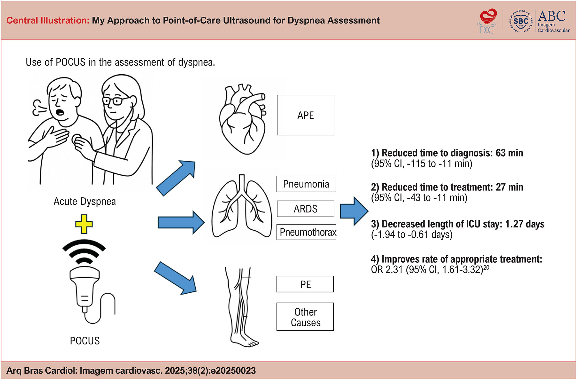
Dyspnea is a common and potentially serious symptom whose traditional diagnostic approach, based on physical examination and complementary tests, may present significant limitations. Point-of-care ultrasound (POCUS) has emerged as an effective tool for the rapid and accurate assessment of this symptom, both in outpatient settings and in emergency and critical care environments.

This article provides a practical, illustrated guide to the integrated application of POCUS in the evaluation of dyspnea, with an emphasis on distinguishing cardiac from pulmonary causes. It describes techniques for acquiring cardiac, pulmonary, and venous ultrasound images, highlights key sonographic findings, and outlines a step-by-step approach to clinical application. Lung POCUS has demonstrated high sensitivity in detecting differential diagnoses of dyspnea. Its integration with echocardiography and lower limb ultrasound further refines diagnosis by identifying specific clinical patterns such as acute pulmonary edema (APE), pulmonary embolism (PE), and acute respiratory distress syndrome (ARDS).

The structured use of POCUS represents a significant advancement in the modern physical exam, enabling faster and more accurate bedside clinical decision-making. Its adoption should be encouraged among cardiologists, intensivists, and healthcare professionals working in critical care settings.

  • Dyspnea
  • Echocardiography
  • Ultrasonography
Read Full Article
Creative Commons Attribution (CC BY) Creative Commons license
27/06/2025 DOI: 10.36660/abcimg.20250023i Views 1
Figure 1
Original Article

Assessment of Interrater Reliability in Point-of-Care Ultrasound for Assessing Congestion in Cardiovascular Intensive Care

Marina Petersen Saadi et al. Guilherme Pinheiro Machado Gustavo Paes Silvano João Pedro da Rosa Barbato … Renato Ferraz Almeida Fernando Luis Scolari Guilherme Heiden Telo Anderson Donelli da Silveira

Abstract

Background:

The assessment of congestion is critical for managing patients with cardiovascular conditions, including heart failure (HF). Traditional methods often lack sensitivity, whereas point-of-care ultrasound (POCUS) provides an objective bedside alternative.

Objective:

To evaluate the interrater reliability (IRR) of key POCUS variables used to assess hemodynamic, pulmonary, and venous congestion.

Methods:

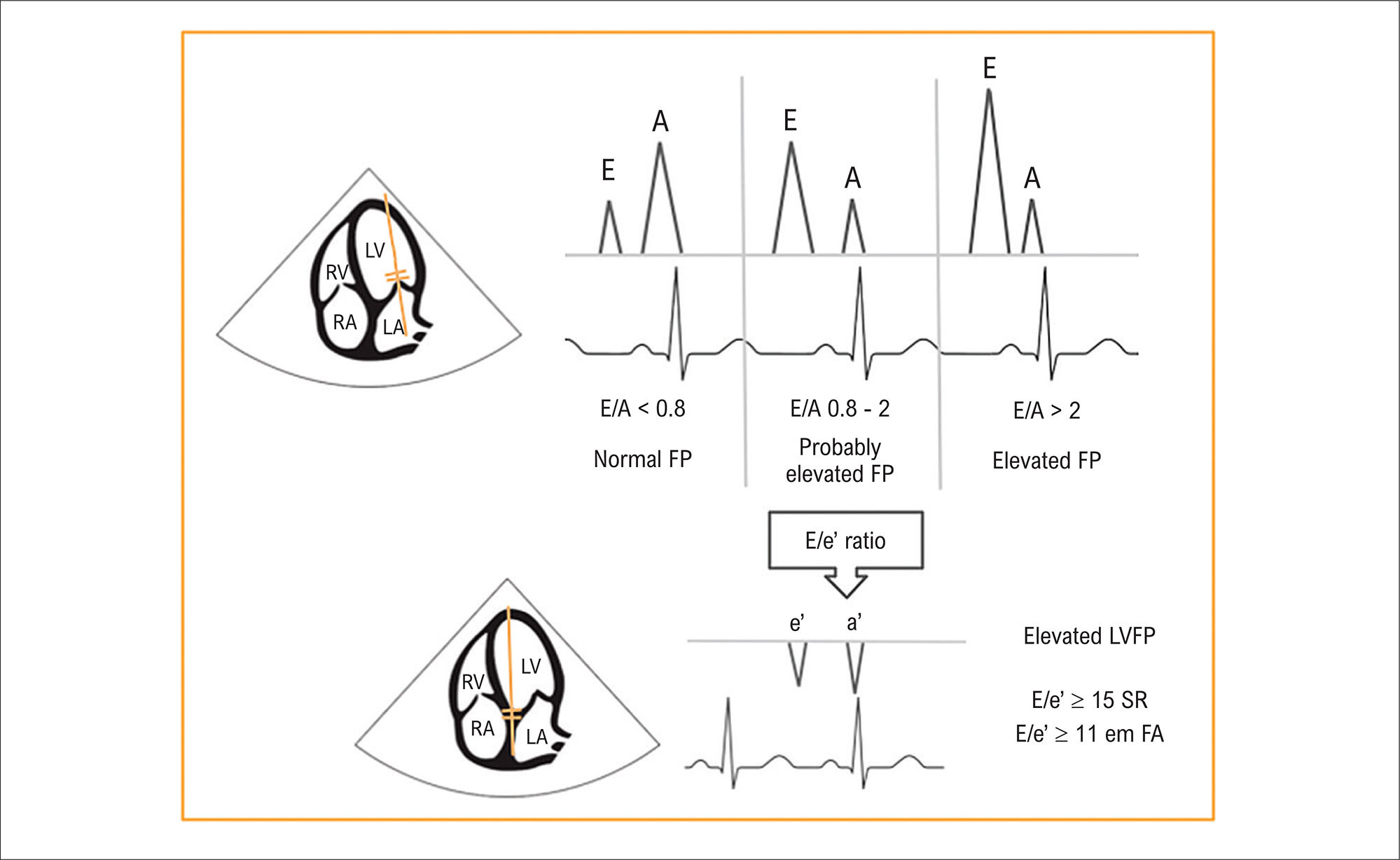
This single-center, prospective study was conducted from January to June 2023 in a cardiovascular intensive care unit (CICU) in Brazil. Adult patients underwent standardized POCUS examinations. Three trained investigators independently assessed lung ultrasound (LUS) (B-lines), left ventricular filling pressures (LVFP) (E/A and E/e′ ratios), inferior vena cava (IVC) measurements, hepatic and portal vein Doppler flow, and modified Venous Excess Ultrasound (mVExUS) Score. IRR was analyzed using intraclass correlation coefficient (ICC).

Results:

A total of 23 patients were included, with a median age of 65 years, each undergoing three independent POCUS examinations (69 total assessments). LUS and IVC measurements showed excellent IRR (ICC 0.903). Hepatic and portal vein flows demonstrated good IRR (ICCs 0.808 and 0.796, respectively). mVExUS grading achieved the highest IRR (ICC 0.957). The E wave showed excellent IRR (ICC 0.934), while the A wave and e′ velocity had lower IRRs (0.512 and 0.399, respectively). E/e′ and E/A ratios demonstrated moderate-to-good IRR (ICCs 0.662 and 0.852, respectively). The median exam duration was 10 minutes.

Conclusion:

POCUS variables demonstrated high reproducibility, particularly for LUS and mVExUS. The reproducibility of LUS and mVExUS was higher than that of LVFP parameters, suggesting they may be more reliable than traditional measures such as E/e′. These findings support the use of POCUS for standardized assessment of congestion. Further studies are needed to validate its prognostic value.

  • Heart Failure
  • Intensive Care Units
  • Ultrasonics
  • VExUs, modified VExUS
Read Full Article
Creative Commons Attribution (CC BY) Creative Commons license
27/06/2025 DOI: 10.36660/abcimg.20250022i Views 2
Figure 1
Original Article

Conventional and Partially ECG-Gated Triple Rule-Out Computed Tomography Angiography with Extension to Abdominal Aorta: Comparative Radiation Dose and Imaging Quality

Pamela Bertolazzi et al. Carla Franco Greco Silva Leonardo Iunes Fernando Freitas de Oliveira … Fabio Payão Pereira Publio Cesar Cavalcanti Viana Isac Castro Natally Horvat José Arimateia Batista Araújo-Filho

Abstract

Background:

Triple rule-out (TRO) computed tomography angiography (CTA) is an ECG-gated protocol that enables the simultaneous evaluation of the coronary arteries, thoracic aorta, and pulmonary arteries in a single scan. It is especially useful for patients in the emergency department with low to moderate risk of acute coronary syndrome, particularly when aortic dissection and pulmonary embolism are also considered in the differential diagnosis.

Objective:

This study aimed to compare two TRO protocols, fully electrocardiogram (ECG)-gated (protocol A) and partially ECG-gated (protocol B), both including coverage of the abdominal aorta, in terms of radiation dose and image quality. Such a comparison of protocols can be useful when iterative reconstruction algorithms are not available and a manual transition between partially ECG-synchronized protocols is required.

Methods:

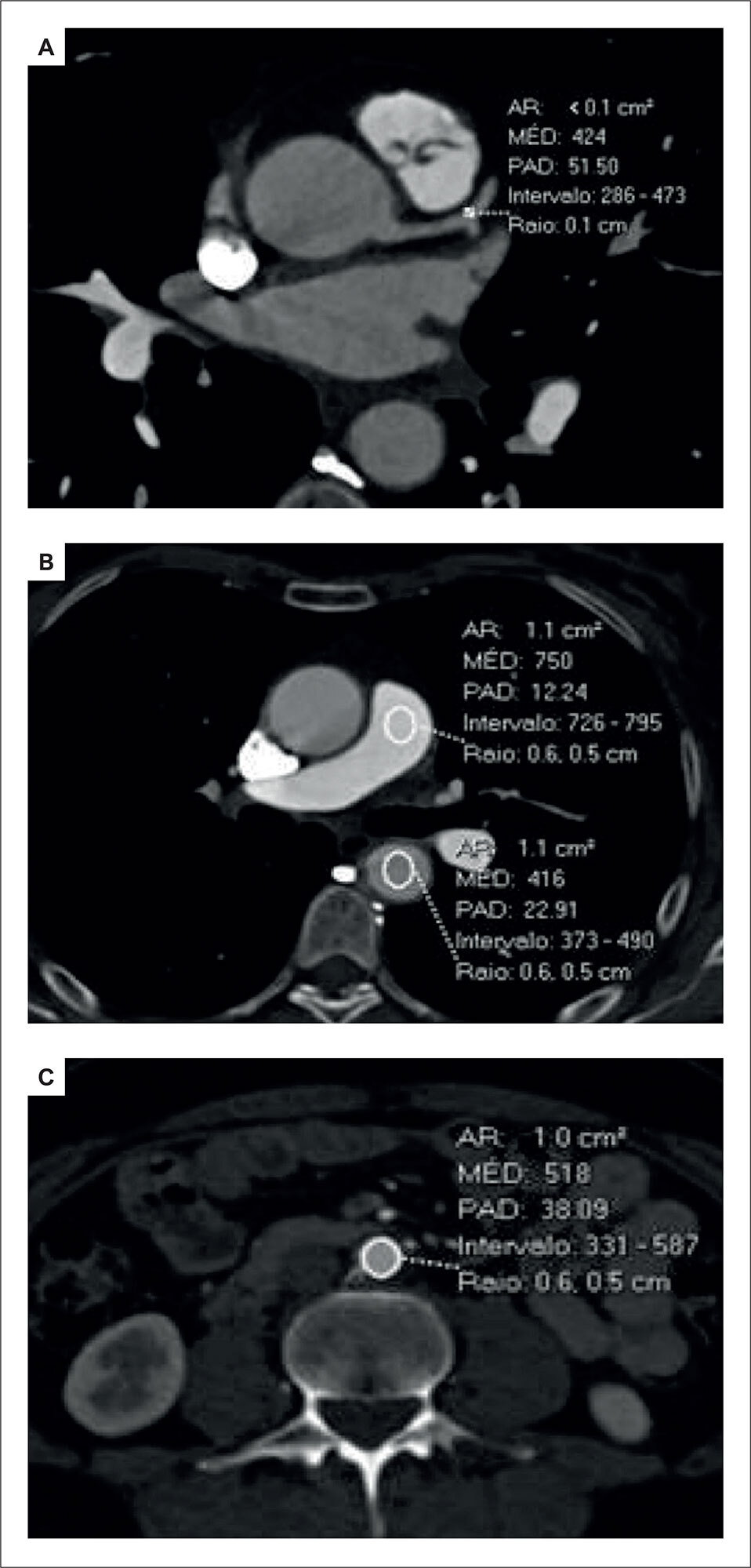
Radiation dose was evaluated using dose-length product (DLP), effective dose (ED), and virtual dose. Attenuation values were measured in the coronary and pulmonary arteries, as well as in the descending and abdominal aorta. Image quality and vessel conspicuity were assessed using a 5-point Likert scale.

Results:

A total of 56 patients were included. Protocol B demonstrated significantly lower radiation exposure compared to protocol A across all metrics: median DLP (1.1 mSv [interquartile range: 0.9 to 1.1] versus 2.2 mSv [interquartile range: 1.6 to 2.8]), ED (17.0 mSv [interquartile range: 14.3 to 18.1] versus 32.6 mSv [interquartile range: 24.4 to 42.7]), and virtual dose (16.2 [interquartile range: 9.3 to 20.4] versus 34.7 [interquartile range: 19.9 to 43.5]); all differences were statistically significant (p < 0.001). There were no significant differences in attenuation measurements or qualitative image assessment between the two protocols.

Conclusion:

Partially ECG-gated TRO CTA provides comparable image quality to the fully ECG-gated technique while significantly reducing radiation exposure, becoming a more radiation dose-efficient alternative when iterative reconstruction algorithms are not available and manual transition is necessary.

  • Drug Tapering
  • Electrocardiography
  • Radiation Dose-Response Relationship
Read Full Article
Creative Commons Attribution (CC BY) Creative Commons license
27/06/2025 DOI: 10.36660/abcimg.20250025i
Figure 1
Review Article

My Approach to Intracardiac Echocardiography During Atrial Fibrillation Ablation

Simone Nascimento dos Santos et al. Benhur Davi Henz Maria Eduarda Leite da Silva Luiz Roberto Leite da Silva

Abstract

Atrial fibrillation (AF) is the most common arrhythmia encountered in clinical practice and is associated with serious complications such as stroke and heart failure. The prevalence of AF is increasing significantly due to population aging and related comorbidities. In addition, advances in technology — particularly the detection of AF through wearable devices such as smartwatches — have enabled earlier diagnosis. Intracardiac echocardiography (ICE) is a valuable imaging modality in electrophysiology and hemodynamic labs and is well-supported by the literature.– Since the early studies conducted at the Mayo Clinic,, ICE has rapidly become an indispensable tool for assessing cardiac anatomy. ICE enables real-time visualization of catheter positioning, tissue contact, its relationship to arrhythmogenic substrates and ablation targets, lesion formation, and procedural complications. Several recent studies have demonstrated the benefits of ICE in complex ablations, including reduced procedure times and complication rates, such as lower risks of cardiac tamponade and mortality, along with higher success rates (Central Illustration).– ICE-guided procedures are increasingly used in clinical practice across a variety of settings. They are considered feasible, safe, and associated with decreased fluoroscopy exposure — or even complete fluoroless procedures, as implemented by several groups in Brazil and worldwide.,– This article provides a step-by-step “My Approach to” guide on how to perform ICE during AF ablation.

  • Atrial Fibrillation
  • Catheter Ablation
  • Intracardiac Echocardiography
Read Full Article
Creative Commons Attribution (CC BY) Creative Commons license
17/06/2025 DOI: 10.36660/abcimg.20250021i Views 4
Editorial

Intracardiac Echocardiography: Current Status, Clinical Benefits, and Future Perspectives

Renner Augusto Raposo Pereira et al. Daniel Moreira Costa Moura

Intracardiac echocardiography (ICE) is an ultrasound imaging technique capable of providing real-time, high-quality visualization of cardiac structures and their adjacent tissues through a catheter with a transducer inserted directly into the heart chambers, typically via transfemoral venous access. By allowing direct and precise visualization of intracardiac anatomy, ICE has been increasingly used to guide structural interventions and catheter ablations for cardiac arrhythmias, as well as to enable immediate detection of intraoperative complications.

Initial clinical investigations began in the 1960s, focusing on the assessment of atrial septal defects. Since then, ICE has evolved significantly. The introduction of the phased-array transducer, the addition of color Doppler functionality, and advancements in manipulation systems have enabled its widespread clinical application. The latest version of phased-array ICE features a position sensor at the catheter tip, allowing real-time integration with three-dimensional electroanatomic mapping systems used in complex ablations. The ability to precisely maneuver the catheter, combined with its versatile positioning, makes ICE a valuable tool for anatomic navigation and real-time monitoring during complex interventions.

[…]

  • Cardiac Electrophysiology
  • Echocardiography
  • Patient Safety
Read Full Article
Creative Commons Attribution (CC BY) Creative Commons license
17/06/2025 DOI: 10.36660/abcimg.20250031i
Figure 1
Image

Idiopathic Multiple Coronary Artery Aneurysms

Alberto Vera et al. Virginia Álvarez

Case presentation

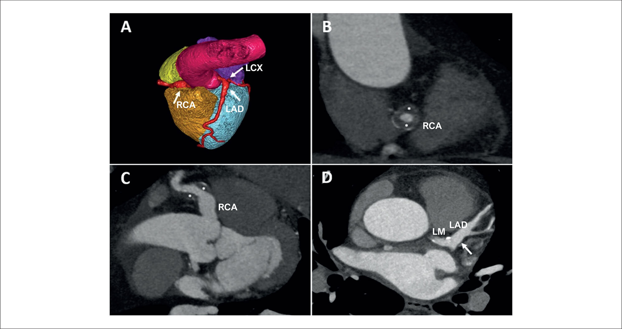
A 54-year-old patient with no previous medical history was evaluated in the cardiology clinic for chest pain with atypical characteristics. Due to a pretest probability of ischemic heart disease of 20%, a coronary computed tomography scan was performed, which showed a coronary aneurysm (10 × 11 mm) from the origin of the right coronary artery (RCA) to the mid-segment (, white arrow; ). In the RCA mid-segment, there was partial circumferential thrombosis with 50% lumen stenosis ( and ; , white asterisks). The rest of the RCA showed no coronary artery obstruction. A 9 × 9 mm aneurysm was also observed in the left main coronary artery extending to the bifurcation and proximal portion of the left anterior descending coronary artery, without significant lesions ( and , white arrows; ). A 5 × 6 mm aneurysm was also observed in the proximal left circumflex artery, without significant coronary lesions (, white arrow; ). The study was completed with a regadenoson stress cardiac magnetic resonance that was negative for inducible ischemia. Serologies and autoantibodies were also requested, and they were negative. The decision was made to initiate medical treatment with acetylsalicylic acid and atorvastatin. During the 6-month follow-up, the patient did not present coronary outcomes and remained asymptomatic.

A coronary artery aneurysm (CAA) is typically defined as a focal dilation of a coronary artery that exceeds 1.5 times the diameter of an adjacent normal segment, whereas coronary ectasia refers to a diffuse dilation involving at least 50% of the artery’s length. Giant coronary aneurysms, although rare, are classified as those measuring more than 20 mm in diameter. This distinction is clinically significant, as ectasia represents a more extensive and generalized involvement of the artery, whereas aneurysms are focal and may carry a different prognosis and management approach.

[…]

  • Case Reports
  • Coronary Aneurysm
Read Full Article
Creative Commons Attribution (CC BY) Creative Commons license
17/06/2025 DOI: 10.36660/abcimg.20240106i
Assessment of Left Ventricular Function by MAPSE (Mitral Annular Plane Systolic Excursion): Main Clinical Applications
Review Article

Assessment of Left Ventricular Function by MAPSE (Mitral Annular Plane Systolic Excursion): Main Clinical Applications

Andressa Alves de Carvalho et al. Wanessa Alves de Carvalho Luis Fabio Barbosa Botelho Marcelo Dantas Tavares de Melo

Abstract

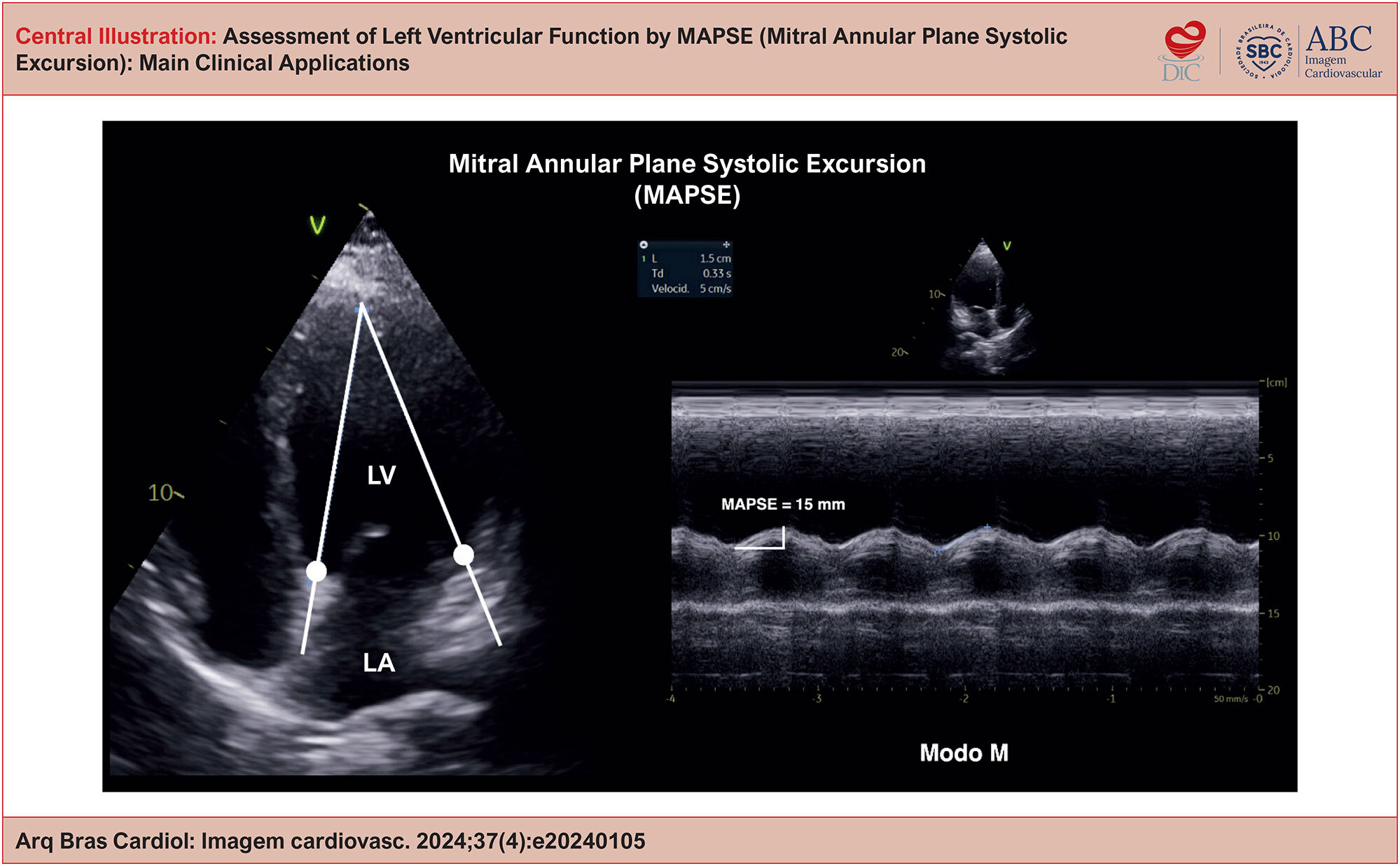
Mitral annular plane systolic excursion (MAPSE), a parameter that can be obtained from transthoracic echocardiography or nuclear magnetic resonance (NMR), is capable of reflecting the longitudinal systolic function of the left ventricle (LV) and may change earlier than the LV ejection fraction (LVEF), estimated by usual methods. Furthermore, it also presents a correlation with global longitudinal strain (GLS), assessed by speckle tracking in two-dimensional (2D) echocardiography. The importance of MAPSE extends to the ease with which it can be obtained and reproduced, especially in patients with poor image quality, for whom LVEF and GLS may be inaccurate. This review intends to clarify the evidence available in the literature on MAPSE, which, despite its applicable usefulness in the context of various heart diseases, is an underused index in clinical practice.

  • Ecocardiografia
  • Função Ventricular
  • Valva Mitral
Read Full Article
Creative Commons Attribution (CC BY) Creative Commons license
28/05/2025 DOI: 10.36660/abcimg.20240105i Views 4
Figure 1
Case Report

Large Heterogeneous Mass in the Right Chambers: A Case Report in Cardio-Oncology

João Pedro Perfeito Frigo et al. Themissa Helena Voss

Abstract

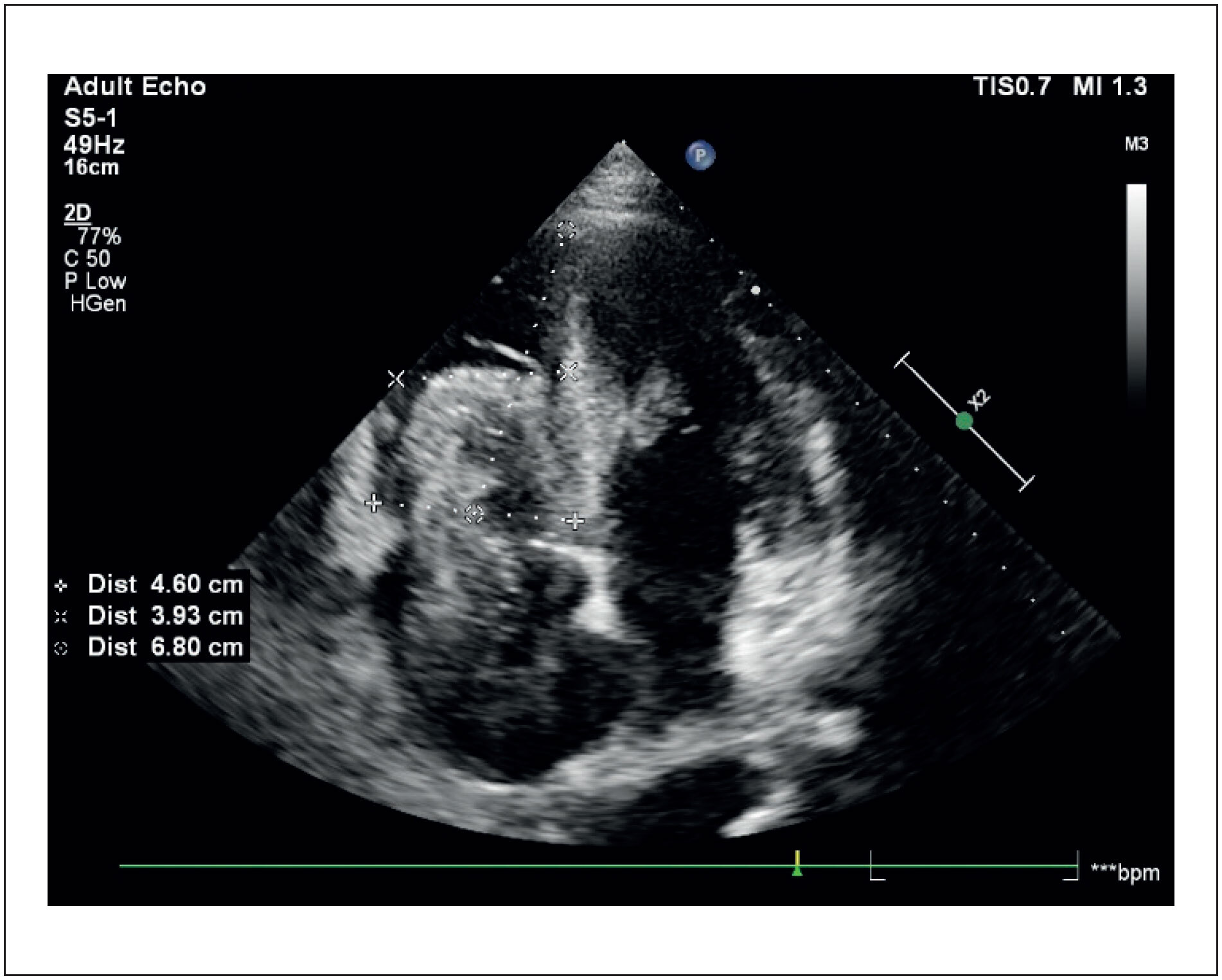
Renal cell carcinoma (RCC) is one of the most lethal cancers. It is frequently associated with the formation of tumor thrombi (TT), an intravascular extension of the tumor. Approximately 4% to 10% of patients with RCC have TT, which can reach the renal vein (10% to 18%), the inferior vena cava (4% to 23%), and, rarely, the right atrium (1%). These thrombi generally accompany advanced tumors, with distant metastases, and they reflect an aggressive biology. Treatment is challenging, especially in metastatic cases, and surgical resection is indicated in the absence of metastases. Anticoagulation remains controversial due to the neoplastic composition of TT. We report the case of a 51-year-old male patient with clear cell RCC and lung and liver metastases treated with pazopanib. The patient presented right heart failure and was diagnosed with extensive TT up to the right atrium, identified by transthoracic echocardiography. In an advanced stage, with clinical deterioration, palliative care was chosen, and the patient died within a few weeks. The diagnosis of TT is crucial to define prognosis and therapeutic strategies. Echocardiography, combined with advanced imaging techniques, plays a fundamental role in differentiating between TT and thromboembolism. This case highlights the importance of a multidisciplinary approach and technological advances for managing complex conditions such as TT, with the goals of early diagnosis and reduced complications.

  • Cardio-Oncology
  • Relatos de Casos
  • Renal Cell Carcinoma
  • Thrombosis
Read Full Article
Creative Commons Attribution (CC BY) Creative Commons license
28/05/2025 DOI: 10.36660/abcimg.20250002i
Central Illustration
Original Article

Classifying the Size of an Atrial Septal Defect According to Echocardiographic Parameters and its Association with the Clinical Presentation in Pediatrics

Lívia de Castro Ribeiro et al. Rose Mary Ferreira Lisboa da Silva Henrique de Assis Fonseca Tonelli Adriana Furletti Machado Guimarães … Fátima Derlene da Rocha Araújo Zilda Maria Alves Meira Sandra Regina Tolentino Castilho Alan Alvarez Conde Lícia Campos Valadares

Abstract

Background

Few studies are using linear echocardiographic measurements for classifying the size of Atrial Septal Defect (ASD).

Objectives

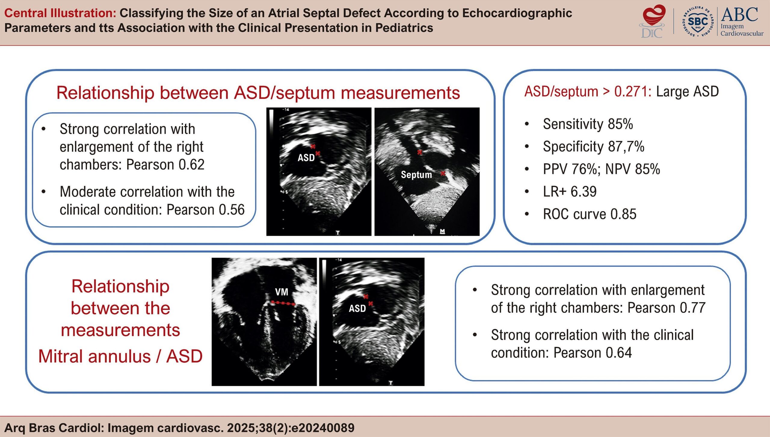
Investigate the relationship between ASD hemodynamic repercussions and ASD diameter to the mitral annulus diameter (MI/ASD) ratio and ASD diameter to the interatrial septum diameter (ASD/septum) ratio, and describe cutoff points to classify the size of the defect.

Methods

An observational, prospective, cross-sectional study, including subjects aged 1 month and 18 years diagnosed with isolated ASD. Hemodynamic repercussion was quantified by clinical evaluation, tricuspid annulus (TA) Z-score, subjective estimation of the enlargement of the right chambers, and the relationship between systemic and pulmonary flows (QP/QS) on echocardiography. Its association with ASD/septum and MI/ASD measurements was also studied. A value of p < 0.05 was considered statistically significant.

Results

Thirty-five subjects, mean age 6.3 years, 69% female, mean ASD 13.5 mm. There was a correlation between MI/ASD and the clinical classification of ASD (Pearson: -0.61; p < 0.001), ASD/septum (Pearson: -0.80; p < 0.001), and QP/QS (-0.76; p < 0.001). There was a correlation between ASD/septum and the clinical classification of ASD (Pearson: 0.56; p < 0.001), QP/QS (0.63; p = 0.001), subjective assessment of the right chambers (0.62; p < 0.001), and a weak correlation with the TA Z-score (0.35; p = 0.04). According to the operating characteristic curve for the stable variable classification of ASD according to the subjective right chamber’s size, an area of 0.85 was obtained for ASD/septum (p = 0.001). The cutoff point of 0.27 for large ASD showed sensitivity of 85%, specificity of 86.7%, positive predictive value (PPV) of 86%, negative predictive value (NPV) of 85.2%, and positive likelihood ratio of 6.39.

Conclusions

ASD/septum was associated with the hemodynamic repercussion of ASD and was useful in detecting large ASD.

  • Atrial Heart Septal Defects
  • Atrial Septum
  • Echocardiography
  • Tricuspid Valve
Read Full Article
Creative Commons Attribution (CC BY) Creative Commons license
15/05/2025 DOI: 10.36660/abcimg.20240089i Views 1
Central Illustration
Original Article

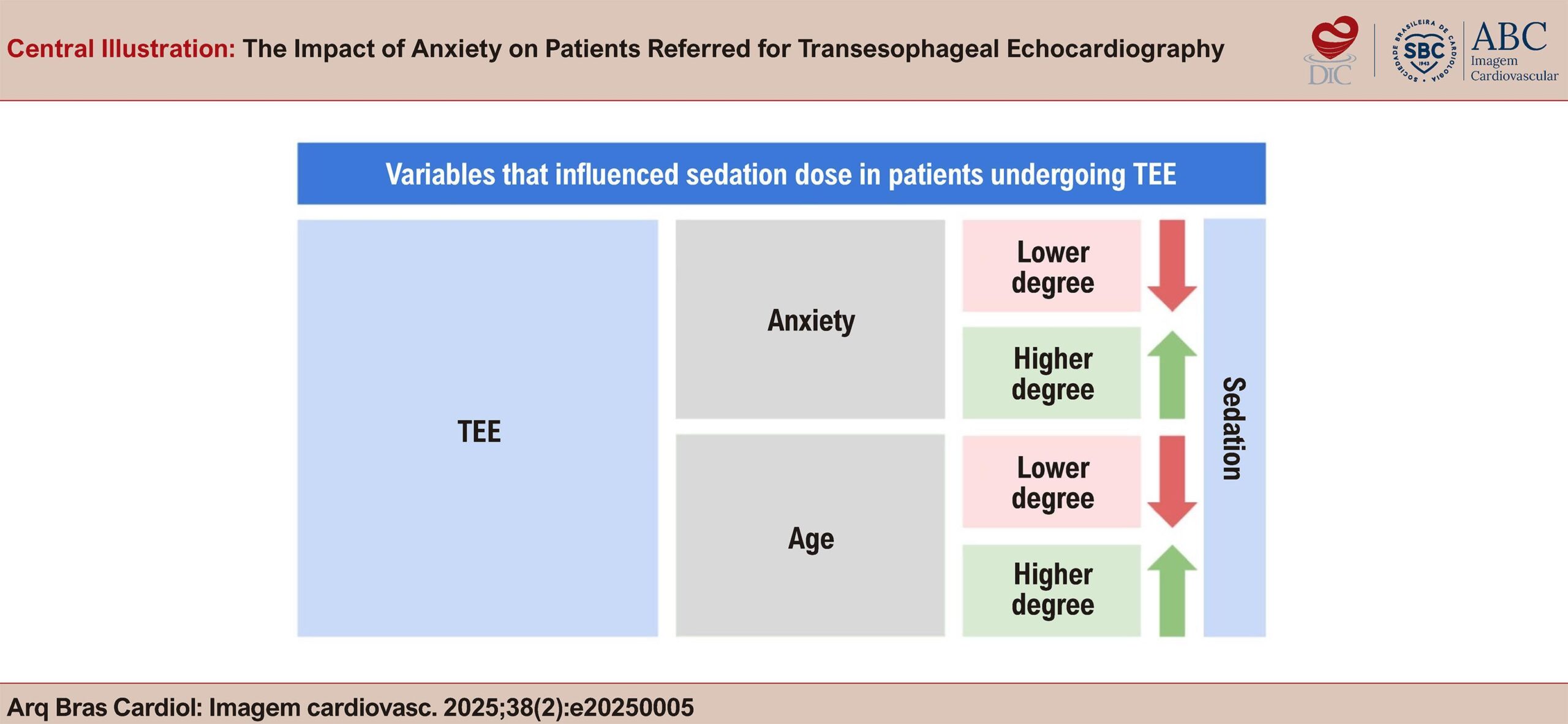
The Impact of Anxiety on Patients Referred for Transesophageal Echocardiography

João Afonso Astolfi Martins et al. Amanda de Vasconcelos Eng Edgar Lira Filho Claudio Henrique Fischer … Claudia Gianini Monaco Alessandra Joslin Oliveira Fernando Rodrigues da Camara Oliveira Marcelo Luiz Campos Vieira Samira Saady Morhy Ana Clara Tude Rodrigues

Abstract

Background

Anxiety is an important factor that influences patient experience during medical procedures. Transesophageal echocardiography (TEE) is an exam that requires sedation, which can exacerbate anxiety and affect its practice.

Objective

To assess variables that influence the degree of anxiety experienced by patients undergoing TEE and its correlation with sedation.

Methods

We assessed patients of both sexes, age >18 years, referred for TEE. We applied an anxiety questionnaire (none, mild, moderate, and high) and collected demographic, clinical, and physiological data. For comparison purposes, patients were divided into the following 2 groups: no/mild anxiety and moderate/severe anxiety.

Results

We studied 63 patients, 41 (66%) of whom were male. The majority were White (87.3%), and the mean age was 52.5 ± 14.7 years. Only 35% of patients had comorbidities, the most frequent being hypertension (63.6%), and 70% patients were undergoing TEE for the first time. The main indication for TEE was valvular disease (30%). The majority of patients (62%) reported some degree of anxiety, as follows: 25 (40%) mild, 10 (16%) moderate, and 4 (6%) severe. Patients with moderate/severe anxiety (p = 0.03) and younger patients (p = 0.001) required higher doses of midazolam for sedation.

Conclusion

Anxiety was common in patients referred for TEE, and it appeared to be less influenced by clinical variables. However, we concluded that more anxious and younger patients required higher doses of sedation.

  • Anxiety
  • Conscious Sedation
  • Echocardiography
Read Full Article
Creative Commons Attribution (CC BY) Creative Commons license
14/05/2025 DOI: 10.36660/abcimg.20250005i Views 2

How to cite this article

EndNote RIS

Electronic Document Format (Vancouver)

Artioli T, Santos BQM, Monteiro DC, Batista LB, Lorenzoni MZ, Jen HG, Vital KM, et al. Coronary Artery Dissection Following Pharmacologic Stress Echocardiography and the Follow-Up of Clinical Management: A Case Report. ABC Imagem Cardiovascular 2025;38(2):e20250034.

Electronic Document Format (ABNT)

Artioli, Thiago; Santos, Bruno Querido Marcondes; Monteiro, Denis Cárdenas; Batista, Layane Bonfante; Lorenzoni, Matheus Zavaris; Jen, Hsu Gwo; Vital, Kelvyn Melo; Vilalva, Kelvin Henrique. Coronary Artery Dissection Following Pharmacologic Stress Echocardiography and the Follow-Up of Clinical Management: A Case Report. ABC Imagem Cardiovascular, v. 38, n. 2, e20250034, Jun. 2025.

Electronic Document Format (APA)

Artioli, T., Santos, B. Q. M., Monteiro, D. C., Batista, L. B., Lorenzoni, M. Z., Jen, H. G., Vital, K. M., & Vilalva, K. H. (2025). Coronary Artery Dissection Following Pharmacologic Stress Echocardiography and the Follow-Up of Clinical Management: A Case Report. ABC Imagem Cardiovascular, 38(2), e20250034.

Electronic Document Format (ISO)

Artioli, Thiago and Santos, Bruno Querido Marcondes and Monteiro, Denis Cárdenas and Batista, Layane Bonfante and Lorenzoni, Matheus Zavaris and Jen, Hsu Gwo and Vital, Kelvyn Melo and Vilalva, Kelvin Henrique. Coronary Artery Dissection Following Pharmacologic Stress Echocardiography and the Follow-Up of Clinical Management: A Case Report. ABC Imagem Cardiovascular [online]. 2025, vol. 38, n. 2, [cited 2026-10-14], e20250034. Available from: <https://journals.cardiol.br/en/article/coronary-artery-dissection-following-pharmacologic-stress-echocardiography-and-the-follow-up-of-clinical-management-a-case-report/>. ISSN 2318-8219

EndNote RIS

Electronic Document Format (Vancouver)

Magalhães TA. “Triple Rule-Out”: Including the Abdominal Aorta With a Clear Conscience?. ABC Imagem Cardiovascular 2025;38(2):e20250041.

Electronic Document Format (ABNT)

Magalhães, Tiago A.. “Triple Rule-Out”: Including the Abdominal Aorta With a Clear Conscience?. ABC Imagem Cardiovascular, v. 38, n. 2, e20250041, Jun. 2025.

Electronic Document Format (APA)

Magalhães, T. A. (2025). “Triple Rule-Out”: Including the Abdominal Aorta With a Clear Conscience?. ABC Imagem Cardiovascular, 38(2), e20250041.

Electronic Document Format (ISO)

Magalhães, Tiago A.. “Triple Rule-Out”: Including the Abdominal Aorta With a Clear Conscience?. ABC Imagem Cardiovascular [online]. 2025, vol. 38, n. 2, [cited 2026-10-14], e20250041. Available from: <https://journals.cardiol.br/en/article/triple-rule-out-including-the-abdominal-aorta-with-a-clear-conscience/>. ISSN 2318-8219

EndNote RIS

Electronic Document Format (Vancouver)

Silva SM. My Approach to Diagnose Fabry Disease. ABC Imagem Cardiovascular 2025;38(2):e20240091.

Electronic Document Format (ABNT)

Silva, Sandra Marques e. My Approach to Diagnose Fabry Disease. ABC Imagem Cardiovascular, v. 38, n. 2, e20240091, Jun. 2025.

Electronic Document Format (APA)

Silva, S. M. (2025). My Approach to Diagnose Fabry Disease. ABC Imagem Cardiovascular, 38(2), e20240091.

Electronic Document Format (ISO)

Silva, Sandra Marques e. My Approach to Diagnose Fabry Disease. ABC Imagem Cardiovascular [online]. 2025, vol. 38, n. 2, [cited 2026-10-14], e20240091. Available from: <https://journals.cardiol.br/en/article/my-approach-to-diagnose-fabry-disease/>. ISSN 2318-8219

EndNote RIS

Electronic Document Format (Vancouver)

Ker WS, Yahiro DS, Mangas GM, Salomão IMR, Flor JAAA, Vianna HMS, Mesquita CT, et al. Assessment of Coronary Flow Reserve by Myocardial Perfusion Scintigraphy using CZT Equipment. ABC Imagem Cardiovascular 2025;38(2):e20250020.

Electronic Document Format (ABNT)

Ker, Wilter dos Santos; Yahiro, Davi Shunji; Mangas, Gabriel de Moraes; Salomão, Isabelle Mendes Rodrigues; Flor, José Augusto Antonio Alves; Vianna, Hugo Matheus Silvestre; Mesquita, Cláudio Tinoco. Assessment of Coronary Flow Reserve by Myocardial Perfusion Scintigraphy using CZT Equipment. ABC Imagem Cardiovascular, v. 38, n. 2, e20250020, Jun. 2025.

Electronic Document Format (APA)

Ker, W. S., Yahiro, D. S., Mangas, G. M., Salomão, I. M. R., Flor, J. A. A. A., Vianna, H. M. S., & Mesquita, C. T. (2025). Assessment of Coronary Flow Reserve by Myocardial Perfusion Scintigraphy using CZT Equipment. ABC Imagem Cardiovascular, 38(2), e20250020.

Electronic Document Format (ISO)

Ker, Wilter dos Santos and Yahiro, Davi Shunji and Mangas, Gabriel de Moraes and Salomão, Isabelle Mendes Rodrigues and Flor, José Augusto Antonio Alves and Vianna, Hugo Matheus Silvestre and Mesquita, Cláudio Tinoco. Assessment of Coronary Flow Reserve by Myocardial Perfusion Scintigraphy using CZT Equipment. ABC Imagem Cardiovascular [online]. 2025, vol. 38, n. 2, [cited 2026-10-14], e20250020. Available from: <https://journals.cardiol.br/en/article/assessment-of-coronary-flow-reserve-by-myocardial-perfusion-scintigraphy-using-czt-equipment/>. ISSN 2318-8219

EndNote RIS

Electronic Document Format (Vancouver)

Salgado AA, Bernardo MPL. The Use of POCUS in Daily Practice: Are We Really Ready to Use it Efficiently?. ABC Imagem Cardiovascular 2025;38(2):e20250026.

Electronic Document Format (ABNT)

Salgado, Angelo Antunes; Bernardo, Marcos Paulo Lacerda. The Use of POCUS in Daily Practice: Are We Really Ready to Use it Efficiently?. ABC Imagem Cardiovascular, v. 38, n. 2, e20250026, Jun. 2025.

Electronic Document Format (APA)

Salgado, A. A., & Bernardo, M. P. L. (2025). The Use of POCUS in Daily Practice: Are We Really Ready to Use it Efficiently?. ABC Imagem Cardiovascular, 38(2), e20250026.

Electronic Document Format (ISO)

Salgado, Angelo Antunes and Bernardo, Marcos Paulo Lacerda. The Use of POCUS in Daily Practice: Are We Really Ready to Use it Efficiently?. ABC Imagem Cardiovascular [online]. 2025, vol. 38, n. 2, [cited 2026-10-14], e20250026. Available from: <https://journals.cardiol.br/en/article/the-use-of-pocus-in-daily-practice-are-we-really-ready-to-use-it-efficiently/>. ISSN 2318-8219

EndNote RIS

Electronic Document Format (Vancouver)

Danzmann LC, Kalil E, Kalil SA. New Tools for Optimizing HF Management: The Role of POCUS in the Office. ABC Imagem Cardiovascular 2025;38(2):e20250039.

Electronic Document Format (ABNT)

Danzmann, Luiz Claudio; Kalil, Elisa; Kalil, Soraya Abunader. New Tools for Optimizing HF Management: The Role of POCUS in the Office. ABC Imagem Cardiovascular, v. 38, n. 2, e20250039, Jun. 2025.

Electronic Document Format (APA)

Danzmann, L. C., Kalil, E., & Kalil, S. A. (2025). New Tools for Optimizing HF Management: The Role of POCUS in the Office. ABC Imagem Cardiovascular, 38(2), e20250039.

Electronic Document Format (ISO)

Danzmann, Luiz Claudio and Kalil, Elisa and Kalil, Soraya Abunader. New Tools for Optimizing HF Management: The Role of POCUS in the Office. ABC Imagem Cardiovascular [online]. 2025, vol. 38, n. 2, [cited 2026-10-14], e20250039. Available from: <https://journals.cardiol.br/en/article/new-tools-for-optimizing-hf-management-the-role-of-pocus-in-the-office/>. ISSN 2318-8219

EndNote RIS

Electronic Document Format (Vancouver)

Silva TO, Nascimento SUCN. Do Patients with Homozygosity and Compound Heterozygosity for Variants in the Transthyretin Amyloidosis TTR Gene Have More Severe and Earlier Clinical Conditions?. ABC Imagem Cardiovascular 2025;38(2):e20250027.

Electronic Document Format (ABNT)

Silva, Tonnison de Oliveira; Nascimento, Samuel Ulisses Chaves Nogueira do. Do Patients with Homozygosity and Compound Heterozygosity for Variants in the Transthyretin Amyloidosis TTR Gene Have More Severe and Earlier Clinical Conditions?. ABC Imagem Cardiovascular, v. 38, n. 2, e20250027, Jun. 2025.

Electronic Document Format (APA)

Silva, T. O., & Nascimento, S. U. C. N. (2025). Do Patients with Homozygosity and Compound Heterozygosity for Variants in the Transthyretin Amyloidosis TTR Gene Have More Severe and Earlier Clinical Conditions?. ABC Imagem Cardiovascular, 38(2), e20250027.

Electronic Document Format (ISO)

Silva, Tonnison de Oliveira and Nascimento, Samuel Ulisses Chaves Nogueira do. Do Patients with Homozygosity and Compound Heterozygosity for Variants in the Transthyretin Amyloidosis TTR Gene Have More Severe and Earlier Clinical Conditions?. ABC Imagem Cardiovascular [online]. 2025, vol. 38, n. 2, [cited 2026-10-14], e20250027. Available from: <https://journals.cardiol.br/en/article/do-patients-with-homozygosity-and-compound-heterozygosity-for-variants-in-the-transthyretin-amyloidosis-ttr-gene-have-more-severe-and-earlier-clinical-conditions/>. ISSN 2318-8219

EndNote RIS

Electronic Document Format (Vancouver)

Silva HAGP, Gomes HM. My Approach to Estimate Pulmonary Pressures: Practical Aspects. ABC Imagem Cardiovascular 2025;38(2):e20250028.

Electronic Document Format (ABNT)

Silva, Halsted Alarcão Gomes Pereira da; Gomes, Helder Moura. My Approach to Estimate Pulmonary Pressures: Practical Aspects. ABC Imagem Cardiovascular, v. 38, n. 2, e20250028, Jun. 2025.

Electronic Document Format (APA)

Silva, H. A. G. P. , & Gomes, H. M. (2025). My Approach to Estimate Pulmonary Pressures: Practical Aspects. ABC Imagem Cardiovascular, 38(2), e20250028.

Electronic Document Format (ISO)

Silva, Halsted Alarcão Gomes Pereira da and Gomes, Helder Moura. My Approach to Estimate Pulmonary Pressures: Practical Aspects. ABC Imagem Cardiovascular [online]. 2025, vol. 38, n. 2, [cited 2026-10-14], e20250028. Available from: <https://journals.cardiol.br/en/article/my-approach-to-estimate-pulmonary-pressures-practical-aspects/>. ISSN 2318-8219

EndNote RIS

Electronic Document Format (Vancouver)

Jordão MR, Tavares M, Fernandes F. Mental Traps That Impact Diagnostic Imaging. ABC Imagem Cardiovascular 2025;38(2):e20250405.

Electronic Document Format (ABNT)

Jordão, Maurício Rodrigues; Tavares, Marcelo; Fernandes, Fábio. Mental Traps That Impact Diagnostic Imaging. ABC Imagem Cardiovascular, v. 38, n. 2, e20250405, Jun. 2025.

Electronic Document Format (APA)

Jordão, M. R., Tavares, M., & Fernandes, F. (2025). Mental Traps That Impact Diagnostic Imaging. ABC Imagem Cardiovascular, 38(2), e20250405.

Electronic Document Format (ISO)

Jordão, Maurício Rodrigues and Tavares, Marcelo and Fernandes, Fábio. Mental Traps That Impact Diagnostic Imaging. ABC Imagem Cardiovascular [online]. 2025, vol. 38, n. 2, [cited 2026-10-14], e20250405. Available from: <https://journals.cardiol.br/en/article/mental-traps-that-impact-diagnostic-imaging/>. ISSN 2318-8219

EndNote RIS

Electronic Document Format (Vancouver)

Savoia P, Bello R. Photon-Counting Computed Tomography in Cardiovascular Imaging: Where We Are and What Lies Ahead. ABC Imagem Cardiovascular 2025;38(2):e20250035.

Electronic Document Format (ABNT)

Savoia, Paulo; Bello, Rodrigo. Photon-Counting Computed Tomography in Cardiovascular Imaging: Where We Are and What Lies Ahead. ABC Imagem Cardiovascular, v. 38, n. 2, e20250035, Jun. 2025.

Electronic Document Format (APA)

Savoia, P., & Bello, R. (2025). Photon-Counting Computed Tomography in Cardiovascular Imaging: Where We Are and What Lies Ahead. ABC Imagem Cardiovascular, 38(2), e20250035.

Electronic Document Format (ISO)

Savoia, Paulo and Bello, Rodrigo. Photon-Counting Computed Tomography in Cardiovascular Imaging: Where We Are and What Lies Ahead. ABC Imagem Cardiovascular [online]. 2025, vol. 38, n. 2, [cited 2026-10-14], e20250035. Available from: <https://journals.cardiol.br/en/article/photon-counting-computed-tomography-in-cardiovascular-imaging-where-we-are-and-what-lies-ahead/>. ISSN 2318-8219

EndNote RIS

Electronic Document Format (Vancouver)

Dias VTG, Almeida MF. My Approach To Echocardiographic Evaluation in Pediatric Patients with Sickle Cell Disease. ABC Imagem Cardiovascular 2025;38(2):e20240128.

Electronic Document Format (ABNT)

Dias, Viviane Thome Gonçalves; Almeida, Mirela Frederico de. My Approach To Echocardiographic Evaluation in Pediatric Patients with Sickle Cell Disease. ABC Imagem Cardiovascular, v. 38, n. 2, e20240128, Jun. 2025.

Electronic Document Format (APA)

Dias, V. T. G., & Almeida, M. F. (2025). My Approach To Echocardiographic Evaluation in Pediatric Patients with Sickle Cell Disease. ABC Imagem Cardiovascular, 38(2), e20240128.

Electronic Document Format (ISO)

Dias, Viviane Thome Gonçalves and Almeida, Mirela Frederico de. My Approach To Echocardiographic Evaluation in Pediatric Patients with Sickle Cell Disease. ABC Imagem Cardiovascular [online]. 2025, vol. 38, n. 2, [cited 2026-10-14], e20240128. Available from: <https://journals.cardiol.br/en/article/my-approach-to-echocardiographic-evaluation-in-pediatric-patients-with-sickle-cell-disease/>. ISSN 2318-8219

EndNote RIS

Electronic Document Format (Vancouver)

Vilela AA, Shinzato MH, Valdigem BP, Paladino AT, Correia EB, Assef JE. Hypertrophic Cardiomyopathy: Analysis of Septal Thickness with Gradient Reduction in Patients Undergoing Radiofrequency Septal Ablation. ABC Imagem Cardiovascular 2025;38(2):e20240097.

Electronic Document Format (ABNT)

Vilela, Andrea de Andrade; Shinzato, Mariane Higa; Valdigem, Bruno Pereira; Paladino, Antonio Tito; Correia, Edileide de Barros; Assef, Jorge Eduardo. Hypertrophic Cardiomyopathy: Analysis of Septal Thickness with Gradient Reduction in Patients Undergoing Radiofrequency Septal Ablation. ABC Imagem Cardiovascular, v. 38, n. 2, e20240097, Jun. 2025.

Electronic Document Format (APA)

Vilela, A. A., Shinzato, M. H., Valdigem, B. P., Paladino, A. T., Correia, E. B., & Assef, J. E. (2025). Hypertrophic Cardiomyopathy: Analysis of Septal Thickness with Gradient Reduction in Patients Undergoing Radiofrequency Septal Ablation. ABC Imagem Cardiovascular, 38(2), e20240097.

Electronic Document Format (ISO)

Vilela, Andrea de Andrade and Shinzato, Mariane Higa and Valdigem, Bruno Pereira and Paladino, Antonio Tito and Correia, Edileide de Barros and Assef, Jorge Eduardo. Hypertrophic Cardiomyopathy: Analysis of Septal Thickness with Gradient Reduction in Patients Undergoing Radiofrequency Septal Ablation. ABC Imagem Cardiovascular [online]. 2025, vol. 38, n. 2, [cited 2026-10-14], e20240097. Available from: <https://journals.cardiol.br/en/article/hypertrophic-cardiomyopathy-analysis-of-septal-thickness-with-gradient-reduction-in-patients-undergoing-radiofrequency-septal-ablation/>. ISSN 2318-8219

EndNote RIS

Electronic Document Format (Vancouver)

Souza AC, Carvalho MVSF. Radiofrequency in Obstructive Hypertrophic Cardiomyopathy: The Role of Imaging in the Assessment of Septal Thickness and Gradient Reduction. ABC Imagem Cardiovascular 2025;38(2):e20250024.

Electronic Document Format (ABNT)

Souza, Alexandre Costa; Carvalho, Marcus Vinicius Silva Freire de. Radiofrequency in Obstructive Hypertrophic Cardiomyopathy: The Role of Imaging in the Assessment of Septal Thickness and Gradient Reduction. ABC Imagem Cardiovascular, v. 38, n. 2, e20250024, Jun. 2025.

Electronic Document Format (APA)

Souza, A. C., & Carvalho, M. V. S. F. (2025). Radiofrequency in Obstructive Hypertrophic Cardiomyopathy: The Role of Imaging in the Assessment of Septal Thickness and Gradient Reduction. ABC Imagem Cardiovascular, 38(2), e20250024.

Electronic Document Format (ISO)

Souza, Alexandre Costa and Carvalho, Marcus Vinicius Silva Freire de. Radiofrequency in Obstructive Hypertrophic Cardiomyopathy: The Role of Imaging in the Assessment of Septal Thickness and Gradient Reduction. ABC Imagem Cardiovascular [online]. 2025, vol. 38, n. 2, [cited 2026-10-14], e20250024. Available from: <https://journals.cardiol.br/en/article/radiofrequency-in-obstructive-hypertrophic-cardiomyopathy-the-role-of-imaging-in-the-assessment-of-septal-thickness-and-gradient-reduction/>. ISSN 2318-8219

EndNote RIS

Electronic Document Format (Vancouver)

Ciuffo MP, Mori L, Araújo T, Kowatsch I, Rodrigues ACT. Important Mitral Regurgitation and Ventricular Dysfunction in Hypereosinophilic Syndrome: A Case Report. ABC Imagem Cardiovascular 2025;38(2):e20250029.

Electronic Document Format (ABNT)

Ciuffo, Marcel Pina; Mori, Lucas; Araújo, Tais; Kowatsch, Ingrid; Rodrigues, Ana Clara Tude. Important Mitral Regurgitation and Ventricular Dysfunction in Hypereosinophilic Syndrome: A Case Report. ABC Imagem Cardiovascular, v. 38, n. 2, e20250029, Jun. 2025.

Electronic Document Format (APA)

Ciuffo, M. P., Mori, L., Araújo, T., Kowatsch, I., & Rodrigues, A. C. T. (2025). Important Mitral Regurgitation and Ventricular Dysfunction in Hypereosinophilic Syndrome: A Case Report. ABC Imagem Cardiovascular, 38(2), e20250029.

Electronic Document Format (ISO)

Ciuffo, Marcel Pina and Mori, Lucas and Araújo, Tais and Kowatsch, Ingrid and Rodrigues, Ana Clara Tude. Important Mitral Regurgitation and Ventricular Dysfunction in Hypereosinophilic Syndrome: A Case Report. ABC Imagem Cardiovascular [online]. 2025, vol. 38, n. 2, [cited 2026-10-14], e20250029. Available from: <https://journals.cardiol.br/en/article/important-mitral-regurgitation-and-ventricular-dysfunction-in-hypereosinophilic-syndrome-a-case-report/>. ISSN 2318-8219

EndNote RIS

Electronic Document Format (Vancouver)

Brentegani A, Solis FAE, Paulis M, Vieira MLC. My Approach to Point-of-Care Ultrasound for Dyspnea Assessment. ABC Imagem Cardiovascular 2025;38(2):e20250023.

Electronic Document Format (ABNT)

Brentegani, Adriana; Solis, Fernando Arturo Effio; Paulis, Milena de; Vieira, Marcelo Luiz Campos. My Approach to Point-of-Care Ultrasound for Dyspnea Assessment. ABC Imagem Cardiovascular, v. 38, n. 2, e20250023, Jun. 2025.

Electronic Document Format (APA)

Brentegani, A., Solis, F. A. E., Paulis, M. , & Vieira, M. L. C. (2025). My Approach to Point-of-Care Ultrasound for Dyspnea Assessment. ABC Imagem Cardiovascular, 38(2), e20250023.

Electronic Document Format (ISO)

Brentegani, Adriana and Solis, Fernando Arturo Effio and Paulis, Milena de and Vieira, Marcelo Luiz Campos. My Approach to Point-of-Care Ultrasound for Dyspnea Assessment. ABC Imagem Cardiovascular [online]. 2025, vol. 38, n. 2, [cited 2026-10-14], e20250023. Available from: <https://journals.cardiol.br/en/article/my-approach-to-point-of-care-ultrasound-for-dyspnea-assessment/>. ISSN 2318-8219

EndNote RIS

Electronic Document Format (Vancouver)

Saadi MP, Machado GP, Silvano GP, Barbato JPR, Almeida RF, Scolari FL, Telo GH, et al. Assessment of Interrater Reliability in Point-of-Care Ultrasound for Assessing Congestion in Cardiovascular Intensive Care. ABC Imagem Cardiovascular 2025;38(2):e20250022.

Electronic Document Format (ABNT)

Saadi, Marina Petersen; Machado, Guilherme Pinheiro; Silvano, Gustavo Paes; Barbato, João Pedro da Rosa; Almeida, Renato Ferraz; Scolari, Fernando Luis; Telo, Guilherme Heiden; Silveira, Anderson Donelli da. Assessment of Interrater Reliability in Point-of-Care Ultrasound for Assessing Congestion in Cardiovascular Intensive Care. ABC Imagem Cardiovascular, v. 38, n. 2, e20250022, Jun. 2025.

Electronic Document Format (APA)

Saadi, M. P., Machado, G. P., Silvano, G. P., Barbato, J. P. R., Almeida, R. F., Scolari, F. L., Telo, G. H., & Silveira, A. D. (2025). Assessment of Interrater Reliability in Point-of-Care Ultrasound for Assessing Congestion in Cardiovascular Intensive Care. ABC Imagem Cardiovascular, 38(2), e20250022.

Electronic Document Format (ISO)

Saadi, Marina Petersen and Machado, Guilherme Pinheiro and Silvano, Gustavo Paes and Barbato, João Pedro da Rosa and Almeida, Renato Ferraz and Scolari, Fernando Luis and Telo, Guilherme Heiden and Silveira, Anderson Donelli da. Assessment of Interrater Reliability in Point-of-Care Ultrasound for Assessing Congestion in Cardiovascular Intensive Care. ABC Imagem Cardiovascular [online]. 2025, vol. 38, n. 2, [cited 2026-10-14], e20250022. Available from: <https://journals.cardiol.br/en/article/assessment-of-interrater-reliability-in-point-of-care-ultrasound-for-assessing-congestion-in-cardiovascular-intensive-care/>. ISSN 2318-8219

EndNote RIS

Electronic Document Format (Vancouver)

Bertolazzi P, Silva CFG, Iunes L, Oliveira FF, Pereira FP, Viana PCC, Castro I, et al. Conventional and Partially ECG-Gated Triple Rule-Out Computed Tomography Angiography with Extension to Abdominal Aorta: Comparative Radiation Dose and Imaging Quality. ABC Imagem Cardiovascular 2025;38(2):e20250025.

Electronic Document Format (ABNT)

Bertolazzi, Pamela; Silva, Carla Franco Greco; Iunes, Leonardo; Oliveira, Fernando Freitas de; Pereira, Fabio Payão; Viana, Publio Cesar Cavalcanti; Castro, Isac; Horvat, Natally; Araújo-Filho, José Arimateia Batista. Conventional and Partially ECG-Gated Triple Rule-Out Computed Tomography Angiography with Extension to Abdominal Aorta: Comparative Radiation Dose and Imaging Quality. ABC Imagem Cardiovascular, v. 38, n. 2, e20250025, Jun. 2025.

Electronic Document Format (APA)

Bertolazzi, P., Silva, C. F. G., Iunes, L., Oliveira, F. F. , Pereira, F. P., Viana, P. C. C., Castro, I., Horvat, N., & Araújo-Filho, J. A. B. (2025). Conventional and Partially ECG-Gated Triple Rule-Out Computed Tomography Angiography with Extension to Abdominal Aorta: Comparative Radiation Dose and Imaging Quality. ABC Imagem Cardiovascular, 38(2), e20250025.

Electronic Document Format (ISO)

Bertolazzi, Pamela and Silva, Carla Franco Greco and Iunes, Leonardo and Oliveira, Fernando Freitas de and Pereira, Fabio Payão and Viana, Publio Cesar Cavalcanti and Castro, Isac and Horvat, Natally and Araújo-Filho, José Arimateia Batista. Conventional and Partially ECG-Gated Triple Rule-Out Computed Tomography Angiography with Extension to Abdominal Aorta: Comparative Radiation Dose and Imaging Quality. ABC Imagem Cardiovascular [online]. 2025, vol. 38, n. 2, [cited 2026-10-14], e20250025. Available from: <https://journals.cardiol.br/en/article/conventional-and-partially-ecg-gated-triple-rule-out-computed-tomography-angiography-with-extension-to-abdominal-aorta-comparative-radiation-dose-and-imaging-quality/>. ISSN 2318-8219

EndNote RIS

Electronic Document Format (Vancouver)

Santos SN, Henz BD, Silva MEL, Silva LRL. My Approach to Intracardiac Echocardiography During Atrial Fibrillation Ablation. ABC Imagem Cardiovascular 2025;38(2):e20250021.

Electronic Document Format (ABNT)

Santos, Simone Nascimento dos; Henz, Benhur Davi; Silva, Maria Eduarda Leite da; Silva, Luiz Roberto Leite da. My Approach to Intracardiac Echocardiography During Atrial Fibrillation Ablation. ABC Imagem Cardiovascular, v. 38, n. 2, e20250021, Jun. 2025.

Electronic Document Format (APA)

Santos, S. N. , Henz, B. D., Silva, M. E. L. , & Silva, L. R. L. (2025). My Approach to Intracardiac Echocardiography During Atrial Fibrillation Ablation. ABC Imagem Cardiovascular, 38(2), e20250021.

Electronic Document Format (ISO)

Santos, Simone Nascimento dos and Henz, Benhur Davi and Silva, Maria Eduarda Leite da and Silva, Luiz Roberto Leite da. My Approach to Intracardiac Echocardiography During Atrial Fibrillation Ablation. ABC Imagem Cardiovascular [online]. 2025, vol. 38, n. 2, [cited 2026-10-14], e20250021. Available from: <https://journals.cardiol.br/en/article/my-approach-to-intracardiac-echocardiography-during-atrial-fibrillation-ablation/>. ISSN 2318-8219

EndNote RIS

Electronic Document Format (Vancouver)

Pereira RAR, Moura DMC. Intracardiac Echocardiography: Current Status, Clinical Benefits, and Future Perspectives. ABC Imagem Cardiovascular 2025;38(2):e20250031.

Electronic Document Format (ABNT)

Pereira, Renner Augusto Raposo; Moura, Daniel Moreira Costa. Intracardiac Echocardiography: Current Status, Clinical Benefits, and Future Perspectives. ABC Imagem Cardiovascular, v. 38, n. 2, e20250031, Jun. 2025.

Electronic Document Format (APA)

Pereira, R. A. R., & Moura, D. M. C. (2025). Intracardiac Echocardiography: Current Status, Clinical Benefits, and Future Perspectives. ABC Imagem Cardiovascular, 38(2), e20250031.

Electronic Document Format (ISO)

Pereira, Renner Augusto Raposo and Moura, Daniel Moreira Costa. Intracardiac Echocardiography: Current Status, Clinical Benefits, and Future Perspectives. ABC Imagem Cardiovascular [online]. 2025, vol. 38, n. 2, [cited 2026-10-14], e20250031. Available from: <https://journals.cardiol.br/en/article/intracardiac-echocardiography-current-status-clinical-benefits-and-future-perspectives/>. ISSN 2318-8219

EndNote RIS

Electronic Document Format (Vancouver)

Vera A, Álvarez V. Idiopathic Multiple Coronary Artery Aneurysms. ABC Imagem Cardiovascular 2025;38(2):e20240106.

Electronic Document Format (ABNT)

Vera, Alberto; Álvarez, Virginia. Idiopathic Multiple Coronary Artery Aneurysms. ABC Imagem Cardiovascular, v. 38, n. 2, e20240106, Jun. 2025.

Electronic Document Format (APA)

Vera, A., & Álvarez, V. (2025). Idiopathic Multiple Coronary Artery Aneurysms. ABC Imagem Cardiovascular, 38(2), e20240106.

Electronic Document Format (ISO)

Vera, Alberto and Álvarez, Virginia. Idiopathic Multiple Coronary Artery Aneurysms. ABC Imagem Cardiovascular [online]. 2025, vol. 38, n. 2, [cited 2026-10-14], e20240106. Available from: <https://journals.cardiol.br/en/article/idiopathic-multiple-coronary-artery-aneurysms/>. ISSN 2318-8219

EndNote RIS

Electronic Document Format (Vancouver)

Carvalho AA, Carvalho WA, Botelho LFB, Melo MDT. Assessment of Left Ventricular Function by MAPSE (Mitral Annular Plane Systolic Excursion): Main Clinical Applications. ABC Imagem Cardiovascular 2025;38(2):e20240105.

Electronic Document Format (ABNT)

Carvalho, Andressa Alves de; Carvalho, Wanessa Alves de; Botelho, Luis Fabio Barbosa; Melo, Marcelo Dantas Tavares de. Assessment of Left Ventricular Function by MAPSE (Mitral Annular Plane Systolic Excursion): Main Clinical Applications. ABC Imagem Cardiovascular, v. 38, n. 2, e20240105, May. 2025.

Electronic Document Format (APA)

Carvalho, A. A. , Carvalho, W. A. , Botelho, L. F. B., & Melo, M. D. T. (2025). Assessment of Left Ventricular Function by MAPSE (Mitral Annular Plane Systolic Excursion): Main Clinical Applications. ABC Imagem Cardiovascular, 38(2), e20240105.

Electronic Document Format (ISO)

Carvalho, Andressa Alves de and Carvalho, Wanessa Alves de and Botelho, Luis Fabio Barbosa and Melo, Marcelo Dantas Tavares de. Assessment of Left Ventricular Function by MAPSE (Mitral Annular Plane Systolic Excursion): Main Clinical Applications. ABC Imagem Cardiovascular [online]. 2025, vol. 38, n. 2, [cited 2026-10-14], e20240105. Available from: <https://journals.cardiol.br/en/article/assessment-of-left-ventricular-function-by-mapse-mitral-annular-plane-systolic-excursion-main-clinical-applications/>. ISSN 2318-8219

EndNote RIS

Electronic Document Format (Vancouver)

Frigo JPP, Voss TH. Large Heterogeneous Mass in the Right Chambers: A Case Report in Cardio-Oncology. ABC Imagem Cardiovascular 2025;38(2):e20250002.

Electronic Document Format (ABNT)

Frigo, João Pedro Perfeito; Voss, Themissa Helena. Large Heterogeneous Mass in the Right Chambers: A Case Report in Cardio-Oncology. ABC Imagem Cardiovascular, v. 38, n. 2, e20250002, May. 2025.

Electronic Document Format (APA)

Frigo, J. P. P., & Voss, T. H. (2025). Large Heterogeneous Mass in the Right Chambers: A Case Report in Cardio-Oncology. ABC Imagem Cardiovascular, 38(2), e20250002.

Electronic Document Format (ISO)

Frigo, João Pedro Perfeito and Voss, Themissa Helena. Large Heterogeneous Mass in the Right Chambers: A Case Report in Cardio-Oncology. ABC Imagem Cardiovascular [online]. 2025, vol. 38, n. 2, [cited 2026-10-14], e20250002. Available from: <https://journals.cardiol.br/en/article/large-heterogeneous-mass-in-the-right-chambers-a-case-report-in-cardio-oncology/>. ISSN 2318-8219

EndNote RIS

Electronic Document Format (Vancouver)

Ribeiro LC, Silva RMFL, Tonelli HAF, Guimarães AFM, Araújo FDR, Meira ZMA, Castilho SRT, et al. Classifying the Size of an Atrial Septal Defect According to Echocardiographic Parameters and its Association with the Clinical Presentation in Pediatrics. ABC Imagem Cardiovascular 2025;38(2):e20240089.

Electronic Document Format (ABNT)

Ribeiro, Lívia de Castro; Silva, Rose Mary Ferreira Lisboa da; Tonelli, Henrique de Assis Fonseca; Guimarães, Adriana Furletti Machado; Araújo, Fátima Derlene da Rocha; Meira, Zilda Maria Alves; Castilho, Sandra Regina Tolentino; Conde, Alan Alvarez; Valadares, Lícia Campos. Classifying the Size of an Atrial Septal Defect According to Echocardiographic Parameters and its Association with the Clinical Presentation in Pediatrics. ABC Imagem Cardiovascular, v. 38, n. 2, e20240089, May. 2025.

Electronic Document Format (APA)

Ribeiro, L. C., Silva, R. M. F. L. , Tonelli, H. A. F., Guimarães, A. F. M., Araújo, F. D. R., Meira, Z. M. A., Castilho, S. R. T., Conde, A. A., & Valadares, L. C. (2025). Classifying the Size of an Atrial Septal Defect According to Echocardiographic Parameters and its Association with the Clinical Presentation in Pediatrics. ABC Imagem Cardiovascular, 38(2), e20240089.

Electronic Document Format (ISO)

Ribeiro, Lívia de Castro and Silva, Rose Mary Ferreira Lisboa da and Tonelli, Henrique de Assis Fonseca and Guimarães, Adriana Furletti Machado and Araújo, Fátima Derlene da Rocha and Meira, Zilda Maria Alves and Castilho, Sandra Regina Tolentino and Conde, Alan Alvarez and Valadares, Lícia Campos. Classifying the Size of an Atrial Septal Defect According to Echocardiographic Parameters and its Association with the Clinical Presentation in Pediatrics. ABC Imagem Cardiovascular [online]. 2025, vol. 38, n. 2, [cited 2026-10-14], e20240089. Available from: <https://journals.cardiol.br/en/article/classifying-the-size-of-an-atrial-septal-defect-according-to-echocardiographic-parameters-and-its-association-with-the-clinical-presentation-in-pediatrics/>. ISSN 2318-8219

EndNote RIS

Electronic Document Format (Vancouver)

Martins JAA, Eng AV, Lira Filho E, Fischer CH, Monaco CG, Oliveira AJ, Oliveira FRC, et al. The Impact of Anxiety on Patients Referred for Transesophageal Echocardiography. ABC Imagem Cardiovascular 2025;38(2):e20250005.

Electronic Document Format (ABNT)

Martins, João Afonso Astolfi; Eng, Amanda de Vasconcelos; Lira Filho, Edgar; Fischer, Claudio Henrique; Monaco, Claudia Gianini; Oliveira, Alessandra Joslin; Oliveira, Fernando Rodrigues da Camara; Vieira, Marcelo Luiz Campos; Morhy, Samira Saady; Rodrigues, Ana Clara Tude. The Impact of Anxiety on Patients Referred for Transesophageal Echocardiography. ABC Imagem Cardiovascular, v. 38, n. 2, e20250005, May. 2025.

Electronic Document Format (APA)

Martins, J. A. A., Eng, A. V., Lira Filho, E., Fischer, C. H., Monaco, C. G., Oliveira, A. J., Oliveira, F. R. C., Vieira, M. L. C., Morhy, S. S., & Rodrigues, A. C. T. (2025). The Impact of Anxiety on Patients Referred for Transesophageal Echocardiography. ABC Imagem Cardiovascular, 38(2), e20250005.

Electronic Document Format (ISO)

Martins, João Afonso Astolfi and Eng, Amanda de Vasconcelos and Lira Filho, Edgar and Fischer, Claudio Henrique and Monaco, Claudia Gianini and Oliveira, Alessandra Joslin and Oliveira, Fernando Rodrigues da Camara and Vieira, Marcelo Luiz Campos and Morhy, Samira Saady and Rodrigues, Ana Clara Tude. The Impact of Anxiety on Patients Referred for Transesophageal Echocardiography. ABC Imagem Cardiovascular [online]. 2025, vol. 38, n. 2, [cited 2026-10-14], e20250005. Available from: <https://journals.cardiol.br/en/article/the-impact-of-anxiety-on-patients-referred-for-transesophageal-echocardiography/>. ISSN 2318-8219

Sede – São Paulo
Rua Barata Ribeiro, 380 cj 54 Bela Vista
São Paulo, Brasil – CEP: 01308-000
Tel.: +55 (11) 3120-3363
Telefax: +55 (11) 3259-2988
E-mail: depeco@cardiol.br

Sede – Rio de Janeiro
Av. Marechal Câmara, 160
3º andar – Sala: 330 – Centro
Rio de Janeiro – CEP: 20020-907
Tel.: +55 (21) 3478-2700
ou: +55 (21) 99386-3961
E-mail: abcimaging@cardiol.br

  • About the Journal
  • Internal Regulation
  • Editors Section
  • Editorial Board
  • SBC Award

  • Guidelines for Publication
  • Editorial Policy
  • Ethics Policy
  • Policy on the Use of Artificial Intelligence
  • Mandatory Publication Form

Issues

SBC Guidelines