André Malho et al. Renato Silveira Ximenes Nathalie Jeanne Bravo-Valenzuela Edward Araujo Júnior
Heart defects are among the most common congenital malformations, occurring in 4 to 12 per 1,000 live births, depending on the population studied. Congenital heart disease (CHD) is an important cause of mortality related to birth defects. Therefore, prenatal diagnosis of CHD is crucial to improve the prognosis associated with this disease. In this scenario, the development of prenatal cardiac ultrasound screening programs serves as crucial tools that have increased the early detection of CHD. These programs achieve this by adding the ventricular outflow tract and superior mediastinum views (three vessels – 3V and 3V with trachea) to the standard four-chamber (4C) view when scanning the cardiac ultrasound.
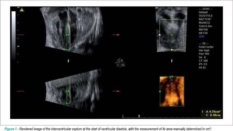
The development of advanced imaging technologies, such as three- and four-dimensional (3D/4D) ultrasound and Spatio-Temporal Image Correlation (STIC), has improved the quality of ultrasound images of the fetal heart with a positive impact on the diagnosis of cardiac malformations. The 4D-STIC technology allows the capture of thousands of cardiac images using a 3D volumetric transducer by a single 7.5 to 15-second slow-motion scan of the 4C view of the fetal heart. The acquired 3D/4D volumes contain a “block” of information of the complete cardiac cycle that can be stored for later off-line navigation of the fetal heart, allowing detailed analysis of cardiac anatomy by the sonographer, or sent via Internet link for analysis by experts in tertiary centers.
[…]