ABC Family Arquivos Brasileiros de Cardiologia International Journal of Cardiovascular Sciences ABC Heart Failure & Cardiomyopathy ABC Imagem Cardiovascular
Português English
ABC Family
Português English
  • About
    • About ABC Family
    • Internal Regulation
    • Editorial Board
    • Brazilian Society of Cardiology (SBC)
  • Issues
  • SBC Guidelines
  • Content
    • Videos
    • Podcasts
    • News
  • About
    • About ABC Family
    • Internal Regulation
    • Editorial Board
    • Brazilian Society of Cardiology (SBC)
  • Issues
  • SBC Guidelines
  • Content
    • Videos
    • Podcasts
    • News

ABC Family

  • ABC Family
  • Arquivos Brasileiros de Cardiologia
  • International Journal of Cardiovascular Sciences
  • ABC Heart Failure & Cardiomyopathy
  • ABC Imagem Cardiovascular
Search in

No content found.

Journals
Ordering
Article types
Others

  1. Home
  2. Keyword: ventricular septum
Congenital Muscular Interventricular Septal Malformation with Complex Anatomical Features
ABC
Image

Congenital Muscular Interventricular Septal Malformation with Complex Anatomical Features

Zafer ışılak et al. Mehmet Uzun Ejder Kardeşoğlu Ömer Uz Uğur Küçük

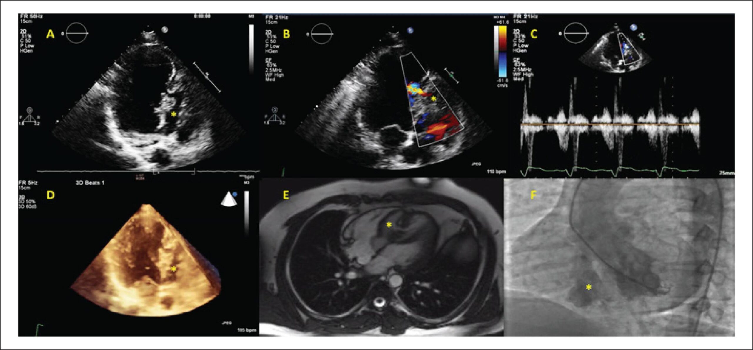
A 21 year-old male patient was admitted with symptoms of exertional dyspnea and palpitation. The physical examination revealed a grade 3/6 systolic murmur, best heard over the left 3-4th intercostal space. Transthoracic echocardiography disclosed a separate chamber (asterisk) in the interventricular septum. The apical portion of the chamber consisted of muscle tissue and the basal portion consisted of membranous and aneurysmatic tissue (Panel A). There was a muscular “tunnel-like” structure connecting the left ventricle and chamber at mid ventricular level. The color and continuous wave Doppler imaging revealed a bidirectional flow across the passage (Panel B, C).

The patient underwent three dimensional transthoracic echocardiographic examinations, which revealed two separate septa. Between these septa there was a third chamber (asterisk). It was connected to both left (via the tunnel at the muscular septum) and right ventricles (via the defect in the membranous septal aneurysm). The membranous septal aneurysm was separated from the left ventricle by a thin membrane, without any passage across it (Panel D). MRI findings were consistent with echocardiographic images (Panel E). Ventriculography and coronary angiography were performed. The coronary arteries were normal; the left ventriculography showed the muscular and membranous/aneurysmatic portions of the malformation (Panel F). The patient was advised to undergo a surgical procedure, but he refused and was discharged with recommendations about control visits.

[…]
  • Heart Defects, Congenital
  • Heart Septal Defects, Ventricular
  • ventricular septum
Read Full Article
Creative Commons Attribution (CC BY) Creative Commons license
01/04/2016 DOI: 10.5935/abc.20160045 Views 1
Reference curve of the fetal ventricular septum area by the STIC method: preliminary study
ABC
Artigo Original

Reference curve of the fetal ventricular septum area by the STIC method: preliminary study

Liliam Cristine Rolo et al. Luciano Marcondes Machado Nardozza Edward Araujo Júnior Christiane Simioni … Marina Maccagnano Zamith Antonio Fernandes Moron

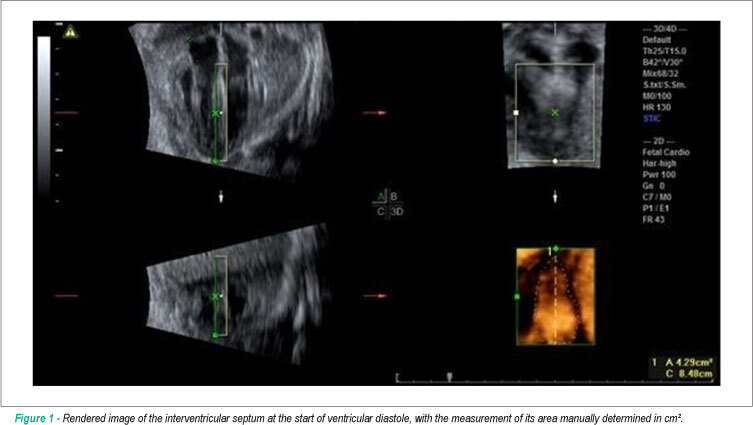
BACKGROUND: Early detection of septal changes such as septal hypertrophy commonly present in fetuses of diabetic mothers would help reduce the high rates of infant mortality. OBJECTIVE: Determine reference ranges for the fetal ventricular septal area through three-dimensional ultrasound (US3D) using the STIC method (Spatio-Temporal Image Correlation). METHODS: We conducted a cross-sectional study with 69 pregnant women between the 18th and 33rd weeks of pregnancy. We used as a reference the four-chamber plane with the ROI (Region of Interest) positioned from the ventricles; the septum area were manually marked. To assess the correlation of the interventricular septum area with gestational age (GA), we constructed scatter plots and calculated Pearson’s correlation coefficient (r), and the adjustment was performed by the coefficient of determination (R²). We calculated averages, medians, standard deviations (sd), as well as maximum and minimum values. To calculate the intraobserver reproducibility, we used the intraclass correlation coefficient (ICC). The interventricular septum thickness was measured and it was correlated with gestational age and the septal area rendered in 52 patients using the ICC. RESULTS: The interventricular septum area was highly correlated with gestational age (r = 0.81), and the average increased from 0.47 cm² in the 18th week to 2.42 cm² in the 33rd of gestation. The intraobserver reproducibility was excellent with ICC = 0.994. No significant correlation was observed between the interventricular septum measurement and the GA (R² = 0.200), as well as there was no correlation with the septal area rendered with ICC = 0.150. CONCLUSION: Reference intervals for the interventricular septum area between the 18th and the 33rd pregnancy week were determined to be highly reproducible.

  • fetal heart
  • Imaging, Three-Dimensional
  • Pregnancy
  • Reference Values
  • ventricular septum
Read Full Article
Creative Commons Attribution (CC BY) Creative Commons license
01/04/2011 DOI: 10.1590/S0066-782X2011005000036 Views 1

How to cite this article

EndNote RIS

Electronic Document Format (Vancouver)

ışılak Z, Uzun M, Kardeşoğlu E, Uz , Küçük U. Congenital Muscular Interventricular Septal Malformation with Complex Anatomical Features. Arquivos Brasileiros de Cardiologia 2016;106(4):349.

Electronic Document Format (ABNT)

ışılak, Zafer; Uzun, Mehmet; Kardeşoğlu, Ejder; Uz, Ömer; Küçük, Uğur. Congenital Muscular Interventricular Septal Malformation with Complex Anatomical Features. Arquivos Brasileiros de Cardiologia, v. 106, n. 4, p. 349-349, Mar. 2016.

Electronic Document Format (APA)

ışılak, Z., Uzun, M., Kardeşoğlu, E., Uz, , & Küçük, U. (2016). Congenital Muscular Interventricular Septal Malformation with Complex Anatomical Features. Arquivos Brasileiros de Cardiologia, 106(4), 349-349.

Electronic Document Format (ISO)

ışılak, Zafer and Uzun, Mehmet and Kardeşoğlu, Ejder and Uz, Ömer and Küçük, Uğur. Congenital Muscular Interventricular Septal Malformation with Complex Anatomical Features. Arquivos Brasileiros de Cardiologia [online]. 2016, vol. 106, n. 4, [cited 2026-10-14], pp. 349-349. Available from: <https://journals.cardiol.br/en/article/congenital-muscular-interventricular-septal-malformation-with-complex-anatomical-features/>. ISSN 0066-782X

EndNote RIS

Electronic Document Format (Vancouver)

Rolo LC, Nardozza LMM, Araujo Júnior E, Simioni C, Zamith MM, Moron AF. Reference curve of the fetal ventricular septum area by the STIC method: preliminary study. Arquivos Brasileiros de Cardiologia 2011;96(5):386-92.

Electronic Document Format (ABNT)

Rolo, Liliam Cristine; Nardozza, Luciano Marcondes Machado; Araujo Júnior, Edward; Simioni, Christiane; Zamith, Marina Maccagnano; Moron, Antonio Fernandes. Reference curve of the fetal ventricular septum area by the STIC method: preliminary study. Arquivos Brasileiros de Cardiologia, v. 96, n. 5, p. 386-392, Mar. 2011.

Electronic Document Format (APA)

Rolo, L. C., Nardozza, L. M. M., Araujo Júnior, E., Simioni, C., Zamith, M. M., & Moron, A. F. (2011). Reference curve of the fetal ventricular septum area by the STIC method: preliminary study. Arquivos Brasileiros de Cardiologia, 96(5), 386-392.

Electronic Document Format (ISO)

Rolo, Liliam Cristine and Nardozza, Luciano Marcondes Machado and Araujo Júnior, Edward and Simioni, Christiane and Zamith, Marina Maccagnano and Moron, Antonio Fernandes. Reference curve of the fetal ventricular septum area by the STIC method: preliminary study. Arquivos Brasileiros de Cardiologia [online]. 2011, vol. 96, n. 5, [cited 2026-10-14], pp. 386-392. Available from: <https://journals.cardiol.br/en/article/reference-curve-of-the-fetal-ventricular-septum-area-by-the-stic-method-preliminary-study/>. ISSN 0066-782X

  • About ABC Family
  • Internal Regulation
  • Editorial Board
  • Brazilian Society of Cardiology (SBC)

Issues

SBC Guidelines

  • Videos
  • Podcasts
  • News