ABC Family Arquivos Brasileiros de Cardiologia International Journal of Cardiovascular Sciences ABC Heart Failure & Cardiomyopathy ABC Imagem Cardiovascular
Português English
ABC Family
Português English
  • About
    • About ABC Family
    • Internal Regulation
    • Editorial Board
    • Brazilian Society of Cardiology (SBC)
  • Issues
  • SBC Guidelines
  • Content
    • Videos
    • Podcasts
    • News
  • About
    • About ABC Family
    • Internal Regulation
    • Editorial Board
    • Brazilian Society of Cardiology (SBC)
  • Issues
  • SBC Guidelines
  • Content
    • Videos
    • Podcasts
    • News

ABC Family

  • ABC Family
  • Arquivos Brasileiros de Cardiologia
  • International Journal of Cardiovascular Sciences
  • ABC Heart Failure & Cardiomyopathy
  • ABC Imagem Cardiovascular
Search in

No content found.

Journals
Ordering
Article types
Others

  1. Home
  2. Keyword: Heart Rupture
Figure 1
ABC
Case Report

A Complicated “One Segment” Myocardial Infarction: The Role of Cardiovascular Imaging

Ana Rita Pereira et al. Ana Rita Almeida Inês Cruz Luis Rocha Lopes … Maria José Loureiro Hélder Pereira

Introduction

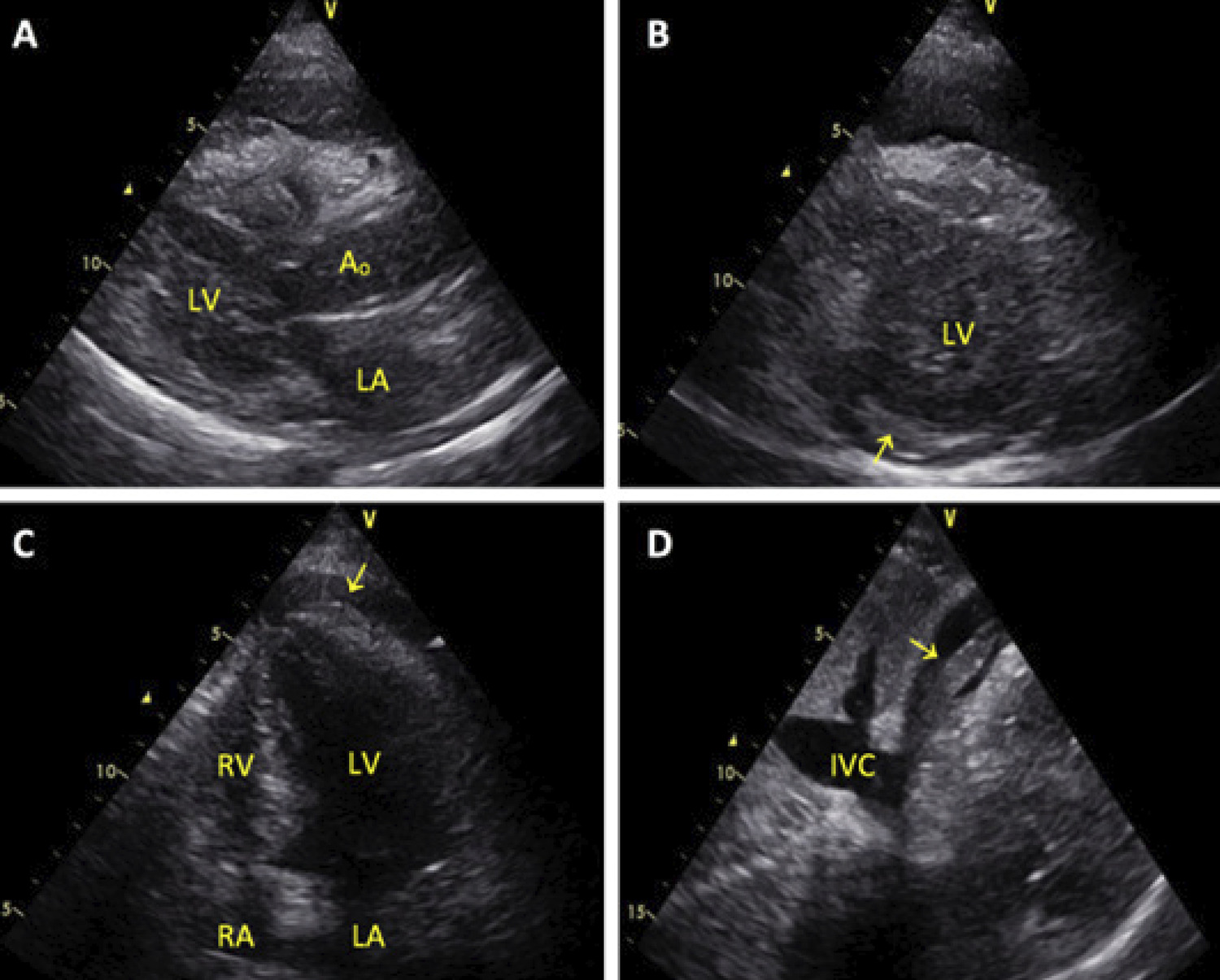
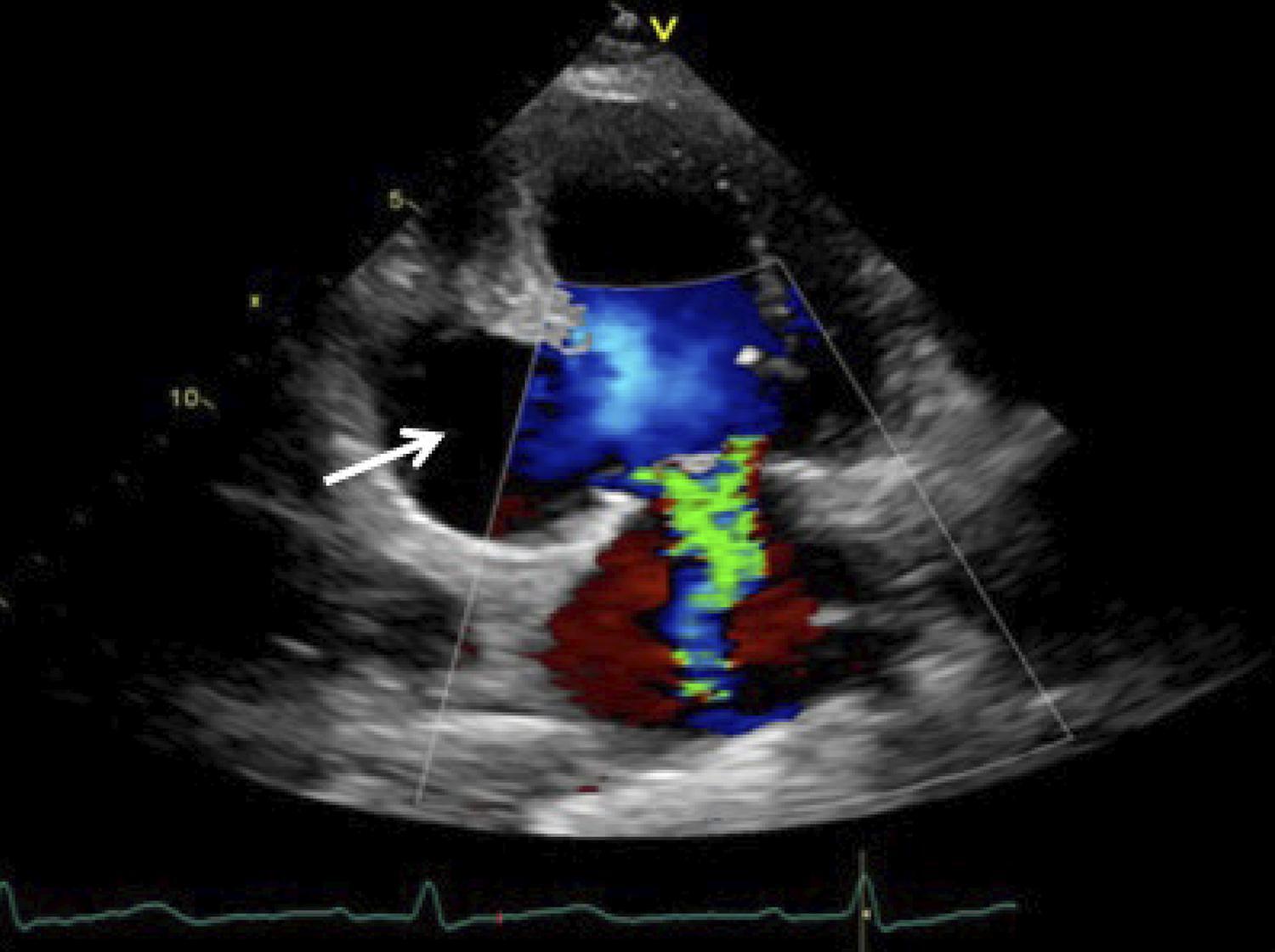
The incidence of mechanical complications (MC) after myocardial infarction (MI) was reduced to less than 1% with the routine use of primary reperfusion therapies.MC are classified as early, including acute and subacute forms, and late or chronic.The former mostly present as cardiogenic shockand the latter may vary from asymptomatic to sudden death.As all of these conditions may have potentially lethal consequences, timely diagnosis and treatment is necessary. –

[…]
  • Aneurysm,False
  • Diagnostic Imaging
  • Echocardiography/methods
  • Heart Rupture
  • Myocardial Infarction
Read Full Article
Creative Commons Attribution (CC BY) Creative Commons license
14/09/2020 DOI: 10.36660/abc.20190323 Views 1
Figure 1
ABC
Image

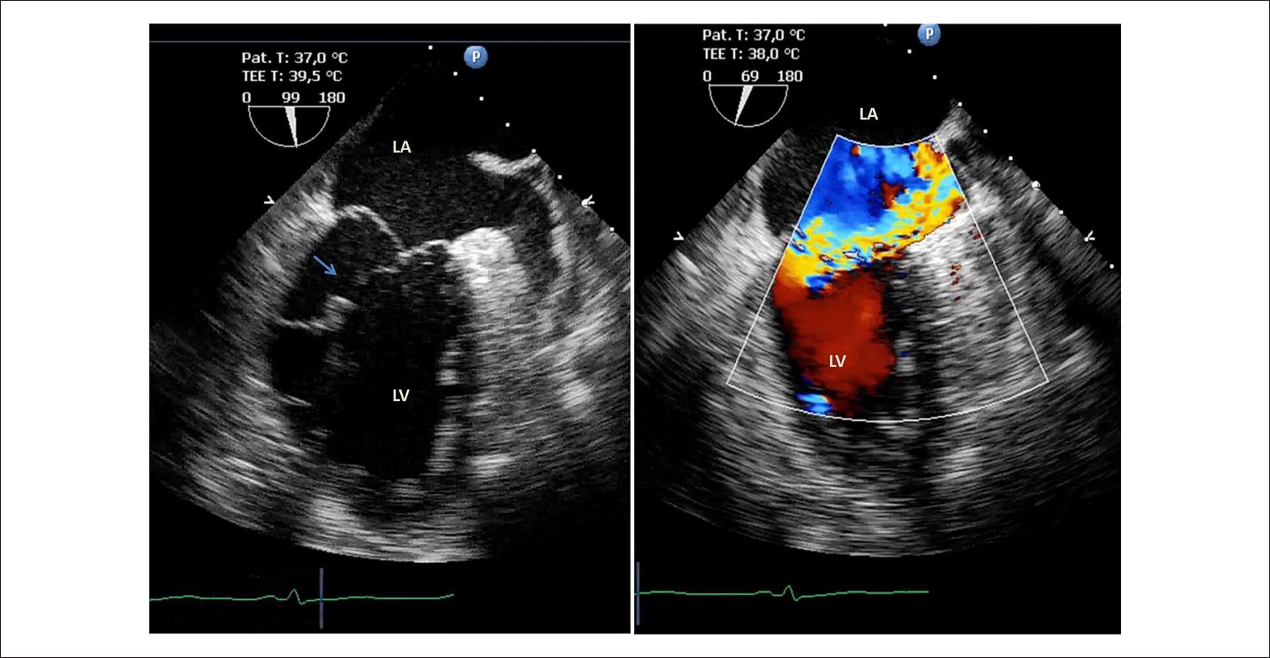
Evolved Inferior Wall Myocardial Infarction with Left Ventricular Pseudoaneurysm: A Diagnostic Dilemma

Sónia Gomes Coelho et al. Clara F. Jorge Pedro B. Carlos Anne Delgado Leopoldina Vicente

Introduction

The left ventricular (LV) pseudoaneurysm (PA) is a rare mechanical complication of acute myocardial infarction (AMI). It results from myocardial rupture, in which the hemorrhagic process is contained by the adherent pericardium. It occurs most commonly in the inferior and posterior ventricular wall, since the rupture of the anterior ventricular wall usually leads to cardiac tamponade and immediate death, while the inferior-posterior face of the heart rests on the diaphragm, facilitating the ventricular cavity containment by the pericardium. – Imaging methods are crucial to establish the diagnosis. Transthoracic (TTE) and transesophageal echocardiography (TEE) allow the definitive diagnosis in 26% and 75% of cases, respectively. , Cardiac magnetic resonance (CMR) imaging is useful in the differential diagnosis of LV PA and aneurysm, with a reported sensitivity of 100%. The presence of late pericardial enhancement in the CMR is highly suggestive of LV PA, which may represent the effect of the passage of blood into the pericardial space at the time of myocardial rupture, with subsequent pericardial inflammation and fibrosis. , ,

[…]

  • Echocardiography/methods
  • Heart Rupture
  • Magnetic Resonance Spectroscopy/ methods
  • Myocardial Infarction/complications
  • Pseudoaneurysm
Read Full Article
Creative Commons Attribution (CC BY) Creative Commons license
29/05/2020 DOI: 10.36660/abc.20200029
Figure 1
ABC
Case Report

Partial Papillary Muscle Rupture after Myocardial Infarction and Early Severe Obstructive Bioprosthetic Valve Thrombosis: an Unusual Combination

Inês Silveira et al. Marta Oliveira Catarina Gomes Sofia Cabral … André Luz Severo Torres

Introduction

Mechanical complications after myocardial infarction (MI) have become uncommon since the introduction of primary angioplasty. They can lead to a rapid clinical deterioration and a fatal outcome, with patient’s survival being depen­dent on their prompt recognition and intervention. We describe a case of two rare mechanical complications: a partial papillary muscle rupture after MI, followed by an early severe obstructive thrombosis of the implanted bioprosthetic valve.

[…]

  • Atrioventricular Block/complications
  • Bioprosthesis
  • Heart Arrest
  • Heart Rupture
  • Myocardial Infarction/complications
  • Pacemaker, Artificial
  • Post-Infarction
  • Thrombolytic Therapy
Read Full Article
Creative Commons Attribution (CC BY) Creative Commons license
01/09/2018 DOI: 10.5935/abc.20180179 Views 2
Frequency of acute plaque ruptures and thin cap atheromas at sites of maximal stenosis
ABC
Artigo Original

Frequency of acute plaque ruptures and thin cap atheromas at sites of maximal stenosis

Fabio Tavora et al. Nathaniel Cresswell Ling Li David Fowler Allen Burke

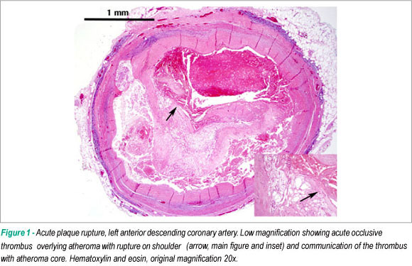
BACKGROUND: There have been few autopsy studies relating sites of thin cap atheroma (TCFA) to sites of acute plaque rupture in culprit arteries, and sites of maximal narrowing in non-culprit arteries. OBJECTIVE: We aimed to quantify and locate the frequency of TCFA related to the sites of maximal stenosis in atherosclerotic plaques. METHODS: We studied 88 hearts in victims of sudden death dying with coronary thrombus overlying acute plaque rupture. Thin cap atheromas were defined as fibrous cap < 65 microns overlying a necrotic core. Percent luminal narrowing was determined at the sites of plaque rupture and thin cap atheromas. RESULTS: There were 81 men and 7 women, mean age 50 years ± 9 SD. The plaque rupture was the site of maximal luminal narrowing in 47% of culprit arteries. TCFAs were present in 67 hearts (83%). Of these, 49 (73%) demonstrated TCFAs in the culprit artery; 17 (25%) in the culprit artery only, 32 (48%) in the culprit artery and in a non-culprit artery, and 18 (27%) only in a non-culprit artery. In non-culprit arteries, TCFAs represented the maximal site of stenosis in 44% of arteries. The acute rupture site is the site of maximal luminal narrowing in the involved vessel in 47% of hearts from patients dying with acute plaque rupture. CONCLUSION: These data may suggest that luminal narrowing is not a reliable marker for TCFA.

  • Atherosclerosis
  • Coronary Artery Disease
  • Coronary stenosis
  • Heart Rupture
Read Full Article
Creative Commons Attribution (CC BY) Creative Commons license
05/05/2010 DOI: 10.1590/S0066-782X2010000200003
Multiple aneurysms of aortocoronary saphenous vein grafts with fatal rupture
ABC
Case Report

Multiple aneurysms of aortocoronary saphenous vein grafts with fatal rupture

Fábio R. Távora et al. Jean Jeudy Allen P. Burke

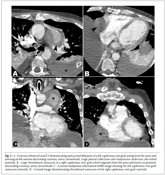
Aortocoronary saphenous vein graft (SVG) aneurysms are rare, and are usually asymptomatic and detected incidentally. Spontaneous rupture of SVG is rare and imaging data are few. We report on a 39-year old man who was admitted to the hospital with hematemesis 10 years after aortocoronary bypass surgery. CT images revealed 3 aortocoronary SVG aneurysms, but failed to detect any rupture. His subsequent death due to rupture of SVG aneurysm was documented at autopsy, illustrating the need for aggressive treatment of symptomatic coronary graft aneurysms.

  • Cardiac Surgery
  • Coronary Aneurysm
  • Coronary Artery Bypass
  • Heart Rupture
  • Myocardial Revascularization
Read Full Article
Creative Commons Attribution (CC BY) Creative Commons license
15/06/2007 DOI: 10.1590/S0066-782X2007000500022 Views 1

How to cite this article

EndNote RIS

Electronic Document Format (Vancouver)

Pereira AR, Almeida AR, Cruz I, Lopes LR, Loureiro MJ, Pereira H. A Complicated “One Segment” Myocardial Infarction: The Role of Cardiovascular Imaging. Arquivos Brasileiros de Cardiologia 2020;115(1 suppl 1):25-30.

Electronic Document Format (ABNT)

Pereira, Ana Rita; Almeida, Ana Rita; Cruz, Inês; Lopes, Luis Rocha; Loureiro, Maria José; Pereira, Hélder. A Complicated “One Segment” Myocardial Infarction: The Role of Cardiovascular Imaging. Arquivos Brasileiros de Cardiologia, v. 115, n. 1 suppl 1, p. 25-30, Sep. 2020.

Electronic Document Format (APA)

Pereira, A. R., Almeida, A. R., Cruz, I., Lopes, L. R., Loureiro, M. J., & Pereira, H. (2020). A Complicated “One Segment” Myocardial Infarction: The Role of Cardiovascular Imaging. Arquivos Brasileiros de Cardiologia, 115(1 suppl 1), 25-30.

Electronic Document Format (ISO)

Pereira, Ana Rita and Almeida, Ana Rita and Cruz, Inês and Lopes, Luis Rocha and Loureiro, Maria José and Pereira, Hélder. A Complicated “One Segment” Myocardial Infarction: The Role of Cardiovascular Imaging. Arquivos Brasileiros de Cardiologia [online]. 2020, vol. 115, n. 1 suppl 1, [cited 2026-10-14], pp. 25-30. Available from: <https://journals.cardiol.br/en/article/a-complicated-one-segment-myocardial-infarction-the-role-of-cardiovascular-imaging/>. ISSN 0066-782X

EndNote RIS

Electronic Document Format (Vancouver)

Coelho SG, Jorge CF, Carlos PB, Delgado A, Vicente L. Evolved Inferior Wall Myocardial Infarction with Left Ventricular Pseudoaneurysm: A Diagnostic Dilemma. Arquivos Brasileiros de Cardiologia 2020;114(4):730-1.

Electronic Document Format (ABNT)

Coelho, Sónia Gomes; Jorge, Clara F.; Carlos, Pedro B.; Delgado, Anne; Vicente, Leopoldina. Evolved Inferior Wall Myocardial Infarction with Left Ventricular Pseudoaneurysm: A Diagnostic Dilemma. Arquivos Brasileiros de Cardiologia, v. 114, n. 4, p. 730-731, May. 2020.

Electronic Document Format (APA)

Coelho, S. G., Jorge, C. F., Carlos, P. B., Delgado, A., & Vicente, L. (2020). Evolved Inferior Wall Myocardial Infarction with Left Ventricular Pseudoaneurysm: A Diagnostic Dilemma. Arquivos Brasileiros de Cardiologia, 114(4), 730-731.

Electronic Document Format (ISO)

Coelho, Sónia Gomes and Jorge, Clara F. and Carlos, Pedro B. and Delgado, Anne and Vicente, Leopoldina. Evolved Inferior Wall Myocardial Infarction with Left Ventricular Pseudoaneurysm: A Diagnostic Dilemma. Arquivos Brasileiros de Cardiologia [online]. 2020, vol. 114, n. 4, [cited 2026-10-14], pp. 730-731. Available from: <https://journals.cardiol.br/en/article/evolved-inferior-wall-myocardial-infarction-with-left-ventricular-pseudoaneurysm-a-diagnostic-dilemma/>. ISSN 0066-782X

EndNote RIS

Electronic Document Format (Vancouver)

Silveira I, Oliveira M, Gomes C, Cabral S, Luz A, Torres S. Partial Papillary Muscle Rupture after Myocardial Infarction and Early Severe Obstructive Bioprosthetic Valve Thrombosis: an Unusual Combination. Arquivos Brasileiros de Cardiologia 2018;111(3):430-3.

Electronic Document Format (ABNT)

Silveira, Inês; Oliveira, Marta; Gomes, Catarina; Cabral, Sofia; Luz, André; Torres, Severo. Partial Papillary Muscle Rupture after Myocardial Infarction and Early Severe Obstructive Bioprosthetic Valve Thrombosis: an Unusual Combination. Arquivos Brasileiros de Cardiologia, v. 111, n. 3, p. 430-433, Aug. 2018.

Electronic Document Format (APA)

Silveira, I., Oliveira, M., Gomes, C., Cabral, S., Luz, A., & Torres, S. (2018). Partial Papillary Muscle Rupture after Myocardial Infarction and Early Severe Obstructive Bioprosthetic Valve Thrombosis: an Unusual Combination. Arquivos Brasileiros de Cardiologia, 111(3), 430-433.

Electronic Document Format (ISO)

Silveira, Inês and Oliveira, Marta and Gomes, Catarina and Cabral, Sofia and Luz, André and Torres, Severo. Partial Papillary Muscle Rupture after Myocardial Infarction and Early Severe Obstructive Bioprosthetic Valve Thrombosis: an Unusual Combination. Arquivos Brasileiros de Cardiologia [online]. 2018, vol. 111, n. 3, [cited 2026-10-14], pp. 430-433. Available from: <https://journals.cardiol.br/en/article/partial-papillary-muscle-rupture-after-myocardial-infarction-and-early-severe-obstructive-bioprosthetic-valve-thrombosis-an-unusual-combination/>. ISSN 0066-782X

EndNote RIS

Electronic Document Format (Vancouver)

Tavora F, Cresswell N, Li , Fowler D, Burke A. Frequency of acute plaque ruptures and thin cap atheromas at sites of maximal stenosis. Arquivos Brasileiros de Cardiologia 2010;94(2):153-9.

Electronic Document Format (ABNT)

Tavora, Fabio; Cresswell, Nathaniel; Li, ng; Fowler, David; Burke, Allen. Frequency of acute plaque ruptures and thin cap atheromas at sites of maximal stenosis. Arquivos Brasileiros de Cardiologia, v. 94, n. 2, p. 153-159, May. 2010.

Electronic Document Format (APA)

Tavora, F., Cresswell, N., Li, , Fowler, D., & Burke, A. (2010). Frequency of acute plaque ruptures and thin cap atheromas at sites of maximal stenosis. Arquivos Brasileiros de Cardiologia, 94(2), 153-159.

Electronic Document Format (ISO)

Tavora, Fabio and Cresswell, Nathaniel and Li, ng and Fowler, David and Burke, Allen. Frequency of acute plaque ruptures and thin cap atheromas at sites of maximal stenosis. Arquivos Brasileiros de Cardiologia [online]. 2010, vol. 94, n. 2, [cited 2026-10-14], pp. 153-159. Available from: <https://journals.cardiol.br/en/article/frequency-of-acute-plaque-ruptures-and-thin-cap-atheromas-at-sites-of-maximal-stenosis/>. ISSN 0066-782X

EndNote RIS

Electronic Document Format (Vancouver)

Távora FR, Jeudy J, Burke AP. Multiple aneurysms of aortocoronary saphenous vein grafts with fatal rupture. Arquivos Brasileiros de Cardiologia 2007;88(5):e107-10.

Electronic Document Format (ABNT)

Távora, Fábio R.; Jeudy, Jean; Burke, Allen P.. Multiple aneurysms of aortocoronary saphenous vein grafts with fatal rupture. Arquivos Brasileiros de Cardiologia, v. 88, n. 5, p. e107-e110, Jun. 2007.

Electronic Document Format (APA)

Távora, F. R., Jeudy, J., & Burke, A. P. (2007). Multiple aneurysms of aortocoronary saphenous vein grafts with fatal rupture. Arquivos Brasileiros de Cardiologia, 88(5), e107-e110.

Electronic Document Format (ISO)

Távora, Fábio R. and Jeudy, Jean and Burke, Allen P.. Multiple aneurysms of aortocoronary saphenous vein grafts with fatal rupture. Arquivos Brasileiros de Cardiologia [online]. 2007, vol. 88, n. 5, [cited 2026-10-14], pp. e107-e110. Available from: <https://journals.cardiol.br/en/article/multiple-aneurysms-of-aortocoronary-saphenous-vein-grafts-with-fatal-rupture/>. ISSN 0066-782X

  • About ABC Family
  • Internal Regulation
  • Editorial Board
  • Brazilian Society of Cardiology (SBC)

Issues

SBC Guidelines

  • Videos
  • Podcasts
  • News