ABC Family Arquivos Brasileiros de Cardiologia International Journal of Cardiovascular Sciences ABC Heart Failure & Cardiomyopathy ABC Imagem Cardiovascular
Português English
ABC Family
Português English
  • About
    • About ABC Family
    • Internal Regulation
    • Editorial Board
    • Brazilian Society of Cardiology (SBC)
  • Issues
  • SBC Guidelines
  • Content
    • Videos
    • Podcasts
    • News
  • About
    • About ABC Family
    • Internal Regulation
    • Editorial Board
    • Brazilian Society of Cardiology (SBC)
  • Issues
  • SBC Guidelines
  • Content
    • Videos
    • Podcasts
    • News

ABC Family

  • ABC Family
  • Arquivos Brasileiros de Cardiologia
  • International Journal of Cardiovascular Sciences
  • ABC Heart Failure & Cardiomyopathy
  • ABC Imagem Cardiovascular
Search in

No content found.

Journals
Ordering
Article types
Others

  1. Home
  2. Keyword: Left Ventricular Hypertrophy
Figure 1
ABCIC
Case Report

Biventricular Noncompaction Cardiomyopathy Associated With Neuropathy in a Young Patient: A Case Report

Mônica de Oliveira Gonçalo et al. André Alexandre dos Santos Gomes Matheus Martins Monteiro Nicolas Babilônia Cavalcanti … Fernando Almeida Bezerra Moisés Abtibol Machado Dênison Clark Corrêa de Miranda Kátia do Nascimento Couceiro João Marcos Bemfica Barbosa Ferreira

Introduction

Ventricular hypertrabeculation, also known as ventricular noncompaction, is a myocardial phenotype characterized by excessive trabeculation and deep intertrabecular recesses caused by incomplete myocardial compaction during embryonic development. The earliest reported cases were associated with congenital heart defects, including ventricular outflow tract obstruction, complex cyanotic congenital heart disease, and coronary artery anomalies.,

The clinical presentation is highly heterogeneous and may include heart failure (HF), arrhythmias, thromboembolic events, and sudden cardiac death. In addition to ventricular structural abnormalities, ventricular hypertrabeculation may be associated with cardiac conduction disorders, atrial arrhythmias, and functional valvular regurgitation (FVR) secondary to cardiac chamber dilatation, all of which contribute to greater clinical complexity and a poorer prognosis.

[…]
  • Case Reports
  • Left Ventricular Hypertrophy
  • Mitral Valve Insufficiency
Read Full Article
Creative Commons Attribution (CC BY) Creative Commons license
03/09/2026 DOI: 10.36660/abcimg.20260078i Views 1
ABC
Viewpoint

The Old Habits Need to be Abandoned and the Term "Overload" Replaced by "High Amplitude Criteria"

José Nunes de Alencar et al. Sandro Pinelli Felicioni Matheus Kiszka Scheffer Mariana Nogueira De Marchi Ljuba Bacharova

Left ventricular overload?

Brazilian electrocardiograms (ECG) reports have long labeled voltage-positive tracings as “left ventricular overload,” a legacy of historical teaching that linked hemodynamic loads to characteristic repolarization patterns. The problem is not the physiology but the nomenclature: overload is a hemodynamic claim, whereas the ECG measures electrical phenomena. Contemporary evidence reframes voltage positivity as an electrical phenotype with variable correlation to left ventricular mass (LVM) and geometry. In this article, we argue that Brazil should retire “overload,” adopt “high-amplitude criteria” as the report label, and explicitly teach the underlying concept of left ventricular electrical remodeling (LVER), so that our language matches what the ECG actually measures.

Phrasing “left ventricular overload” feels pragmatic — if surface ECG is “insensitive” to anatomic hypertrophy, perhaps what it detects is something else, namely “overload.” The evidence base argues otherwise. Electrocardiography records electric fields at the body surface; it does not directly measure mass, geometry, or loading conditions. This distinction is the core of the modern paradigm articulated by Ljuba Bacharova et al.: QRS voltage and morphology reflect an interplay of spatial determinants (source geometry, torso conductivity, electrode distance) and non-spatial determinants (conduction velocity, intercellular coupling, fibrosis, ionic remodeling), such that the same LVM can yield different voltages — and high voltage can arise with normal mass — depending on the myocardium’s electrical properties. The Working Group’s “4th Report” in the Journal of Electrocardiology integrates mechanistic simulation studies with population data and draws three conclusions that are decisive for our terminology. First, increased LVM and geometric type (concentric, eccentric, dilation) are not the main or only determinants of QRS patterns; combinations of anatomy and slowed activation propagation produce the observed spectrum from “pseudo-normal” tracings through high voltage and prolonged intrinsicoid deflection to left bundle branch block. Second, diffuse conduction slowing or midwall conduction abnormalities — without any increase in mass — can generate QRS changes that meet conventional electrocardiographic-left ventricular hypertrophy (LVH) criteria in a normal-sized ventricle. Third, in community cohorts the electrical and anatomic phenotypes frequently diverge, and each carries independent prognostic information.

[…]

  • Electrocardiography
  • Left Ventricular Hypertrophy
Read Full Article
Creative Commons Attribution (CC BY) Creative Commons license
17/07/2026 DOI: 10.36660/abc.20250709i
Accuracy and Prognosis of Electrocardiographic and Echocardiographic Left Ventricular Hypertrophy in Brazilian Adults - ELSA-Brasil Study
ABC
Original Article

Accuracy and Prognosis of Electrocardiographic and Echocardiographic Left Ventricular Hypertrophy in Brazilian Adults – ELSA-Brasil Study

Marcelo M. Pinto-Filho et al. Lillian Guimarães de Faria Murilo Foppa Angela Barreto Santiago Santos … Sandhi Maria Barreto Antonio Luiz Pinho Ribeiro

Abstract

Background

Left ventricular hypertrophy (LVH), defined by increased left ventricular mass, is an independent predictor of cardiovascular mortality. Evidence suggests that LVH identified by electrocardiogram (ECG) and echocardiography represents distinct entities with potentially different prognostic implications.

Objectives

Assess the diagnostic accuracy of electrocardiographic criteria for LVH compared with transthoracic echocardiogram (TTE) and to evaluate their predictive value for fatal cardiovascular outcomes.

Methods

We conducted a cross-sectional diagnostic accuracy analysis and a prospective follow-up within the Longitudinal Study of Adult Health. LVH on ECG was defined by Minnesota Code criteria, and LVH on TTE by increased calculated left ventricular mass. Significance level was defined at 0,05.

Results

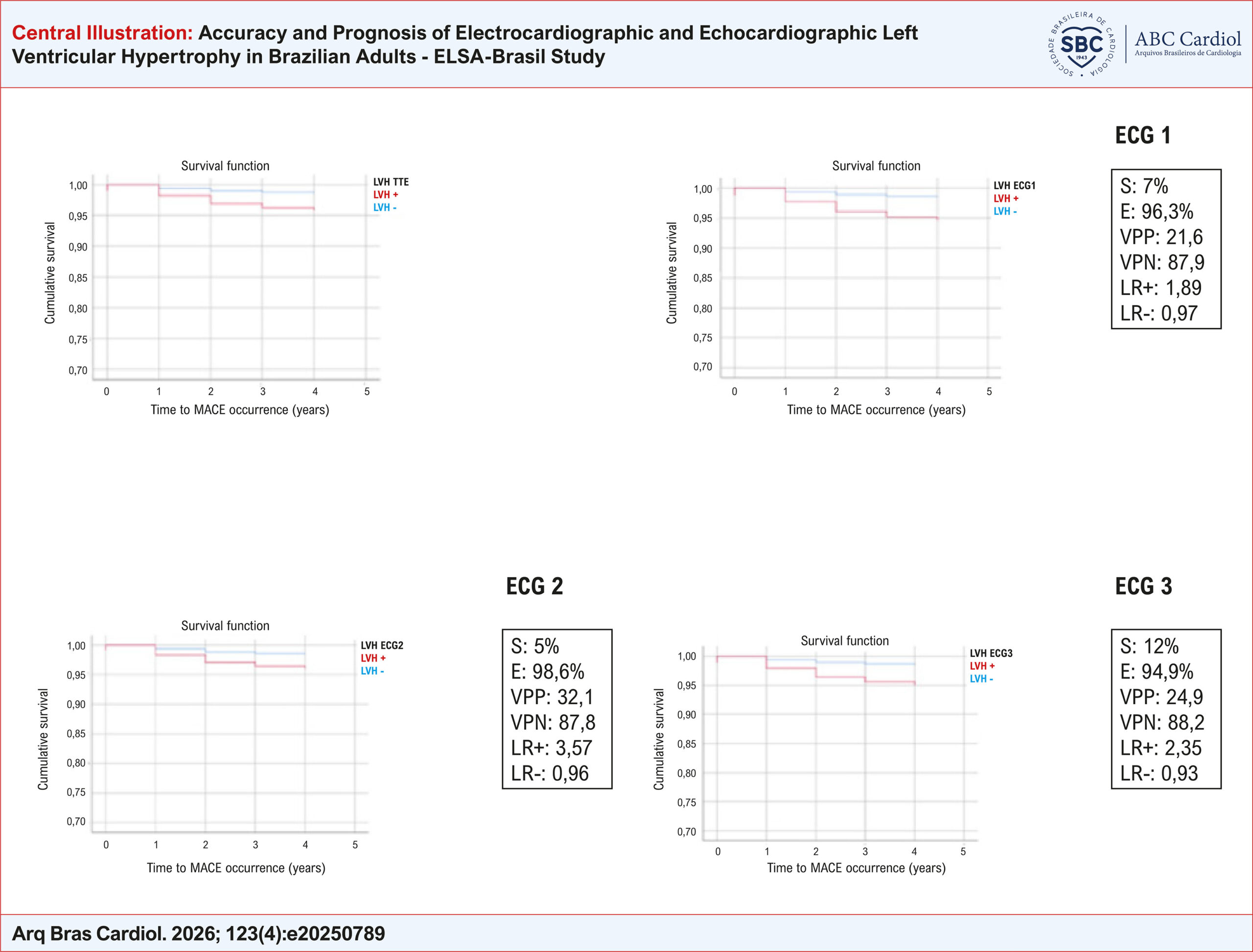
Among 2,849 participants (43.6% male), median age 62 years, 12.5% had LVH by TTE and 5.9% by ECG criteria. ECG-defined LVH showed, from different criteria, very low sensitivity (5–12%) but high specificity (95–99%) compared with TTE. In prognostic analyses, TTE-defined LVH was associated with significant fatal adverse cardiovascular events (FMACE) (HR 3.2, 95% CI 1.7–5.9). ECG-defined LVH also predicted FMACE, with HR close to 3.5 across the different MC criteria in multivariate regression analysis. The predictive capacity remained significant after further adjustment for hypertrophy defined by either of the methods.

Conclusions

Although electrocardiographic criteria demonstrated poor diagnostic accuracy for LVH compared with TTE, both modalities were independently associated with adverse cardiovascular outcomes. These findings suggest that ECG-defined and TTE-defined LVH provide complementary prognostic information and may reflect distinct pathophysiological processes.

  • Electrocardiography
  • Left Ventricular Hypertrophy
  • Longitudinal Studies
  • Prognosis
Read Full Article
Creative Commons Attribution (CC BY) Creative Commons license
12/06/2026 DOI: 10.36660/abc.20250789i Views 1
Figure 1
IJCS
Brief Communication

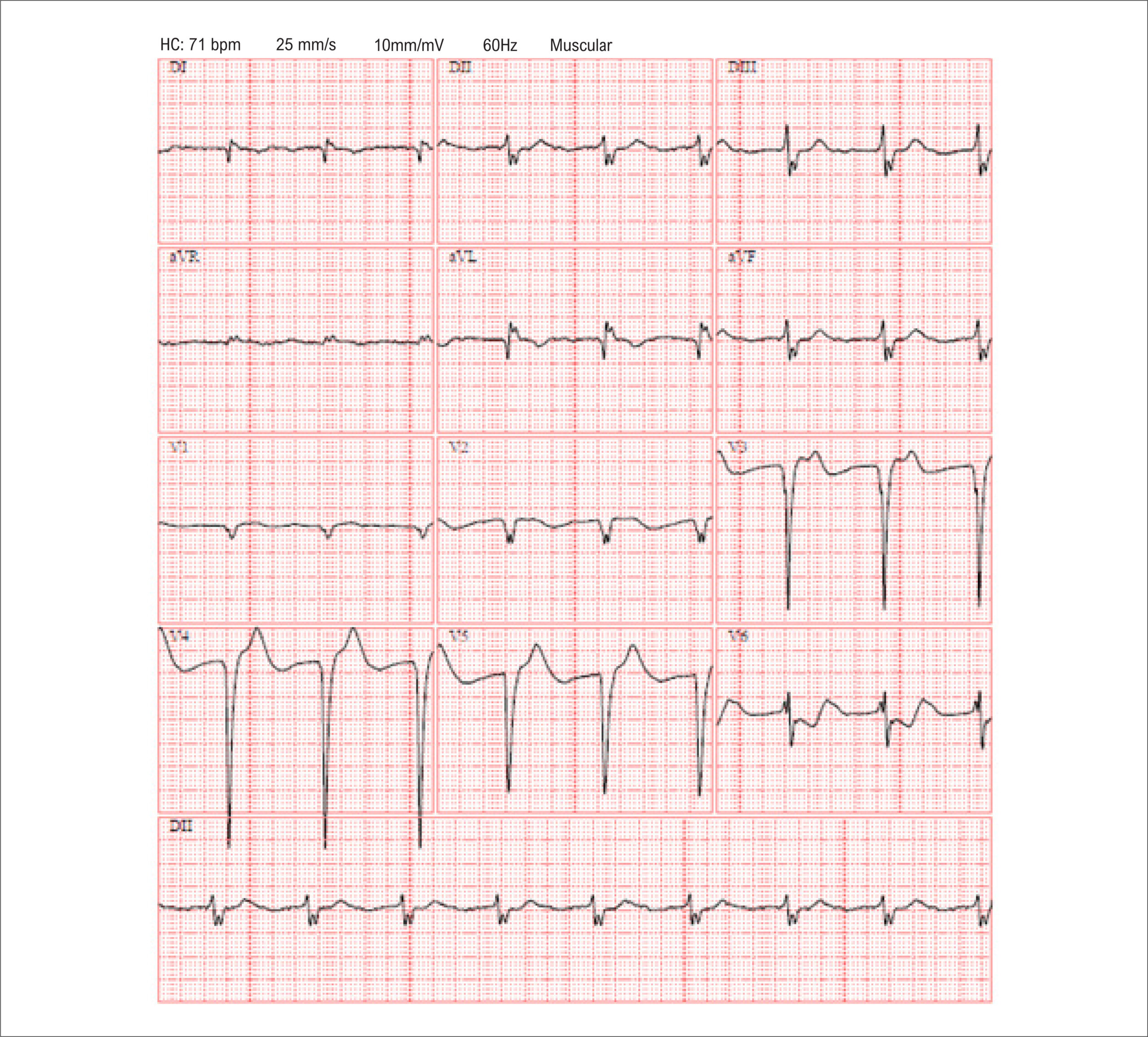
S-Wave Amplitude in Lead V3 Identifies Other Electrocardiography Criteria of Left Ventricular Hypertrophy

Bernardo Augusto Silveira Corrêa et al. Gabriel Pinheiro Soares Alencar e Silva Felipe Campelo de Miranda Marcio Henrique de Jesus Oliveira … Gabriel Scarpioni Barbosa Victor Hudson de Lacerda Borges José Nunes de Alencar

Introduction

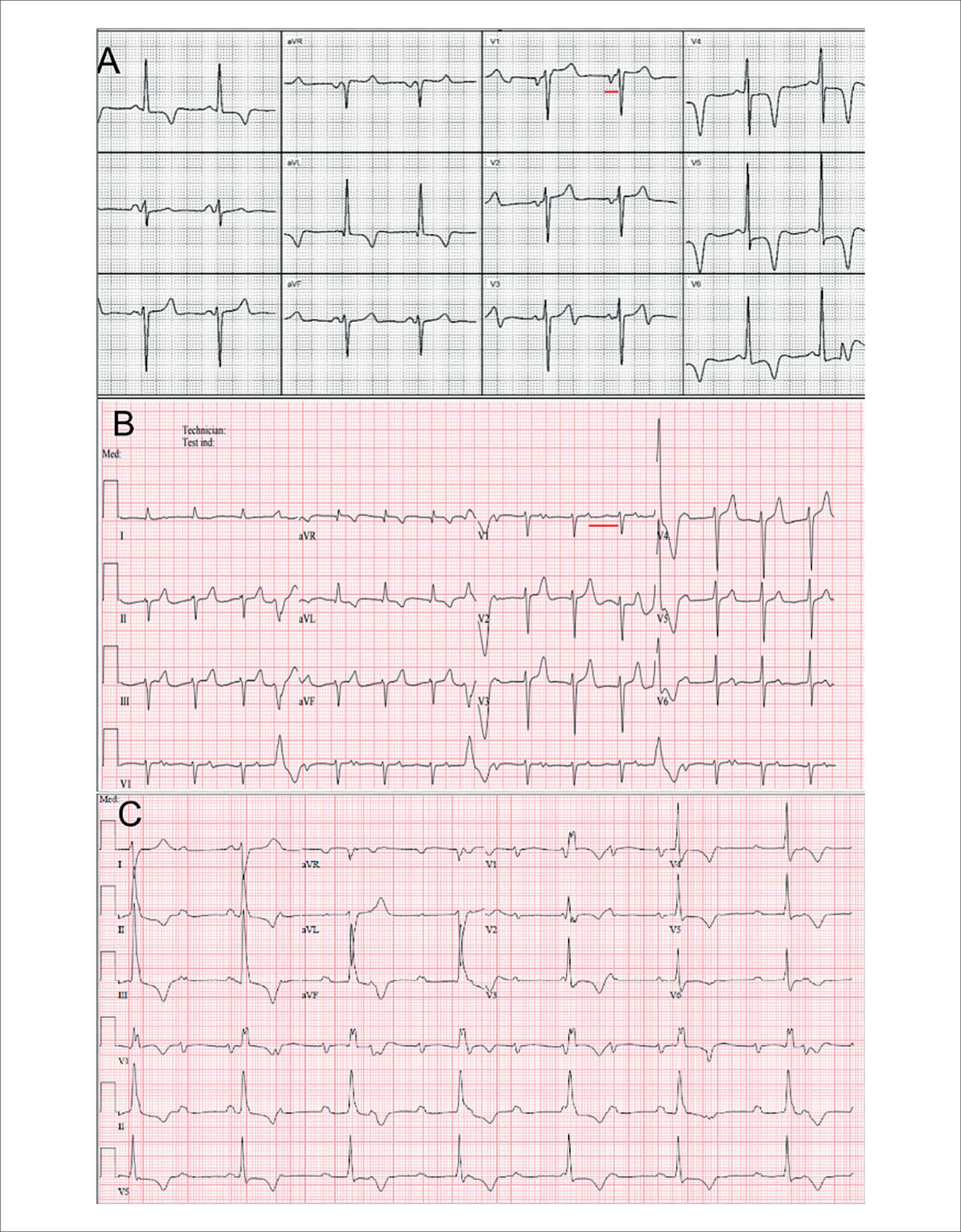
Electrocardiographic (ECG) criteria for detecting left ventricular hypertrophy (LVH) are integral to clinical decision-making. Although these methods are quick to apply, low cost, and widely available, their sensitivity is modest. Multiple voltage-based criteria exist, such as the Sokolow-Lyon, Cornell, and Peguero-Lo Presti criteria, but accurate use often requires familiarity with several indices and can be time-consuming for trainees. Simple, reliable markers could improve the efficiency and consistency with which ECGs are flagged as possibly indicating LVH.

The S-wave amplitude in lead V3 (SV3) is a plausible single-lead marker. It is a component of the Cornell criterion (SV3 + RaVL), and it is closely related to the Peguero-Lo Presti criterion (SV4 plus the deepest precordial S). Because the largest precordial S wave often occurs in V3, a single SV3 measurement could serve as a rapid screening tool to identify the presence of established LVH criteria, especially for those with less experience interpreting ECGs.

[…]

  • Electrocardiography
  • Left Ventricular Hypertrophy
Read Full Article
Creative Commons Attribution (CC BY) Creative Commons license
27/04/2026 DOI: 10.36660/ijcs.20250050
Figure 1
ABC
Research Letter

Late-Onset Fabry Disease: Diagnostic Challenges and Clinical Evolution

Hernán Patricio García Mejía et al. Heleuterio da Conceição Nicolau Madogolele Manuela Cristina Ribeiro Dias Barroso Matheus Carvalho Alves Nogueira … Kevin Rafael de Paula Morales Cristhian Espinoza Romero Luciano Nastari Fábio Fernandes

Introduction

Simultaneous cardiac and renal involvement can result from various systemic conditions, such as hypertension, autoimmune diseases, infiltrative diseases, and hereditary syndromes. Fabry disease (FD) is an X-linked genetic disorder caused by a deficiency or absence of the enzyme α-galactosidase A, leading to the progressive accumulation of glycosphingolipids, especially globotriaosylceramide (Gb3), in various tissues. This accumulation of Gb3 promotes multisystemic dysfunctions, with heterogeneous clinical manifestations that vary according to sex, the degree of residual enzymatic activity, and the form of presentation, classic or non-classic.,

The estimated incidence of FD ranges from 1 in 40,000 to 1 in 117,000 individuals worldwide. In Brazil, the epidemiology of FD remains poorly defined due to the rarity of the condition and underreporting of cases, which reinforces the value of clinical reports such as this one.

[…]

  • Cardiac Conduction System Disease
  • Cardiomyopathies
  • Fabry Disease
  • Left Ventricular Hypertrophy
Read Full Article
Creative Commons Attribution (CC BY) Creative Commons license
17/04/2026 DOI: 10.36660/abc.20250554i Views 3
Left Ventricular Hypertrophy on Electrocardiography and Chronic Kidney Disease: A Case–Control Pilot Study
IJCS
Original Article

Left Ventricular Hypertrophy on Electrocardiography and Chronic Kidney Disease: A Case–Control Pilot Study

Nabil Hajar et al. Nabila Nuralya Joveazhari Shofa Chasani Zulfachmi Wahab … Galih Prakasa Adhyatma Indira Aura Balqis Zeidan Ahmad Fahdalhaq Bintang Adhiyoso Wicaksono

Abstract

Background:

Chronic kidney disease (CKD) is a condition characterized by abnormalities in kidney structure or function lasting more than 3 months. CKD is known to increase the risk of cardiovascular events, which may be predicted by electrocardiography (ECG) abnormalities, such as left ventricular hypertrophy (LVH). ECG is widely used in under-resourced health care settings due to its practicality and accessibility.

Objective:

This study aimed to analyze the relationship between LVH and CKD using ECG.

Methods:

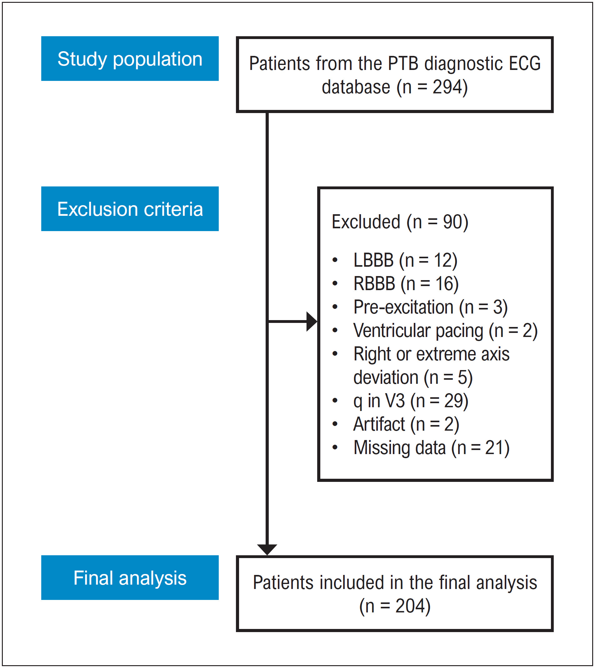
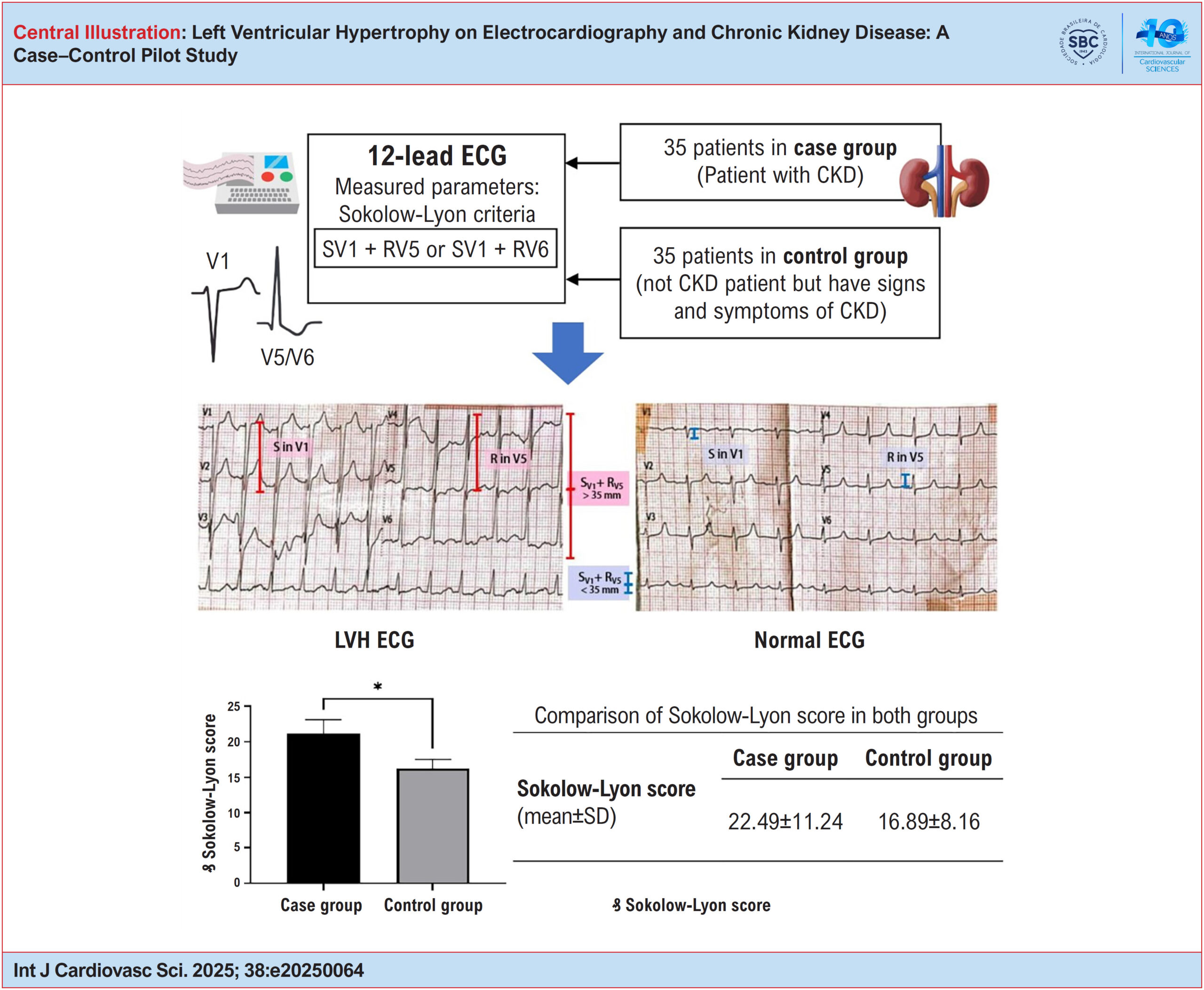
This analytical observational study used a case–control design. The case group included patients diagnosed with CKD, while the control group consisted of patients presenting with signs and symptoms suggestive of CKD but without a confirmed diagnosis. Each group included 35 participants. Simple random sampling was used for the case group, and stratified random sampling for the control group. ECG examinations were performed, and measurements were taken using calipers in millimeters (mm). The Sokolow–Lyon criteria were applied to identify LVH. Data were analyzed using an independent t-test, with statistical significance set at p < 0.05.

Results:

The Sokolow–Lyon score was significantly higher in the case group than in the control group (p = 0.03).

Conclusion:

There is a significant association between LVH, as identified on ECG, and CKD. These findings may assist health professionals in resource-limited settings, where access to ECG is restricted.

  • Cardiovascular Diseases
  • chronic renal insufficiency
  • Electrocardiography
  • Left Ventricular Hypertrophy
Read Full Article
Creative Commons Attribution (CC BY) Creative Commons license
01/09/2025 DOI: 10.36660/ijcs.20250064 Views 2
The Complex Puzzle of Hypertrophic Phenotype: A Practical Approach for the Clinician
ABC
Review Article

The Complex Puzzle of Hypertrophic Phenotype: A Practical Approach for the Clinician

Alex dos Santos Felix et al. Silvio Henrique Barberato Marcelo Dantas Tavares de Melo Sílvia Aguiar Rosa Nuno Cardim

Abstract

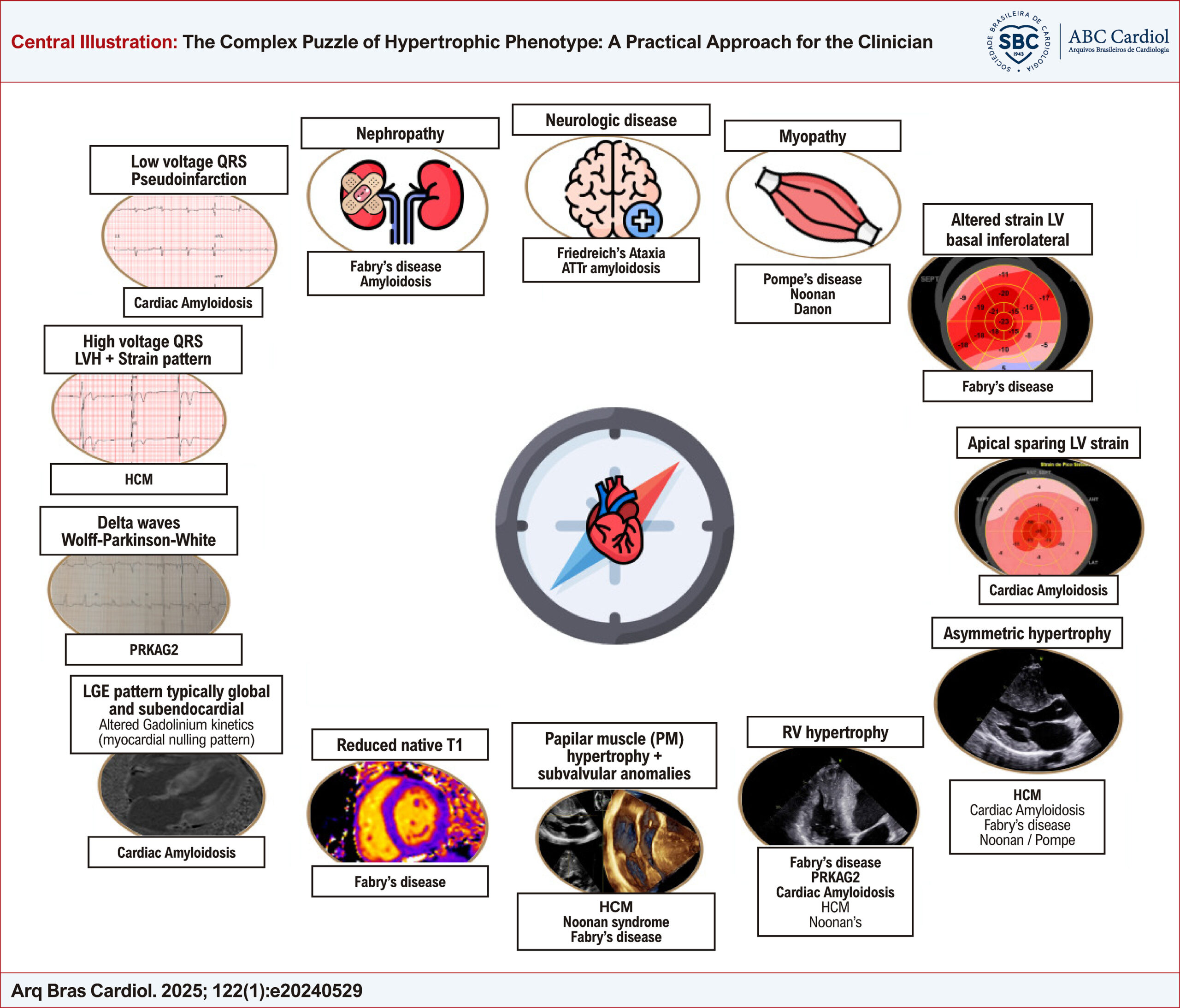
Left ventricular hypertrophy (LVH) represents a frequent observation in clinical practice. Nonetheless, the hypertrophic phenotype emerges as a common manifestation of diverse conditions, thereby presenting a diagnostic conundrum for clinicians. Differentiation among the etiologies of LVH is imperative for therapy decision-making, as different approaches must be implemented for distinct conditions, such as LVH secondary to loading changes, hypertrophic cardiomyopathy (HCM), or HCM mimics.

In some instances, an erroneous or late diagnosis may lead to a progression of the underlying disease with worsening functional capacity, high morbidity and mortality.

The rational use of cardiovascular multimodality imaging is of great importance when carried out in addition to a thorough clinical assessment and correlated with electrocardiographic findings, providing clues to fill the gaps, being, most of the time, the missing piece to solve this challenging puzzle.

An integrative approach is of paramount importance for the evaluation of these patients, as they are often followed by several specialties, with varied systemic manifestations. Although a multidisciplinary team is needed for an optimized follow-up of these patients, the most important player in this journey is the clinician, whose mission is to bring together all the red flags and coordinate all the data for an assertive diagnosis.

The objective of this review is to provide a pragmatic methodology, highlighting important clues for discriminating among the diverse conditions that result in LVH.

  • Cardiac Imaging Techniques
  • Diagnosis
  • Differential Diagnosis
  • Heart Function Tests
  • Left Ventricular Hypertrophy
Read Full Article
Creative Commons Attribution (CC BY) Creative Commons license
04/04/2025 DOI: 10.36660/abc.20240529i Views 1
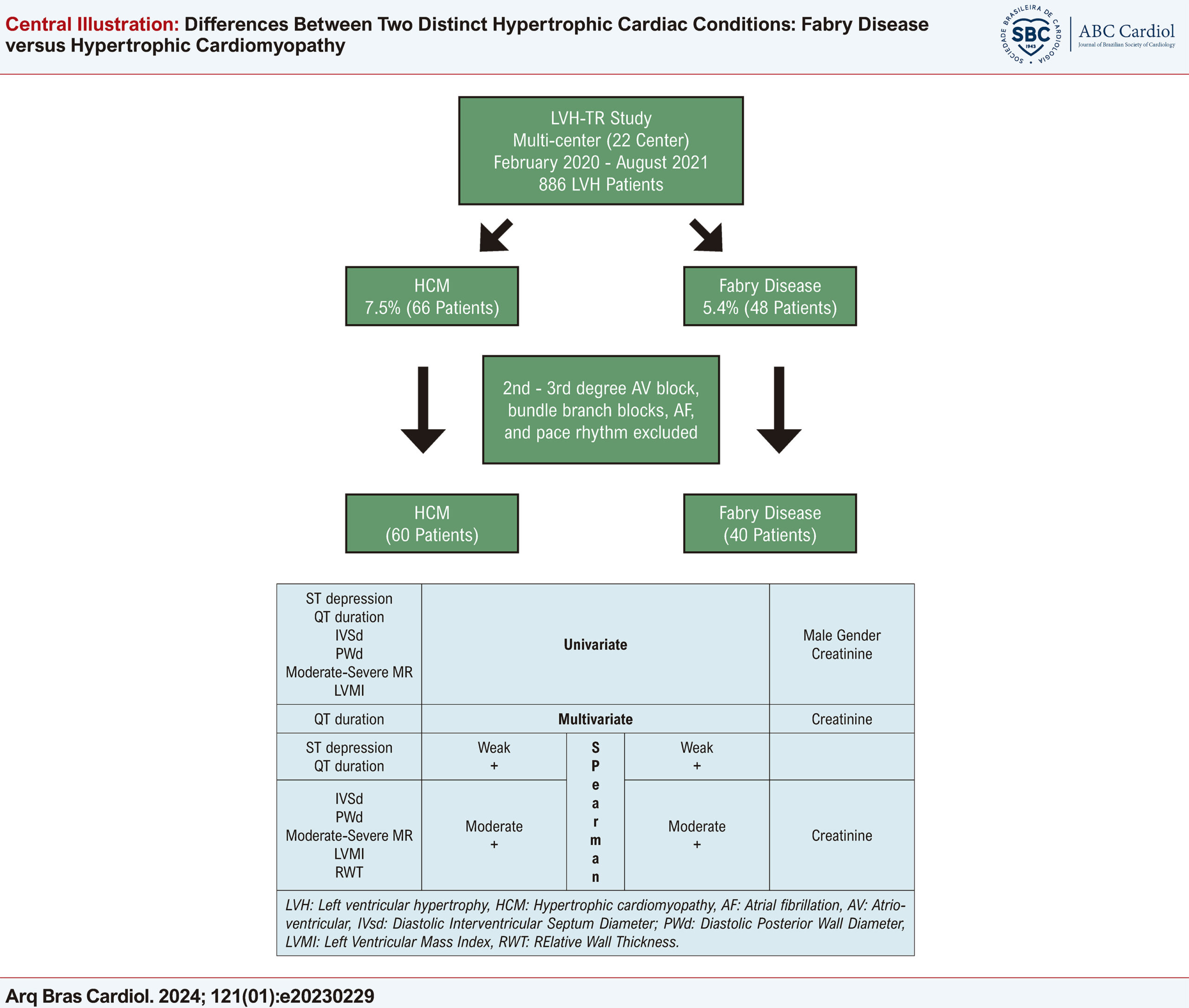
Central Illustration
ABC
Original Article

Differences Between Two Distinct Hypertrophic Cardiac Conditions: Fabry Disease versus Hypertrophic Cardiomyopathy

Onur Akhan et al. Mehmet Kış Tuncay Güzel Mehdi Zoghi

Abstract

Background

Hypertrophic cardiomyopathy (HCM) and Fabry disease (FD) are genetically inherited diseases with left ventricular hypertrophy (LVH) phenotype characteristics that cause adverse cardiac outcomes.

Objectives

To investigate the demographic, clinical, biochemical, electrocardiographic (ECG), and echocardiographic (ECHO) differences between HCM and FD.

Methods

60 HCM and 40 FD patients were analyzed retrospectively as a subanalysis of the ‘LVH-TR study’ after excluding patients with atrial fibrillation, pace rhythm, bundle branch blocks, and second and third-degree atrioventricular (AV) blocks. The significance level was accepted as <0.05.

Results

Male gender (p=0.048) and creatinine (p=0.010) are significantly higher in favor of FD; however, ST depression (p=0.028), QT duration (p=0.041), interventricular septum thickness (IVSd) (p=0.003), posterior wall thickness (PWd) (p=0.009), moderate-severe mitral regurgitation (MR) (p=0.013), and LV mass index (LVMI) (p=0.041) are significantly higher in favor of HCM in the univariate analyses. In multivariate analysis, statistical significance only continues in creatinine (p=0.018) and QT duration (0.045). FD was positively correlated with creatinine (rho=0.287, p=0.004) and HCM was positively correlated with PWd (rho=0.306, p=0.002), IVSd (rho=0.395, p<0.001), moderate-severe MR (rho=0.276, p<0.005), LVMI (rho=0.300, p=0.002), relative wall thickness (RWT) (rho=0.271, p=0.006), QT duration (rho=0.213, p=0.034) and ST depression (rho=0.222, p=0.026).

Conclusion

Specific biochemical, ECG, and ECHO characteristics can aid in the differentiation and early diagnosis of HCM and FD.

  • Cardiomyopathies
  • Fabry Disease
  • Hypertrophic Cardiomyopathy
  • Left Ventricular Hypertrophy
Read Full Article
Creative Commons Attribution (CC BY) Creative Commons license
05/02/2024 DOI: 10.36660/abc.20230229
Figure 1
ABC
Original Article

Atrial Mechanics in Hypertrophic Cardiomyopathy: Discriminating between Ventricular Hypertrophy and Fibrosis

Patrícia Marques-Alves et al. João André Ferreira André Azul Freitas José Paulo Almeida … Rui Baptista Graça Castro Rui Martins Paulo Donato Maria João Ferreira Lino Gonçalves

Abstract

Background

Hypertrophic cardiomyopathy (HCM) and left ventricular hypertrophy (LVH) secondary to systemic hypertension (HTN) may be associated with left atrial (LA) functional abnormalities.

Objectives

We aimed to characterize LA mechanics in HCM and HTN and determine any correlation with the extent of left ventricular (LV) fibrosis measured by cardiac magnetic resonance (CMR) in HCM patients.

Methods

Two-dimensional speckle tracking-derived longitudinal LA function was acquired from apical views in 60 HCM patients, 60 HTN patients, and 34 age-matched controls. HCM patients also underwent CMR, with measurement of late gadolinium enhancement (LGE) extension. Association with LA strain parameters was analyzed. Statistical significance was set at p<0.05.

Results

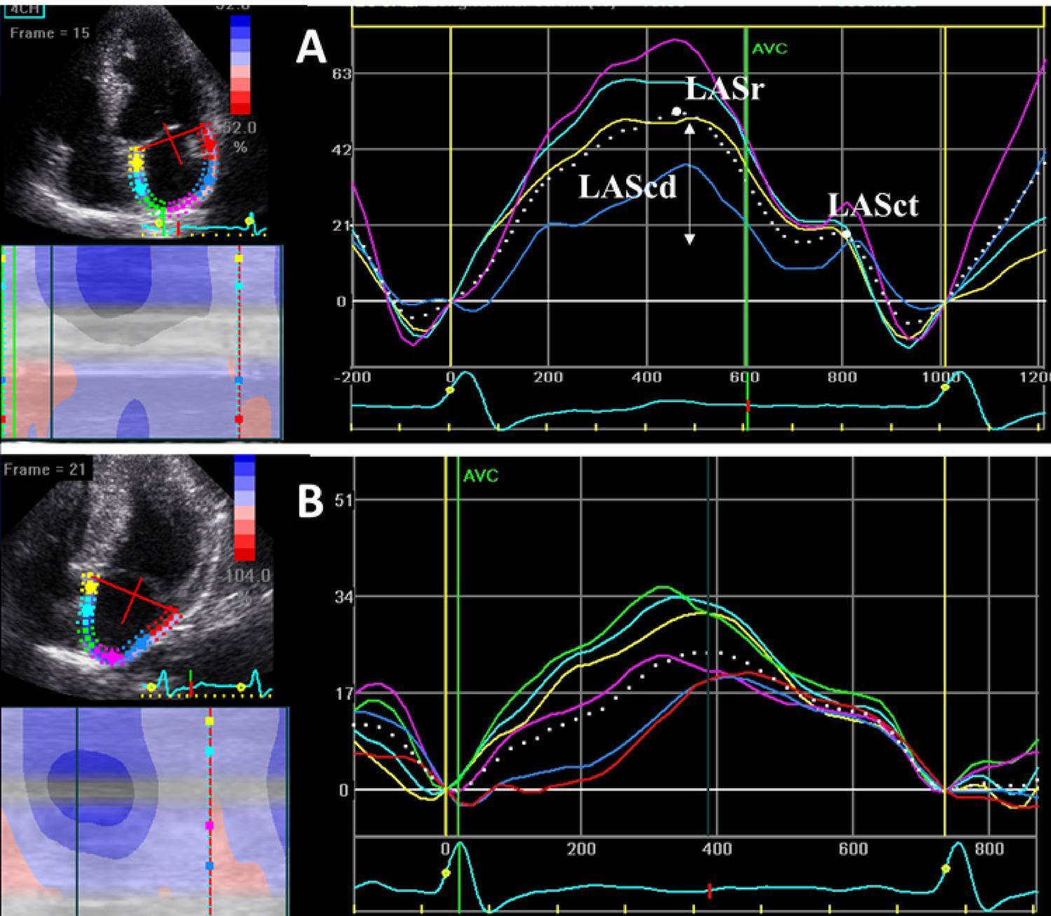
Mean LV ejection fraction was not different between the groups. The E/e’ ratio was impaired in the HCM group and preserved in the control group. LA mechanics was significantly reduced in HCM, compared to the HTN group. LA strain rate in reservoir (LASRr) and in contractile (LASRct) phases were the best discriminators of HCM, with an area under the curve (AUC) of 0.8, followed by LA strain in reservoir phase (LASr) (AUC 0.76). LASRr and LASR-ct had high specificity (89% and 91%, respectively) and LASr had sensitivity of 80%. A decrease in 2.79% of LA strain rate in conduit phase (LASRcd) predicted an increase of 1cm in LGE extension (r2=0.42, β 2.79, p=0.027).

Conclusions

LASRr and LASRct were the best discriminators for LVH secondary to HCM. LASRcd predicted the degree of LV fibrosis assessed by CMR. These findings suggest that LA mechanics is a potential predictor of disease severity in HCM.

  • Cardiomyopathy, Hypertrophic
  • Echocardiography/methods
  • Hypertension
  • Left Ventricular Hypertrophy
  • Magnetic Resonance Spectroscopy/ methods
Read Full Article
Creative Commons Attribution (CC BY) Creative Commons license
21/02/2022 DOI: 10.36660/abc.20200890 Views 1
Figure 1
ABC
Original Article

Cost-Effectiveness Analysis of Evolocumab Therapy in Patients at High Risk of Cardiovascular Events in the Context of the Brazilian Unified Health System

Luiza Latado et al. Rodrigo Morel Vieira de Melo Sóstenes Mistro Adriana Lopes Latado … Harrison Floriano do Nascimento Yasmin Menezes Lira Natalia Ferreira Cardoso de Oliveira Yuri de Santana Galindo Tainá Viana Luiz Carlos Santana Passos

Abstract

Background:

Hypertrophic cardiomyopathy (HCM) and left ventricular hypertrophy (LVH) secondary to systemic hypertension (HTN) may be associated with left atrial (LA) functional abnormalities.

Objectives:

We aimed to characterize LA mechanics in HCM and HTN and determine any correlation with the extent of left ventricular (LV) fibrosis measured by cardiac magnetic resonance (CMR) in HCM patients.

Methods:

Two-dimensional speckle tracking-derived longitudinal LA function was acquired from apical views in 60 HCM patients, 60 HTN patients, and 34 age-matched controls. HCM patients also underwent CMR, with measurement of late gadolinium enhancement (LGE) extension. Association with LA strain parameters was analyzed. Statistical significance was set at p<0.05.

Results:

Mean LV ejection fraction was not different between the groups. The E/e’ ratio was impaired in the HCM group and preserved in the control group. LA mechanics was significantly reduced in HCM, compared to the HTN group. LA strain rate in reservoir (LASRr) and in contractile (LASRct) phases were the best discriminators of HCM, with an area under the curve (AUC) of 0.8, followed by LA strain in reservoir phase (LASr) (AUC 0.76). LASRr and LASR-ct had high specificity (89% and 91%, respectively) and LASr had sensitivity of 80%. A decrease in 2.79% of LA strain rate in conduit phase (LASRcd) predicted an increase of 1cm in LGE extension (r2=0.42, β 2.79, p=0.027).

Conclusions:

LASRr and LASRct were the best discriminators for LVH secondary to HCM. LASRcd predicted the degree of LV fibrosis assessed by CMR. These findings suggest that LA mechanics is a potential predictor of disease severity in HCM.

  • Cardiomyopathy, Hypertrophic
  • Echocardiography/methods
  • Hypertension
  • Left Ventricular Hypertrophy
  • Magnetic Resonance Spectroscopy/ methods
Read Full Article
Creative Commons Attribution (CC BY) Creative Commons license
22/11/2021 DOI: 10.36660/abc.20200690 Views 1
Figure 1
ABC
Original Article

MCP-1 Levels are Associated with Cardiac Remodeling but not with Resistant Hypertension

Alessandra Mileni Versuti Ritter et al. Ana Paula Cabral de Faria Andrea Sabbatini Nathalia Batista Corrêa … Veridiana Brunelli Rodrigo Modolo Heitor Moreno

Abstract

Background:

Hypertension is a chronic, low-grade inflammation process associated with the release of cytokines and development of target organ damage. Deregulated monocyte chemoattractant protein-1 (MCP-1) levels have been associated with high blood pressure and cardiovascular complications; however, the mechanisms involved are complex and not fully understood.

Objective:

This study aimed to compare the levels of MCP-1 in patients with resistant (RH) versus mild-to-moderate (HTN) hypertension and their association with the presence or absence of left ventricular hypertrophy (LVH) in all hypertensive subjects.

Methods:

We enrolled 256 hypertensive subjects: 120 RH and 136 HTN, investigating the relationship between circulating MCP-1 levels and blood pressure, biochemical data, hematologic profile, and cardiac damage within the RH and HTN groups. Plasma MCP-1 levels were measured by ELISA and LVH was assessed by echocardiography.

Results:

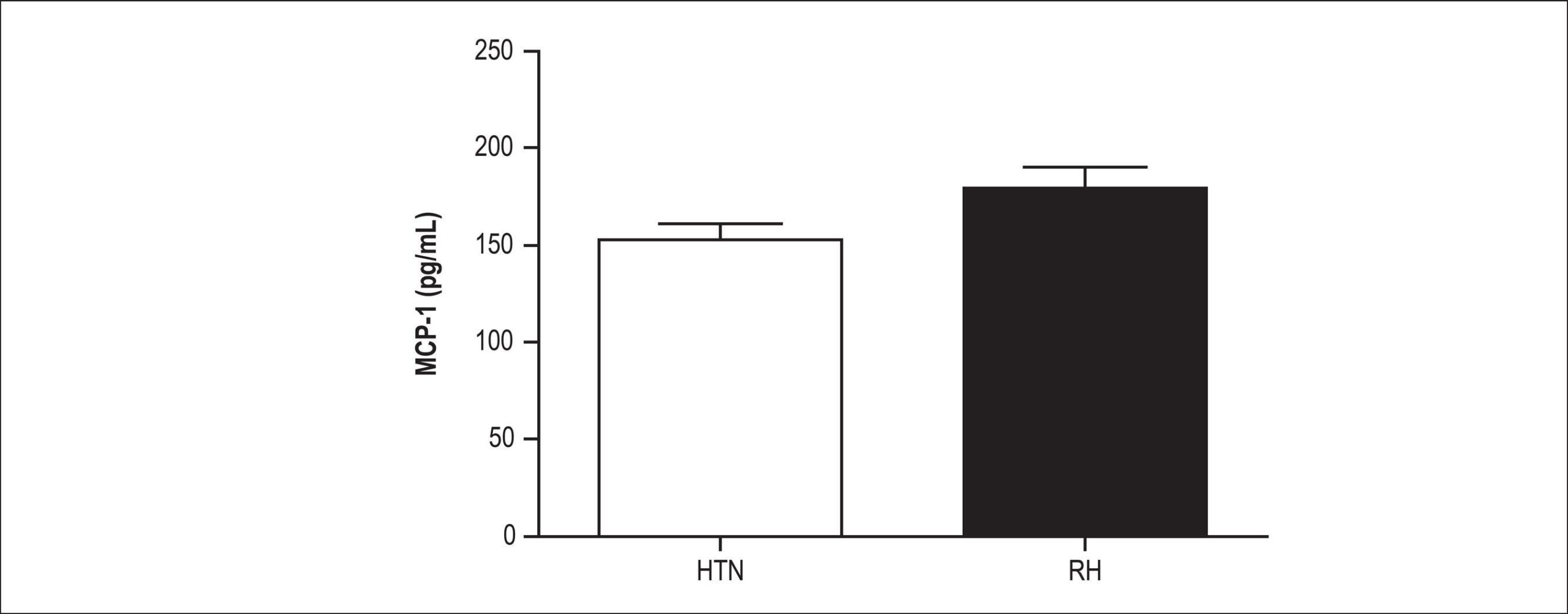
We found no difference in MCP-1 levels between RH and HTN subjects. On the other hand, we encountered lower MCP-1 levels in patients with LVH (105 pg/mL [100 – 260 pg/mL] versus 136 pg/mL (100 – 200 pg/mL), p = 0.005, respectively] compared with those without LVH. A logistic regression model adjusted for body mass index (BMI), age, race, aldosterone levels, and presence of diabetes and RH demonstrated that median levels of MCP-1 (2.55 pg/mL [1.22 – 5.2 pg/mL], p = 0.01) were independently associated with LVH in the entire hypertensive population.

Conclusion:

Since MCP-1 levels were similar in both RH and HTN subjects and decreased in hypertensive patients with existing LVH, our study suggests a possible downregulation in MCP-1 levels in hypertensive individuals with LVH, regardless of hypertension strata.

  • Cytokines
  • Left Ventricular Hypertrophy
  • Monocyte Chemoattractant Proteins
  • Refractory Hypertension
Read Full Article
Creative Commons Attribution (CC BY) Creative Commons license
30/03/2017 DOI: 10.5935/abc.20170033
Echocardiography evaluation for asymptomatic patients with severe obesity
ABC
Artigo Original

Echocardiography evaluation for asymptomatic patients with severe obesity

Isaura Elaine Gonçalves Moreira Rocha et al. Edgar Guimarães Victor Maria Cynthia Braga Odwaldo Barbosa e Silva Mônica de Moraes Chaves Becker

OBJECTIVE: To study the systolic and diastolic function of asymptomatic patients with severe obesity using a Doppler echocardiography. METHODS: Thirty candidates for bariatric surgery, with an average BMI of 49.2 ± 8.8 Kg/m² and no previous history of heart disease were evaluated through transthoracic echocardiography. RESULTS: Enlarged left chambers were observed in 42.9% of the sample, diastolic dysfunction in 54.6% and left ventricular hypertrophy in 82.1%, of which 50% of the cases presented the geometric pattern of eccentric hypertrophy. Indexation of left ventricular mass to height resulted in a significantly higher number of diagnoses for hypertrophy than indexation to body surface area (p = 0.0053), demonstrating that this index is more appropriate to determine ventricular hypertrophy in obese people. Correlations between left ventricular hypertrophy with obesity duration and pressure levels were positive as well as correlations between body mass index and diastolic dysfunction indicators. CONCLUSION: This study demonstrated that echocardiograms performed on asymptomatic severely obese patients can detect alterations in the cardiac structure that are common in cases of obesity cardiomyopathy and can be associated with the development of heart failure, arrhythmias and sudden death, enabling the identification of patients with greater cardiovascular risk.

  • Echocardiography
  • Left Ventricular Hypertrophy
  • obesity cardiomyopathy
  • severe obesity
Read Full Article
Creative Commons Attribution (CC BY) Creative Commons license
04/04/2007 DOI: 10.1590/S0066-782X2007000100009 Views 2
Left Ventricular Hypertrophy Evaluation in Obese Hypertensive Patients: Effect of Left Ventricular Mass Index Criteria
ABC
Original Article

Left Ventricular Hypertrophy Evaluation in Obese Hypertensive Patients: Effect of Left Ventricular Mass Index Criteria

Eduardo Cantoni Rosa et al. Valdir Ambrósio Moysés Ricardo Cintra Sesso Frida Liane Plavnik … Fernando Flexa Ribeiro Nárcia E. B. Kohlmann Artur Beltrame Ribeiro Maria Tereza Zanella Osvaldo Kohlmann Jr.

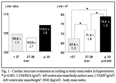
PURPOSE: To evaluate left ventricular mass (LVM) index in hypertensive and normotensive obese individuals. METHODS: Using M mode echocardiography, 544 essential hypertensive and 106 normotensive patients were evaluated, and LVM was indexed for body surface area (LVM/BSA) and for height² (LVM/h²). The 2 indexes were then compared in both populations, in subgroups stratified according to body mass index (BMI): or = 30kg/m². RESULTS: The BSA index does not allow identification of significant differences between BMI subgroups. Indexing by height² provides significantly increased values for high BMI subgroups in normotensive and hypertensive populations. CONCLUSION: Left ventricular hypertrophy (LVH) has been underestimated in the obese with the use of LVM/BSA because this index considers obesity as a physiological variable. Indexing by height² allows differences between BMI subgroups to become apparent and seems to be more appropriate for detecting LVH in obese populations.

  • Essential Hypertension
  • Left Ventricular Hypertrophy
  • left ventricular mass index
  • Obesity
Read Full Article
Creative Commons Attribution (CC BY) Creative Commons license
31/01/2007 DOI: 10.1590/S0066-782X2002000400001 Views 1
Detection of incipient left ventricular hypertrophy in mild to moderate arterial hypertension with normal electrocardiogram and echocardiogram: a new use for signal-averaged electrocardiography
ABC
Original Article

Detection of incipient left ventricular hypertrophy in mild to moderate arterial hypertension with normal electrocardiogram and echocardiogram: a new use for signal-averaged electrocardiography

Paulo Ginefra et al. Eduardo C. Barbosa P. R. Benchimol-Barbosa Alfredo S. Bomfim … Sílvia H. Boghossian Angelo A. Salgado Flávia G. Brasil Elizabete A. Freitas Francisco M. Albanesi Filho

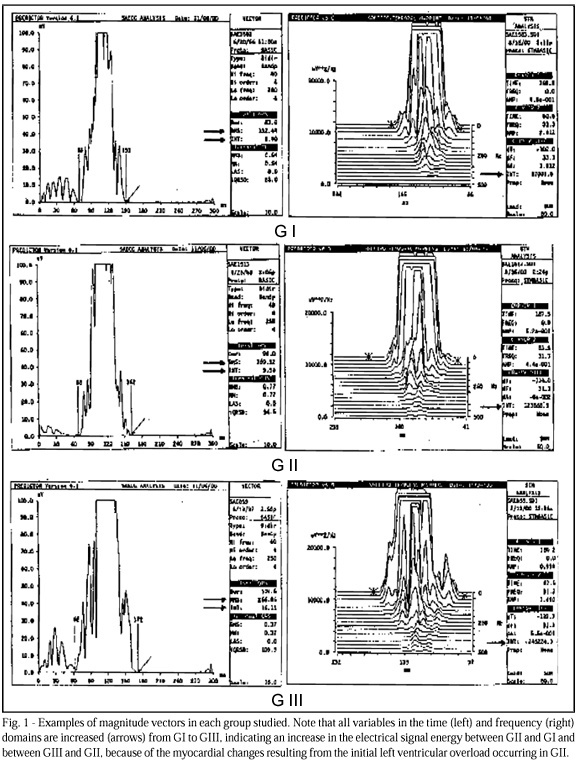
OBJECTIVE: To assess signal-averaged electrocardiogram (SAECG) for diagnosing incipient left ventricular hypertrophy (LVH). METHODS: A study with 115 individuals was carried out. The individuals were divided as follows: GI – 38 healthy individuals; GII – 47 individuals with mild to moderate hypertension and normal findings on echocardiogram and ECG; and GIII – 30 individuals with hypertension and documented LVH. The magnitude vector of the SAECG was analyzed with the high-pass cutoff frequency of 40 Hz through the bidirectional four-pole Butterworth high-pass digital filter. The mean quadratic root of the total QRS voltage (RMST) and the two-dimensional integral of the QRS area of the spectro-temporal map were analyzed between 0 and 30 Hz for the frequency domain (Int FD), and between 40 and 250 Hz for the time domain (Int TD). The electrocardiographic criterion for LVH was based on the Cornell Product. Left ventricular mass was calculated with the Devereux formula. RESULTS: All parameters analyzed increased from GI to GIII, except for Int FD (GII vs GIII) and RMST log (GII vs GIII). Int TD showed greater accuracy for detecting LVH with an appropriate cutoff > 8 (sensitivity of 55%, specificity of 81%). Positive values (> 8) were found in 56.5% of the G II patients and in 18.4% of the GI patients (p< 0.0005). CONCLUSION: SAECG can be used in the early diagnosis of LVH in hypertensive patients with normal ECG and echocardiogram.

  • arterial hypertension
  • Left Ventricular Hypertrophy
  • signal-averaged electrocardiogram
Read Full Article
Creative Commons Attribution (CC BY) Creative Commons license
31/01/2007 DOI: 10.1590/S0066-782X2003000900007
Left ventricular mass in patients with heart failure
ABC
Artigo Original

Left ventricular mass in patients with heart failure

Marcello Ricardo Paulista Markus et al. Humberto Felício Gonçalves de Freitas Paulo Roberto Chizzola Gisela Tunes da Silva … Antonio Carlos Pedroso de Lima Alfredo José Mansur

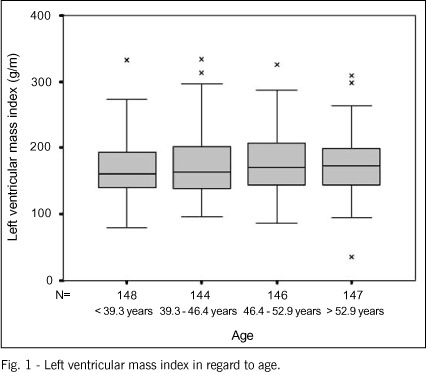
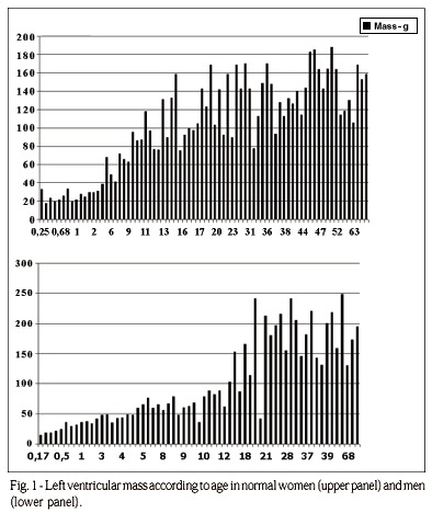
OBJECTIVE: To assess left ventricular mass in patients with heart failure and its correlations with other clinical variables and prognosis. METHODS: The study comprised 587 patients aged from 13.8 years to 68.9 years, 461 (78.5%) being males and 126 (21.5%) females. Left ventricular mass was estimated by using M-mode echocardiography and was indexed by height. RESULTS: The left ventricular mass index ranged from 35.3 g/m to 333.5 g/m and increased with age. The left ventricular mass index was greater in males (mean, 175.7 g/m) than in females (mean, 165.7 g/m). The left ventricular mass index was greater in patients with hypertensive cardiomyopathy (mean of 188.1 g/m), with idiopathic dilated cardiomyopathy (mean, 177.7 g/m) and with cardiomyopathies of other etiologies (mean, 175.1 g/m) than in patients with chagasic (mean, 164.3 g/m) or ischemic (mean, 162 g/m) cardiomyopathy. The left ventricular mass index in patients with heart failure showed a correlation with age, sex, etiology, and left atrial diameter. The correlation with left ventricular ejection fraction was negative: the increase in the left ventricular mass index was associated with a reduction in ejection fraction. The relative risk of death was 1.22 for each 50-g/m increase in the left ventricular mass index. CONCLUSIONS: The estimate of left ventricular mass may contribute to the prognostic assessment of patients with heart failure.

  • Heart Failure
  • Left Ventricular Hypertrophy
  • left ventricular mass
  • Prognosis
Read Full Article
Creative Commons Attribution (CC BY) Creative Commons license
13/09/2004 DOI: 10.1590/S0066-782X2004001500006 Views 1
QRS Voltage-Duration Product in the Identification of Left Ventricular Hypertrophy in Spontaneously Hypertensive Rats
ABC
Original Article

QRS Voltage-Duration Product in the Identification of Left Ventricular Hypertrophy in Spontaneously Hypertensive Rats

Ljuba Bacharova et al. Jan Kyselovic Jan Klimas

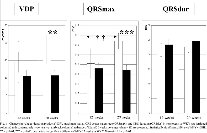
OBJECTIVE – Evaluation of the performance of the QRS voltage-duration product (VDP) for detection of left ventricular hypertrophy (LVH) in spontaneously hypertensive rats (SHR). METHODS – Orthogonal electrocardiograms (ECG) were recorded in male SHR at the age of 12 and 20 weeks, when systolic blood pressure (sBP) reached the average values of 165±3 mmHg and 195±12 mmHg, respectively. Age- and sex- matched normotensive Wistar Kyoto (WKY) rats were used as controls. VDP was calculated as a product of maximum QRS spatial vector magnitude and QRS duration. Left ventricular mass (LVM) was weighed after rats were sacrificed. RESULTS – LVM in SHR at 12 and 20 weeks of age (0.86±0.05 g and 1.05±0.07 g, respectively) was significantly higher as compared with that in WKY (0.65±0.07 g and 0.70±0.02 g). The increase in LVM closely correlated with the sBP increase. VDP did not reflect the increase in LVM in SHR. VDP was lower in SHR as compared with that in WKY, and the difference was significant at the age of 20 weeks (18.2mVms compared with 10.7mVms, p<0.01). On the contrary, a significant increase in the VDP was observed in the control WKY at the age of 20 weeks without changes in LVM. The changes in VDP were influenced mainly by the changes in QRSmax. CONCLUSION – LVM was not the major determinant of QRS voltage changes and consequently of the VDP. These data point to the importance of the nonspatial determinants of the recorded QRS voltage in terms of the solid angle theory.

  • Electrocardiography
  • Left Ventricular Hypertrophy
  • QRS voltage-duration product
  • spontaneously hypertensive rat
Read Full Article
Creative Commons Attribution (CC BY) Creative Commons license
10/09/2002 DOI: 10.1590/S0066-782X2002001100006 Views 1
An analysis of electrocardiographic criteria for determining left ventricular hypertrophy
ABC
Original Article

An analysis of electrocardiographic criteria for determining left ventricular hypertrophy

Carlos Alberto Gasperin et al. Helio Germiniani Carlos Roberto Facin Admar Moraes de Souza Cláudio Leinig Pereira da Cunha

OBJECTIVE: To determine the most sensitive criterion for the detection of left ventricular hypertrophy according to echocardiographically defined left ventricular mass. METHODS: The Sokolow-Lyon voltage, Sokolow-Lyon-Rappaport, Cornell voltage duration product, White-Bock, and Romhilt-Estes point scoring criteria were compared with left ventricular mass index, corrected for body surface, obtained from the echocardiograms of 306 outpatients (176 females, 130 males), of all age groups. RESULTS: The Cornell voltage duration product criteria index had the greatest sensitivity in women (54.90%), and the Sokolow-Lyon-Rappaport index was most sensitive in men (73.53%). When applied to men at the same voltage amplitude (20mm) as that in women, the Cornell index showed increased sensitivity relative to the conventional index (28mm) of 67.65% (P<=0.01) and a sensitivity similar to that of the Sokolow-Lyon-Rappaport index, with higher specificity (P<=0.01). The White-Bock and Romhilt-Estes criteria were the least sensitive in men and women, despite their high specificity. The electrocardiographic criteria were more efficient when dilatation predominated over left ventricular hypertrophy. CONCLUSION: The Cornell index had greater sensitivity in women, and the Sokolow-Lyon-Rappaport index was more sensitive in men. When applied to men at the same voltage amplitude as that of women, the Cornell index had an increase in sensitivity similar to that of the Sokolow-Lyon-Rappaport index.

  • electrocardiogram
  • Hypertrophy
  • Left Ventricular Hypertrophy
Read Full Article
Creative Commons Attribution (CC BY) Creative Commons license
19/02/2002 DOI: 10.1590/S0066-782X2002000100006 Views 1
ABC
Original Article

Distribution of cardiac geometric patterns on echocardiography in essential hypertension. Impact of two criteria of stratification

Eduardo Cantoni Rosa et al. Valdir Ambrósio Moisés Ricardo Cintra Sesso Nárcia E. B. Kohlmann … Frida Liane Plavnik Maria Teresa Zanella Artur Beltrame Ribeiro Osvaldo Kohlmann Júnior

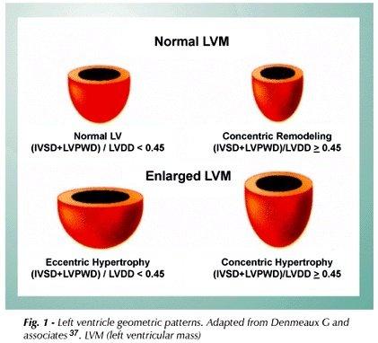
PURPOSE: To evaluate 2 left ventricular mass index (LVMI) normality criteria for the prevalence of left ventricular geometric patterns in a hypertensive population ( HT ) . METHODS: 544 essential hypertensive patients, were evaluated by echocardiography, and different left ventricular hypertrophy criteria were applied: 1 – classic : men – 134 g/m² and women – 110 g/m² ; 2- obtained from the 95th percentil of LVMI from a normotensive population (NT). RESULTS: The prevalence of 4 left ventricular geometric patterns, respectively for criteria 1 and 2, were: normal geometry – 47.7% and 39.3%; concentric remodelying – 25.4% and 14.3%; concentric hypertrophy – 18.4% and 27.7% and excentric hypertrophy – 8.8% and 16.7%, which confered abnormal geometry to 52.6% and 60.7% of hypertensive. The comparative analysis between NT and normal geometry hypertensive group according to criteria 1, detected significative stuctural differences,”( *p < 0.05):LVMI- 78.4 ± 1.50 vs 85.9 ±0.95 g/m² *; posterior wall thickness -8.5 ± 0.1 vs 8.9 ± 0.05 mm*; left atrium – 33.3 ± 0.41 vs 34.7 ± 0.30 mm *. With criteria 2, significative structural differences between the 2 groups were not observed. CONCLUSION: The use of a reference population based criteria, increased the abnormal left ventricular geometry prevalence in hypertensive patients and seemed more appropriate for left ventricular hypertrophy detection and risk stratification.

  • criteria of hypertrophy
  • Essential Hypertension
  • Left Ventricular Hypertrophy
Read Full Article
Creative Commons Attribution (CC BY) Creative Commons license
08/01/2002 DOI: 10.1590/S0066-782X2001000500002 Views 1
Protective effect of left ventricular hypertrophy in right coronary artery occlusions
ABC
Original Article

Protective effect of left ventricular hypertrophy in right coronary artery occlusions

Marne de Freitas Gomes et al. Carlos Antônio Mascia Gottschall

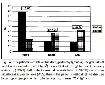
OBJECTIVE: To test the hypothesis that left ventricular hypertrophy (LVH) reduces the electrocardiographic and functional effects of right coronary artery occlusion. METHODS: We analysed 215 patients (166 males and 49 women,age of 58.9±10.6 years), with occlusion of the right coronary artery without other associated lesions. There was no significant difference (p>0.05) in age and gender distribution between the 78 patients with LVH (left ventricular mass >100g/m²) (Group A) when compared with the 137 patients without LVH (left ventricular mass <100g/m²) (Group B). RESULTS: The electrocardiographic finding of transmural necrosis was more often found in group B patients than in group A patients (56.9% and 30.8%, respectively; p<0.05). The left ventricular function parameters of group A were better than those of group B: the ratio end-diastolic pressure/systolic pressure (EDP/SP) (A: 0.108±0.036; B: 0.121±0.050; p<0.05); the end-diastolic volume index (A: 75.9±31.3ml/m²; B: 88.0±31.0ml/m²; p<0.01); the end-systolic volume index (A: 16.0±10.0ml/m²; B: 27.0 ±20.0ml/m²; p<0.001); the ejection fraction (A 78.6±10.8%; B 67.7±17.9%; p<0.001); the anteroinferior shortening (A: 43.9±10.3%; B: 35.1±12.8%; p<0.001). A higher degree of coronary tortuosity was observed in group A than in group B (78.2% and 24.1%; p<0.001) and also a more frequent absent or minimal diaphragmatic hypokinetic area (A: 80.8%; B: 54.0%; p<0.05). CONCLUSION: LVH reduces the effects of myocardial sequela and protects LV function when right coronary occlusion develops.

  • Acute Myocardial Infarction
  • ischemic cardiopathy
  • Left Ventricular Hypertrophy
Read Full Article
Creative Commons Attribution (CC BY) Creative Commons license
08/01/2002 DOI: 10.1590/S0066-782X1999000200004 Views 1
Echocardiographic assessment of the different left ventricular geometric patterns in hypertensive patients
ABC
Original Article

Echocardiographic assessment of the different left ventricular geometric patterns in hypertensive patients

Delma Maria Cunha et al. Ademir Batista da Cunha Wolney de Andrade Martins Luís Augusto de Freitas Pinheiro … Luís José Martins Romêo Álvaro Vilela de Moraes Fernando Portugal Morcerf

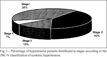
OBJECTIVE: To identiy left ventricular geometric patterns in hypertensive patients on echocardiography, and to correlate those patterns with casual blood pressure measurements and with the parameters obtained on a 24-hour ambulatory blood pressure monitoring. METHODS: We studied sixty hypertensive patients, grouped according to the Joint National Committee stages of hypertension.. Using the single- and two-dimensional Doppler Echocardiography, we analyzed the left ventricular mass and the geometric patterns through the correlation of left ventricular mass index and relative wall thickness. On ambulatory blood pressure monitoring we assessed the means and pressure loads in the different geometric patterns detected on echocardiography RESULTS: We identified three left ventricular geometric patterns: 1) concentric hypertrophy, in 25% of the patients; 2) concentric remodeling, in 25%; and 3) normal geometry, in 50%. Casual systolic blood pressure was higher in the group with concentric hypertrophy than in the other groups (p=0.001). Mean systolic pressure in the 24h, daytime and nighttime periods was also higher in patients with concentric hypertrophy, as compared to the other groups (p=0.003, p=0.004 and p=0.007). Daytime systolic load and nighttime diastolic load were higher in patients with concentric hypertrophy ( p=0.004 and p=0.01, respectively). CONCLUSIONS: Left ventricular geometric patterns show significant correlation with casual systolic blood pressure, and with means and pressure loads on ambulatory blood pressure monitoring.

  • Echocardiography
  • Hypertension
  • Left Ventricular Hypertrophy
Read Full Article
Creative Commons Attribution (CC BY) Creative Commons license
02/02/2001 DOI: 10.1590/S0066-782X2001000100003 Views 2
ABC
Artigo Original

Electro-echocardiographic correlation in the diagnosis of left ventricular hypertrophy

Hamilton Domingos et al. Josane Couto Elias Luzio Gualberto Nogueira de Leles Leandro Sauer Luiz Alberto Ovando

PURPOSE: To compare the efficacy of four electrocardiographic criteria: Sokolov, Gubner, Cornell and Romhilt indexes, in the diagnosis of left ventricular hypertrophy (LVH) in hypertensive patients. METHODS: LVH was analyzed in the eletrocardiogram of 30 ambulatory patients presenting with systemic arterial hypertension, classified as mild, moderate and severe, according to the following indexes: Sokolov >35mm, Gubner >22mm, Romhilt >5 points and Cornell >20mm for women and 28mm for men. Sensitivity, specificity, diagnostic accuracy and other diagnostic variables were determined. Mass index of the left ventricle, >98g/m² for women and >120g/m² for men, obtained by echocardiography, was considered the gold standard for the diagnosis of LVH. RESULTS: When electrocardiographic criteria were considered separately, the Sokolov index showed the highest accuracy, with a sensitivity of 40%, diagnostic accuracy of 50% and specificity of 100%; the second most accurate index was Gubner, and Romhilt and Cornell indexes followed. When at least one of the indexes was positive, the sensitivity was 52% and diagnostic accuracy was 57%. CONCLUSION: The four electrocardiographic indexes were not diagnostic of LVH, when analyzed either separately or together. Considering the high prevalence of this pathological condition, we conclude that a more accurate diagnostic method should be used in this diagnosis.

  • Electrocardiography
  • Hypertension
  • Left Ventricular Hypertrophy
Read Full Article
Creative Commons Attribution (CC BY) Creative Commons license
07/12/2000 DOI: 10.1590/S0066-782X1998000700007 Views 1

How to cite this article

EndNote RIS

Electronic Document Format (Vancouver)

Gonçalo MO, Gomes AAS, Monteiro MM, Cavalcanti NB, Bezerra FA, Machado MA, Miranda DCC, et al. Biventricular Noncompaction Cardiomyopathy Associated With Neuropathy in a Young Patient: A Case Report. ABC Imagem Cardiovascular 2026;39(3):e20260078.

Electronic Document Format (ABNT)

Gonçalo, Mônica de Oliveira; Gomes, André Alexandre dos Santos; Monteiro, Matheus Martins; Cavalcanti, Nicolas Babilônia; Bezerra, Fernando Almeida; Machado, Moisés Abtibol; Miranda, Dênison Clark Corrêa de; Couceiro, Kátia do Nascimento; Ferreira, João Marcos Bemfica Barbosa. Biventricular Noncompaction Cardiomyopathy Associated With Neuropathy in a Young Patient: A Case Report. ABC Imagem Cardiovascular, v. 39, n. 3, e20260078, Sep. 2026.

Electronic Document Format (APA)

Gonçalo, M. O., Gomes, A. A. S., Monteiro, M. M., Cavalcanti, N. B., Bezerra, F. A., Machado, M. A., Miranda, D. C. C. , Couceiro, K. N., & Ferreira, J. M. B. B. (2026). Biventricular Noncompaction Cardiomyopathy Associated With Neuropathy in a Young Patient: A Case Report. ABC Imagem Cardiovascular, 39(3), e20260078.

Electronic Document Format (ISO)

Gonçalo, Mônica de Oliveira and Gomes, André Alexandre dos Santos and Monteiro, Matheus Martins and Cavalcanti, Nicolas Babilônia and Bezerra, Fernando Almeida and Machado, Moisés Abtibol and Miranda, Dênison Clark Corrêa de and Couceiro, Kátia do Nascimento and Ferreira, João Marcos Bemfica Barbosa. Biventricular Noncompaction Cardiomyopathy Associated With Neuropathy in a Young Patient: A Case Report. ABC Imagem Cardiovascular [online]. 2026, vol. 39, n. 3, [cited 2026-10-14], e20260078. Available from: <https://journals.cardiol.br/en/article/biventricular-noncompaction-cardiomyopathy-associated-with-neuropathy-in-a-young-patient-a-case-report/>. ISSN 2318-8219

EndNote RIS

Electronic Document Format (Vancouver)

Alencar JN, Felicioni SP, Scheffer MK, Marchi MND, Bacharova L. The Old Habits Need to be Abandoned and the Term “Overload” Replaced by “High Amplitude Criteria”. Arquivos Brasileiros de Cardiologia 2026;123(5):e20250709.

Electronic Document Format (ABNT)

Alencar, José Nunes de; Felicioni, Sandro Pinelli; Scheffer, Matheus Kiszka; Marchi, Mariana Nogueira De; Bacharova, Ljuba. The Old Habits Need to be Abandoned and the Term “Overload” Replaced by “High Amplitude Criteria”. Arquivos Brasileiros de Cardiologia, v. 123, n. 5, e20250709, Jul. 2026.

Electronic Document Format (APA)

Alencar, J. N. , Felicioni, S. P., Scheffer, M. K., Marchi, M. N. D., & Bacharova, L. (2026). The Old Habits Need to be Abandoned and the Term “Overload” Replaced by “High Amplitude Criteria”. Arquivos Brasileiros de Cardiologia, 123(5), e20250709.

Electronic Document Format (ISO)

Alencar, José Nunes de and Felicioni, Sandro Pinelli and Scheffer, Matheus Kiszka and Marchi, Mariana Nogueira De and Bacharova, Ljuba. The Old Habits Need to be Abandoned and the Term “Overload” Replaced by “High Amplitude Criteria”. Arquivos Brasileiros de Cardiologia [online]. 2026, vol. 123, n. 5, [cited 2026-10-14], e20250709. Available from: <https://journals.cardiol.br/en/article/the-old-habits-need-to-be-abandoned-and-the-term-overload-replaced-by-high-amplitude-criteria/>. ISSN 0066-782X

EndNote RIS

Electronic Document Format (Vancouver)

Pinto-Filho MM, Faria LG, Foppa M, Santos ABS, Barreto SM, Ribeiro ALP. Accuracy and Prognosis of Electrocardiographic and Echocardiographic Left Ventricular Hypertrophy in Brazilian Adults – ELSA-Brasil Study. Arquivos Brasileiros de Cardiologia 2026;123(4):e20250789.

Electronic Document Format (ABNT)

Pinto-Filho, Marcelo M.; Faria, Lillian Guimarães de; Foppa, Murilo; Santos, Angela Barreto Santiago; Barreto, Sandhi Maria; Ribeiro, Antonio Luiz Pinho. Accuracy and Prognosis of Electrocardiographic and Echocardiographic Left Ventricular Hypertrophy in Brazilian Adults – ELSA-Brasil Study. Arquivos Brasileiros de Cardiologia, v. 123, n. 4, e20250789, Jun. 2026.

Electronic Document Format (APA)

Pinto-Filho, M. M., Faria, L. G. , Foppa, M., Santos, A. B. S., Barreto, S. M., & Ribeiro, A. L. P. (2026). Accuracy and Prognosis of Electrocardiographic and Echocardiographic Left Ventricular Hypertrophy in Brazilian Adults – ELSA-Brasil Study. Arquivos Brasileiros de Cardiologia, 123(4), e20250789.

Electronic Document Format (ISO)

Pinto-Filho, Marcelo M. and Faria, Lillian Guimarães de and Foppa, Murilo and Santos, Angela Barreto Santiago and Barreto, Sandhi Maria and Ribeiro, Antonio Luiz Pinho. Accuracy and Prognosis of Electrocardiographic and Echocardiographic Left Ventricular Hypertrophy in Brazilian Adults – ELSA-Brasil Study. Arquivos Brasileiros de Cardiologia [online]. 2026, vol. 123, n. 4, [cited 2026-10-14], e20250789. Available from: <https://journals.cardiol.br/en/article/accuracy-and-prognosis-of-electrocardiographic-and-echocardiographic-left-ventricular-hypertrophy-in-brazilian-adults-elsa-brasil-study/>. ISSN 0066-782X

EndNote RIS

Electronic Document Format (Vancouver)

Corrêa BAS, Silva GPSA, Miranda FC, Oliveira MHJ, Barbosa GS, Borges VHL, Alencar JN, et al. S-Wave Amplitude in Lead V3 Identifies Other Electrocardiography Criteria of Left Ventricular Hypertrophy. International Journal of Cardiovascular Sciences 2026;39:e20250050.

Electronic Document Format (ABNT)

Corrêa, Bernardo Augusto Silveira; Silva, Gabriel Pinheiro Soares Alencar e; Miranda, Felipe Campelo de; Oliveira, Marcio Henrique de Jesus; Barbosa, Gabriel Scarpioni; Borges, Victor Hudson de Lacerda; Alencar, José Nunes de. S-Wave Amplitude in Lead V3 Identifies Other Electrocardiography Criteria of Left Ventricular Hypertrophy. International Journal of Cardiovascular Sciences, v. 39, e20250050, Apr. 2026.

Electronic Document Format (APA)

Corrêa, B. A. S., Silva, G. P. S. A. , Miranda, F. C. , Oliveira, M. H. J., Barbosa, G. S., Borges, V. H. L., & Alencar, J. N. (2026). S-Wave Amplitude in Lead V3 Identifies Other Electrocardiography Criteria of Left Ventricular Hypertrophy. International Journal of Cardiovascular Sciences, 39, e20250050.

Electronic Document Format (ISO)

Corrêa, Bernardo Augusto Silveira and Silva, Gabriel Pinheiro Soares Alencar e and Miranda, Felipe Campelo de and Oliveira, Marcio Henrique de Jesus and Barbosa, Gabriel Scarpioni and Borges, Victor Hudson de Lacerda and Alencar, José Nunes de. S-Wave Amplitude in Lead V3 Identifies Other Electrocardiography Criteria of Left Ventricular Hypertrophy. International Journal of Cardiovascular Sciences [online]. 2026, vol. 39, [cited 2026-10-14], e20250050. Available from: <https://journals.cardiol.br/en/article/s-wave-amplitude-in-lead-v3-identifies-other-electrocardiography-criteria-of-left-ventricular-hypertrophy/>. ISSN 2359-4802

EndNote RIS

Electronic Document Format (Vancouver)

Mejía HPG, Madogolele HCN, Barroso MCRD, Nogueira MCA, Morales KRP, Romero CE, Nastari L, et al. Late-Onset Fabry Disease: Diagnostic Challenges and Clinical Evolution. Arquivos Brasileiros de Cardiologia 2026;123(2):e20250554.

Electronic Document Format (ABNT)

Mejía, Hernán Patricio García; Madogolele, Heleuterio da Conceição Nicolau; Barroso, Manuela Cristina Ribeiro Dias; Nogueira, Matheus Carvalho Alves; Morales, Kevin Rafael de Paula; Romero, Cristhian Espinoza; Nastari, Luciano; Fernandes, Fábio. Late-Onset Fabry Disease: Diagnostic Challenges and Clinical Evolution. Arquivos Brasileiros de Cardiologia, v. 123, n. 2, e20250554, Apr. 2026.

Electronic Document Format (APA)

Mejía, H. P. G., Madogolele, H. C. N., Barroso, M. C. R. D., Nogueira, M. C. A., Morales, K. R. P., Romero, C. E., Nastari, L., & Fernandes, F. (2026). Late-Onset Fabry Disease: Diagnostic Challenges and Clinical Evolution. Arquivos Brasileiros de Cardiologia, 123(2), e20250554.

Electronic Document Format (ISO)

Mejía, Hernán Patricio García and Madogolele, Heleuterio da Conceição Nicolau and Barroso, Manuela Cristina Ribeiro Dias and Nogueira, Matheus Carvalho Alves and Morales, Kevin Rafael de Paula and Romero, Cristhian Espinoza and Nastari, Luciano and Fernandes, Fábio. Late-Onset Fabry Disease: Diagnostic Challenges and Clinical Evolution. Arquivos Brasileiros de Cardiologia [online]. 2026, vol. 123, n. 2, [cited 2026-10-14], e20250554. Available from: <https://journals.cardiol.br/en/article/late-onset-fabry-disease-diagnostic-challenges-and-clinical-evolution/>. ISSN 0066-782X

EndNote RIS

Electronic Document Format (Vancouver)

Hajar N, Joveazhari NN, Chasani S, Wahab Z, Adhyatma GP, Balqis IA, Fahdalhaq ZA, et al. Left Ventricular Hypertrophy on Electrocardiography and Chronic Kidney Disease: A Case–Control Pilot Study. International Journal of Cardiovascular Sciences 2025;38:e20250064.

Electronic Document Format (ABNT)

Hajar, Nabil; Joveazhari, Nabila Nuralya; Chasani, Shofa; Wahab, Zulfachmi; Adhyatma, Galih Prakasa; Balqis, Indira Aura; Fahdalhaq, Zeidan Ahmad; Wicaksono, Bintang Adhiyoso. Left Ventricular Hypertrophy on Electrocardiography and Chronic Kidney Disease: A Case–Control Pilot Study. International Journal of Cardiovascular Sciences, v. 38, e20250064, Aug. 2025.

Electronic Document Format (APA)

Hajar, N., Joveazhari, N. N., Chasani, S., Wahab, Z., Adhyatma, G. P., Balqis, I. A., Fahdalhaq, Z. A., & Wicaksono, B. A. (2025). Left Ventricular Hypertrophy on Electrocardiography and Chronic Kidney Disease: A Case–Control Pilot Study. International Journal of Cardiovascular Sciences, 38, e20250064.

Electronic Document Format (ISO)

Hajar, Nabil and Joveazhari, Nabila Nuralya and Chasani, Shofa and Wahab, Zulfachmi and Adhyatma, Galih Prakasa and Balqis, Indira Aura and Fahdalhaq, Zeidan Ahmad and Wicaksono, Bintang Adhiyoso. Left Ventricular Hypertrophy on Electrocardiography and Chronic Kidney Disease: A Case–Control Pilot Study. International Journal of Cardiovascular Sciences [online]. 2025, vol. 38, [cited 2026-10-14], e20250064. Available from: <https://journals.cardiol.br/en/article/left-ventricular-hypertrophy-on-electrocardiography-and-chronic-kidney-disease-a-case-control-pilot-study/>. ISSN 2359-4802

EndNote RIS

Electronic Document Format (Vancouver)

Felix AS, Barberato SH, Melo MDT, Rosa SA, Cardim N. The Complex Puzzle of Hypertrophic Phenotype: A Practical Approach for the Clinician. Arquivos Brasileiros de Cardiologia 2025;122(1):e20240529.

Electronic Document Format (ABNT)

Felix, Alex dos Santos; Barberato, Silvio Henrique; Melo, Marcelo Dantas Tavares de; Rosa, Sílvia Aguiar; Cardim, Nuno. The Complex Puzzle of Hypertrophic Phenotype: A Practical Approach for the Clinician. Arquivos Brasileiros de Cardiologia, v. 122, n. 1, e20240529, Apr. 2025.

Electronic Document Format (APA)

Felix, A. S., Barberato, S. H., Melo, M. D. T. , Rosa, S. A., & Cardim, N. (2025). The Complex Puzzle of Hypertrophic Phenotype: A Practical Approach for the Clinician. Arquivos Brasileiros de Cardiologia, 122(1), e20240529.

Electronic Document Format (ISO)

Felix, Alex dos Santos and Barberato, Silvio Henrique and Melo, Marcelo Dantas Tavares de and Rosa, Sílvia Aguiar and Cardim, Nuno. The Complex Puzzle of Hypertrophic Phenotype: A Practical Approach for the Clinician. Arquivos Brasileiros de Cardiologia [online]. 2025, vol. 122, n. 1, [cited 2026-10-14], e20240529. Available from: <https://journals.cardiol.br/en/article/the-complex-puzzle-of-hypertrophic-phenotype-a-practical-approach-for-the-clinician/>. ISSN 0066-782X

EndNote RIS

Electronic Document Format (Vancouver)

Akhan O, Kış M, Güzel T, Zoghi M. Differences Between Two Distinct Hypertrophic Cardiac Conditions: Fabry Disease versus Hypertrophic Cardiomyopathy. Arquivos Brasileiros de Cardiologia 2024;121(1):e20230229.

Electronic Document Format (ABNT)

Akhan, Onur; Kış, Mehmet; Güzel, Tuncay; Zoghi, Mehdi. Differences Between Two Distinct Hypertrophic Cardiac Conditions: Fabry Disease versus Hypertrophic Cardiomyopathy. Arquivos Brasileiros de Cardiologia, v. 121, n. 1, e20230229, Feb. 2024.

Electronic Document Format (APA)

Akhan, O., Kış, M., Güzel, T., & Zoghi, M. (2024). Differences Between Two Distinct Hypertrophic Cardiac Conditions: Fabry Disease versus Hypertrophic Cardiomyopathy. Arquivos Brasileiros de Cardiologia, 121(1), e20230229.

Electronic Document Format (ISO)

Akhan, Onur and Kış, Mehmet and Güzel, Tuncay and Zoghi, Mehdi. Differences Between Two Distinct Hypertrophic Cardiac Conditions: Fabry Disease versus Hypertrophic Cardiomyopathy. Arquivos Brasileiros de Cardiologia [online]. 2024, vol. 121, n. 1, [cited 2026-10-14], e20230229. Available from: <https://journals.cardiol.br/en/article/differences-between-two-distinct-hypertrophic-cardiac-conditions-fabry-disease-versus-hypertrophic-cardiomyopathy/>. ISSN 0066-782X

EndNote RIS

Electronic Document Format (Vancouver)

Marques-Alves P, Ferreira JA, Freitas AA, Almeida JP, Baptista R, Castro G, Martins R, et al. Atrial Mechanics in Hypertrophic Cardiomyopathy: Discriminating between Ventricular Hypertrophy and Fibrosis. Arquivos Brasileiros de Cardiologia 2022;118(1):77-8.

Electronic Document Format (ABNT)

Marques-Alves, Patrícia; Ferreira, João André; Freitas, André Azul; Almeida, José Paulo; Baptista, Rui; Castro, Graça; Martins, Rui; Donato, Paulo; Ferreira, Maria João; Gonçalves, Lino. Atrial Mechanics in Hypertrophic Cardiomyopathy: Discriminating between Ventricular Hypertrophy and Fibrosis. Arquivos Brasileiros de Cardiologia, v. 118, n. 1, p. 77-87, Feb. 2022.

Electronic Document Format (APA)

Marques-Alves, P., Ferreira, J. A., Freitas, A. A., Almeida, J. P., Baptista, R., Castro, G., Martins, R., Donato, P., Ferreira, M. J., & Gonçalves, L. (2022). Atrial Mechanics in Hypertrophic Cardiomyopathy: Discriminating between Ventricular Hypertrophy and Fibrosis. Arquivos Brasileiros de Cardiologia, 118(1), 77-87.

Electronic Document Format (ISO)

Marques-Alves, Patrícia and Ferreira, João André and Freitas, André Azul and Almeida, José Paulo and Baptista, Rui and Castro, Graça and Martins, Rui and Donato, Paulo and Ferreira, Maria João and Gonçalves, Lino. Atrial Mechanics in Hypertrophic Cardiomyopathy: Discriminating between Ventricular Hypertrophy and Fibrosis. Arquivos Brasileiros de Cardiologia [online]. 2022, vol. 118, n. 1, [cited 2026-10-14], pp. 77-87. Available from: <https://journals.cardiol.br/en/article/atrial-mechanics-in-hypertrophic-cardiomyopathy-discriminating-between-ventricular-hypertrophy-and-fibrosis/>. ISSN 0066-782X

EndNote RIS

Electronic Document Format (Vancouver)

Latado L, Melo RMV, Mistro S, Latado AL, Nascimento HF, Lira YM, Oliveira NFC, et al. Cost-Effectiveness Analysis of Evolocumab Therapy in Patients at High Risk of Cardiovascular Events in the Context of the Brazilian Unified Health System. Arquivos Brasileiros de Cardiologia 2021;117(5):988-96.

Electronic Document Format (ABNT)

Latado, Luiza; Melo, Rodrigo Morel Vieira de; Mistro, Sóstenes; Latado, Adriana Lopes; Nascimento, Harrison Floriano do; Lira, Yasmin Menezes; Oliveira, Natalia Ferreira Cardoso de; Galindo, Yuri de Santana; Viana, Tainá; Passos, Luiz Carlos Santana. Cost-Effectiveness Analysis of Evolocumab Therapy in Patients at High Risk of Cardiovascular Events in the Context of the Brazilian Unified Health System. Arquivos Brasileiros de Cardiologia, v. 117, n. 5, p. 988-996, Nov. 2021.

Electronic Document Format (APA)

Latado, L., Melo, R. M. V. , Mistro, S., Latado, A. L., Nascimento, H. F. , Lira, Y. M., Oliveira, N. F. C. , Galindo, Y. S., Viana, T., & Passos, L. C. S. (2021). Cost-Effectiveness Analysis of Evolocumab Therapy in Patients at High Risk of Cardiovascular Events in the Context of the Brazilian Unified Health System. Arquivos Brasileiros de Cardiologia, 117(5), 988-996.

Electronic Document Format (ISO)

Latado, Luiza and Melo, Rodrigo Morel Vieira de and Mistro, Sóstenes and Latado, Adriana Lopes and Nascimento, Harrison Floriano do and Lira, Yasmin Menezes and Oliveira, Natalia Ferreira Cardoso de and Galindo, Yuri de Santana and Viana, Tainá and Passos, Luiz Carlos Santana. Cost-Effectiveness Analysis of Evolocumab Therapy in Patients at High Risk of Cardiovascular Events in the Context of the Brazilian Unified Health System. Arquivos Brasileiros de Cardiologia [online]. 2021, vol. 117, n. 5, [cited 2026-10-14], pp. 988-996. Available from: <https://journals.cardiol.br/en/article/cost-effectiveness-analysis-of-evolocumab-therapy-in-patients-at-high-risk-of-cardiovascular-events-in-the-context-of-the-brazilian-unified-health-system/>. ISSN 0066-782X

EndNote RIS

Electronic Document Format (Vancouver)

Ritter AMV, Faria APC, Sabbatini A, Corrêa NB, Brunelli V, Modolo R, Moreno H, et al. MCP-1 Levels are Associated with Cardiac Remodeling but not with Resistant Hypertension. Arquivos Brasileiros de Cardiologia 2017;108(4):331-8.

Electronic Document Format (ABNT)

Ritter, Alessandra Mileni Versuti; Faria, Ana Paula Cabral de; Sabbatini, Andrea; Corrêa, Nathalia Batista; Brunelli, Veridiana; Modolo, Rodrigo; Moreno, Heitor. MCP-1 Levels are Associated with Cardiac Remodeling but not with Resistant Hypertension. Arquivos Brasileiros de Cardiologia, v. 108, n. 4, p. 331-338, Mar. 2017.

Electronic Document Format (APA)

Ritter, A. M. V., Faria, A. P. C. , Sabbatini, A., Corrêa, N. B., Brunelli, V., Modolo, R., & Moreno, H. (2017). MCP-1 Levels are Associated with Cardiac Remodeling but not with Resistant Hypertension. Arquivos Brasileiros de Cardiologia, 108(4), 331-338.

Electronic Document Format (ISO)

Ritter, Alessandra Mileni Versuti and Faria, Ana Paula Cabral de and Sabbatini, Andrea and Corrêa, Nathalia Batista and Brunelli, Veridiana and Modolo, Rodrigo and Moreno, Heitor. MCP-1 Levels are Associated with Cardiac Remodeling but not with Resistant Hypertension. Arquivos Brasileiros de Cardiologia [online]. 2017, vol. 108, n. 4, [cited 2026-10-14], pp. 331-338. Available from: <https://journals.cardiol.br/en/article/mcp-1-levels-are-associated-with-cardiac-remodeling-but-not-with-resistant-hypertension/>. ISSN 0066-782X

EndNote RIS

Electronic Document Format (Vancouver)

Rocha IEGM, Victor EG, Braga MC, Silva OB, Becker MMC. Echocardiography evaluation for asymptomatic patients with severe obesity. Arquivos Brasileiros de Cardiologia 2007;88(1):52-8.

Electronic Document Format (ABNT)

Rocha, Isaura Elaine Gonçalves Moreira; Victor, Edgar Guimarães; Braga, Maria Cynthia; Silva, Odwaldo Barbosa e; Becker, Mônica de Moraes Chaves. Echocardiography evaluation for asymptomatic patients with severe obesity. Arquivos Brasileiros de Cardiologia, v. 88, n. 1, p. 52-58, Apr. 2007.

Electronic Document Format (APA)

Rocha, I. E. G. M., Victor, E. G., Braga, M. C., Silva, O. B. , & Becker, M. M. C. (2007). Echocardiography evaluation for asymptomatic patients with severe obesity. Arquivos Brasileiros de Cardiologia, 88(1), 52-58.

Electronic Document Format (ISO)

Rocha, Isaura Elaine Gonçalves Moreira and Victor, Edgar Guimarães and Braga, Maria Cynthia and Silva, Odwaldo Barbosa e and Becker, Mônica de Moraes Chaves. Echocardiography evaluation for asymptomatic patients with severe obesity. Arquivos Brasileiros de Cardiologia [online]. 2007, vol. 88, n. 1, [cited 2026-10-14], pp. 52-58. Available from: <https://journals.cardiol.br/en/article/echocardiography-evaluation-for-asymptomatic-patients-with-severe-obesity/>. ISSN 0066-782X

EndNote RIS

Electronic Document Format (Vancouver)

Rosa EC, Moysés VA, Sesso RC, Plavnik FL, Ribeiro FF, Kohlmann NEB, Ribeiro AB, et al. Left Ventricular Hypertrophy Evaluation in Obese Hypertensive Patients: Effect of Left Ventricular Mass Index Criteria. Arquivos Brasileiros de Cardiologia 2007;78(4):347-51.

Electronic Document Format (ABNT)

Rosa, Eduardo Cantoni; Moysés, Valdir Ambrósio; Sesso, Ricardo Cintra; Plavnik, Frida Liane; Ribeiro, Fernando Flexa; Kohlmann, Nárcia E. B.; Ribeiro, Artur Beltrame; Zanella, Maria Tereza; Kohlmann Jr., Osvaldo. Left Ventricular Hypertrophy Evaluation in Obese Hypertensive Patients: Effect of Left Ventricular Mass Index Criteria. Arquivos Brasileiros de Cardiologia, v. 78, n. 4, p. 347-351, Jan. 2007.

Electronic Document Format (APA)

Rosa, E. C., Moysés, V. A., Sesso, R. C., Plavnik, F. L., Ribeiro, F. F., Kohlmann, N. E. B., Ribeiro, A. B., Zanella, M. T., & Kohlmann Jr., O. (2007). Left Ventricular Hypertrophy Evaluation in Obese Hypertensive Patients: Effect of Left Ventricular Mass Index Criteria. Arquivos Brasileiros de Cardiologia, 78(4), 347-351.

Electronic Document Format (ISO)

Rosa, Eduardo Cantoni and Moysés, Valdir Ambrósio and Sesso, Ricardo Cintra and Plavnik, Frida Liane and Ribeiro, Fernando Flexa and Kohlmann, Nárcia E. B. and Ribeiro, Artur Beltrame and Zanella, Maria Tereza and Kohlmann Jr., Osvaldo. Left Ventricular Hypertrophy Evaluation in Obese Hypertensive Patients: Effect of Left Ventricular Mass Index Criteria. Arquivos Brasileiros de Cardiologia [online]. 2007, vol. 78, n. 4, [cited 2026-10-14], pp. 347-351. Available from: <https://journals.cardiol.br/en/article/left-ventricular-hypertrophy-evaluation-in-obese-hypertensive-patients-effect-of-left-ventricular-mass-index-criteria/>. ISSN 0066-782X

EndNote RIS

Electronic Document Format (Vancouver)

Ginefra P, Barbosa EC, Benchimol-Barbosa PR, Bomfim AS, Boghossian SH, Salgado AA, Brasil FG, et al. Detection of incipient left ventricular hypertrophy in mild to moderate arterial hypertension with normal electrocardiogram and echocardiogram: a new use for signal-averaged electrocardiography. Arquivos Brasileiros de Cardiologia 2007;81(1):79-84.

Electronic Document Format (ABNT)

Ginefra, Paulo; Barbosa, Eduardo C.; Benchimol-Barbosa, P. R.; Bomfim, Alfredo S.; Boghossian, Sílvia H.; Salgado, Angelo A.; Brasil, Flávia G.; Freitas, Elizabete A.; Albanesi Filho, Francisco M.. Detection of incipient left ventricular hypertrophy in mild to moderate arterial hypertension with normal electrocardiogram and echocardiogram: a new use for signal-averaged electrocardiography. Arquivos Brasileiros de Cardiologia, v. 81, n. 1, p. 79-84, Jan. 2007.

Electronic Document Format (APA)

Ginefra, P., Barbosa, E. C., Benchimol-Barbosa, P. R., Bomfim, A. S., Boghossian, S. H., Salgado, A. A., Brasil, F. G., Freitas, E. A., & Albanesi Filho, F. M. (2007). Detection of incipient left ventricular hypertrophy in mild to moderate arterial hypertension with normal electrocardiogram and echocardiogram: a new use for signal-averaged electrocardiography. Arquivos Brasileiros de Cardiologia, 81(1), 79-84.

Electronic Document Format (ISO)

Ginefra, Paulo and Barbosa, Eduardo C. and Benchimol-Barbosa, P. R. and Bomfim, Alfredo S. and Boghossian, Sílvia H. and Salgado, Angelo A. and Brasil, Flávia G. and Freitas, Elizabete A. and Albanesi Filho, Francisco M.. Detection of incipient left ventricular hypertrophy in mild to moderate arterial hypertension with normal electrocardiogram and echocardiogram: a new use for signal-averaged electrocardiography. Arquivos Brasileiros de Cardiologia [online]. 2007, vol. 81, n. 1, [cited 2026-10-14], pp. 79-84. Available from: <https://journals.cardiol.br/en/article/detection-of-incipient-left-ventricular-hypertrophy-in-mild-to-moderate-arterial-hypertension-with-normal-electrocardiogram-and-echocardiogram-a-new-use-for-signal-averaged-electrocardiography/>. ISSN 0066-782X

EndNote RIS

Electronic Document Format (Vancouver)

Markus MRP, Freitas HFG, Chizzola PR, Silva GT, Lima ACP, Mansur AJ. Left ventricular mass in patients with heart failure. Arquivos Brasileiros de Cardiologia 2004;83(3):227-31.

Electronic Document Format (ABNT)

Markus, Marcello Ricardo Paulista; Freitas, Humberto Felício Gonçalves de; Chizzola, Paulo Roberto; Silva, Gisela Tunes da; Lima, Antonio Carlos Pedroso de; Mansur, Alfredo José. Left ventricular mass in patients with heart failure. Arquivos Brasileiros de Cardiologia, v. 83, n. 3, p. 227-231, Sep. 2004.

Electronic Document Format (APA)

Markus, M. R. P., Freitas, H. F. G. , Chizzola, P. R., Silva, G. T. , Lima, A. C. P. , & Mansur, A. J. (2004). Left ventricular mass in patients with heart failure. Arquivos Brasileiros de Cardiologia, 83(3), 227-231.

Electronic Document Format (ISO)

Markus, Marcello Ricardo Paulista and Freitas, Humberto Felício Gonçalves de and Chizzola, Paulo Roberto and Silva, Gisela Tunes da and Lima, Antonio Carlos Pedroso de and Mansur, Alfredo José. Left ventricular mass in patients with heart failure. Arquivos Brasileiros de Cardiologia [online]. 2004, vol. 83, n. 3, [cited 2026-10-14], pp. 227-231. Available from: <https://journals.cardiol.br/en/article/left-ventricular-mass-in-patients-with-heart-failure/>. ISSN 0066-782X

EndNote RIS

Electronic Document Format (Vancouver)

Bacharova L, Kyselovic J, Klimas J. QRS Voltage-Duration Product in the Identification of Left Ventricular Hypertrophy in Spontaneously Hypertensive Rats. Arquivos Brasileiros de Cardiologia 2002;79(2):143-8.

Electronic Document Format (ABNT)

Bacharova, Ljuba; Kyselovic, Jan; Klimas, Jan. QRS Voltage-Duration Product in the Identification of Left Ventricular Hypertrophy in Spontaneously Hypertensive Rats. Arquivos Brasileiros de Cardiologia, v. 79, n. 2, p. 143-148, Sep. 2002.

Electronic Document Format (APA)

Bacharova, L., Kyselovic, J., & Klimas, J. (2002). QRS Voltage-Duration Product in the Identification of Left Ventricular Hypertrophy in Spontaneously Hypertensive Rats. Arquivos Brasileiros de Cardiologia, 79(2), 143-148.

Electronic Document Format (ISO)

Bacharova, Ljuba and Kyselovic, Jan and Klimas, Jan. QRS Voltage-Duration Product in the Identification of Left Ventricular Hypertrophy in Spontaneously Hypertensive Rats. Arquivos Brasileiros de Cardiologia [online]. 2002, vol. 79, n. 2, [cited 2026-10-14], pp. 143-148. Available from: <https://journals.cardiol.br/en/article/qrs-voltage-duration-product-in-the-identification-of-left-ventricular-hypertrophy-in-spontaneously-hypertensive-rats/>. ISSN 0066-782X

EndNote RIS

Electronic Document Format (Vancouver)

Gasperin CA, Germiniani H, Facin CR, Souza AM, Cunha CLP. An analysis of electrocardiographic criteria for determining left ventricular hypertrophy. Arquivos Brasileiros de Cardiologia 2002;78(1):72-8.

Electronic Document Format (ABNT)

Gasperin, Carlos Alberto; Germiniani, Helio; Facin, Carlos Roberto; Souza, Admar Moraes de; Cunha, Cláudio Leinig Pereira da. An analysis of electrocardiographic criteria for determining left ventricular hypertrophy. Arquivos Brasileiros de Cardiologia, v. 78, n. 1, p. 72-82, Feb. 2002.

Electronic Document Format (APA)

Gasperin, C. A., Germiniani, H., Facin, C. R., Souza, A. M. , & Cunha, C. L. P. (2002). An analysis of electrocardiographic criteria for determining left ventricular hypertrophy. Arquivos Brasileiros de Cardiologia, 78(1), 72-82.

Electronic Document Format (ISO)

Gasperin, Carlos Alberto and Germiniani, Helio and Facin, Carlos Roberto and Souza, Admar Moraes de and Cunha, Cláudio Leinig Pereira da. An analysis of electrocardiographic criteria for determining left ventricular hypertrophy. Arquivos Brasileiros de Cardiologia [online]. 2002, vol. 78, n. 1, [cited 2026-10-14], pp. 72-82. Available from: <https://journals.cardiol.br/en/article/an-analysis-of-electrocardiographic-criteria-for-determining-left-ventricular-hypertrophy/>. ISSN 0066-782X

EndNote RIS

Electronic Document Format (Vancouver)

Rosa EC, Moisés VA, Sesso RC, Kohlmann NEB, Plavnik FL, Zanella MT, Ribeiro AB, et al. Distribution of cardiac geometric patterns on echocardiography in essential hypertension. Impact of two criteria of stratification. Arquivos Brasileiros de Cardiologia 2002;76(5):362-8.

Electronic Document Format (ABNT)

Rosa, Eduardo Cantoni; Moisés, Valdir Ambrósio; Sesso, Ricardo Cintra; Kohlmann, Nárcia E. B.; Plavnik, Frida Liane; Zanella, Maria Teresa; Ribeiro, Artur Beltrame; Kohlmann Júnior, Osvaldo. Distribution of cardiac geometric patterns on echocardiography in essential hypertension. Impact of two criteria of stratification. Arquivos Brasileiros de Cardiologia, v. 76, n. 5, p. 362-368, Jan. 2002.

Electronic Document Format (APA)

Rosa, E. C., Moisés, V. A., Sesso, R. C., Kohlmann, N. E. B., Plavnik, F. L., Zanella, M. T., Ribeiro, A. B., & Kohlmann Júnior, O. (2002). Distribution of cardiac geometric patterns on echocardiography in essential hypertension. Impact of two criteria of stratification. Arquivos Brasileiros de Cardiologia, 76(5), 362-368.

Electronic Document Format (ISO)

Rosa, Eduardo Cantoni and Moisés, Valdir Ambrósio and Sesso, Ricardo Cintra and Kohlmann, Nárcia E. B. and Plavnik, Frida Liane and Zanella, Maria Teresa and Ribeiro, Artur Beltrame and Kohlmann Júnior, Osvaldo. Distribution of cardiac geometric patterns on echocardiography in essential hypertension. Impact of two criteria of stratification. Arquivos Brasileiros de Cardiologia [online]. 2002, vol. 76, n. 5, [cited 2026-10-14], pp. 362-368. Available from: <https://journals.cardiol.br/en/article/distribution-of-cardiac-geometric-patterns-on-echocardiography-in-essential-hypertension-impact-of-two-criteria-of-stratification/>. ISSN 0066-782X

EndNote RIS

Electronic Document Format (Vancouver)

Gomes MF, Gottschall CAM. Protective effect of left ventricular hypertrophy in right coronary artery occlusions. Arquivos Brasileiros de Cardiologia 2002;72(2):161-70.

Electronic Document Format (ABNT)

Gomes, Marne de Freitas; Gottschall, Carlos Antônio Mascia. Protective effect of left ventricular hypertrophy in right coronary artery occlusions. Arquivos Brasileiros de Cardiologia, v. 72, n. 2, p. 161-170, Jan. 2002.

Electronic Document Format (APA)

Gomes, M. F., & Gottschall, C. A. M. (2002). Protective effect of left ventricular hypertrophy in right coronary artery occlusions. Arquivos Brasileiros de Cardiologia, 72(2), 161-170.

Electronic Document Format (ISO)

Gomes, Marne de Freitas and Gottschall, Carlos Antônio Mascia. Protective effect of left ventricular hypertrophy in right coronary artery occlusions. Arquivos Brasileiros de Cardiologia [online]. 2002, vol. 72, n. 2, [cited 2026-10-14], pp. 161-170. Available from: <https://journals.cardiol.br/article/protective-effect-of-left-ventricular-hypertrophy-in-right-coronary-artery-occlusions/>. ISSN 0066-782X

EndNote RIS

Electronic Document Format (Vancouver)

Cunha DM, Cunha AB, Martins WA, Pinheiro LAF, Romêo LJM, Moraes V, Morcerf FP, et al. Echocardiographic assessment of the different left ventricular geometric patterns in hypertensive patients. Arquivos Brasileiros de Cardiologia 2001;76(1):22-8.

Electronic Document Format (ABNT)

Cunha, Delma Maria; Cunha, Ademir Batista da; Martins, Wolney de Andrade; Pinheiro, Luís Augusto de Freitas; Romêo, Luís José Martins; Moraes, Álvaro Vilela de; Morcerf, Fernando Portugal. Echocardiographic assessment of the different left ventricular geometric patterns in hypertensive patients. Arquivos Brasileiros de Cardiologia, v. 76, n. 1, p. 22-28, Feb. 2001.

Electronic Document Format (APA)

Cunha, D. M., Cunha, A. B. , Martins, W. A., Pinheiro, L. A. F., Romêo, L. J. M., Moraes, V. , & Morcerf, F. P. (2001). Echocardiographic assessment of the different left ventricular geometric patterns in hypertensive patients. Arquivos Brasileiros de Cardiologia, 76(1), 22-28.

Electronic Document Format (ISO)

Cunha, Delma Maria and Cunha, Ademir Batista da and Martins, Wolney de Andrade and Pinheiro, Luís Augusto de Freitas and Romêo, Luís José Martins and Moraes, Álvaro Vilela de and Morcerf, Fernando Portugal. Echocardiographic assessment of the different left ventricular geometric patterns in hypertensive patients. Arquivos Brasileiros de Cardiologia [online]. 2001, vol. 76, n. 1, [cited 2026-10-14], pp. 22-28. Available from: <https://journals.cardiol.br/en/article/echocardiographic-assessment-of-the-different-left-ventricular-geometric-patterns-in-hypertensive-patients/>. ISSN 0066-782X

EndNote RIS

Electronic Document Format (Vancouver)

Domingos H, Luzio JCE, Leles GN, Sauer L, Ovando LA. Electro-echocardiographic correlation in the diagnosis of left ventricular hypertrophy. Arquivos Brasileiros de Cardiologia 2000;71(1):31-5.

Electronic Document Format (ABNT)

Domingos, Hamilton; Luzio, Josane Couto Elias; Leles, Gualberto Nogueira de; Sauer, Leandro; Ovando, Luiz Alberto. Electro-echocardiographic correlation in the diagnosis of left ventricular hypertrophy. Arquivos Brasileiros de Cardiologia, v. 71, n. 1, p. 31-35, Dec. 2000.

Electronic Document Format (APA)

Domingos, H., Luzio, J. C. E., Leles, G. N. , Sauer, L., & Ovando, L. A. (2000). Electro-echocardiographic correlation in the diagnosis of left ventricular hypertrophy. Arquivos Brasileiros de Cardiologia, 71(1), 31-35.

Electronic Document Format (ISO)

Domingos, Hamilton and Luzio, Josane Couto Elias and Leles, Gualberto Nogueira de and Sauer, Leandro and Ovando, Luiz Alberto. Electro-echocardiographic correlation in the diagnosis of left ventricular hypertrophy. Arquivos Brasileiros de Cardiologia [online]. 2000, vol. 71, n. 1, [cited 2026-10-14], pp. 31-35. Available from: <https://journals.cardiol.br/en/article/electro-echocardiographic-correlation-in-the-diagnosis-of-left-ventricular-hypertrophy/>. ISSN 0066-782X

  • About ABC Family
  • Internal Regulation
  • Editorial Board
  • Brazilian Society of Cardiology (SBC)

Issues

SBC Guidelines

  • Videos
  • Podcasts
  • News