ABC Family Arquivos Brasileiros de Cardiologia International Journal of Cardiovascular Sciences ABC Heart Failure & Cardiomyopathy ABC Imagem Cardiovascular
Português English
ABC Family
Português English
  • About
    • About ABC Family
    • Internal Regulation
    • Editorial Board
    • Brazilian Society of Cardiology (SBC)
  • Issues
  • SBC Guidelines
  • Content
    • Videos
    • Podcasts
    • News
  • About
    • About ABC Family
    • Internal Regulation
    • Editorial Board
    • Brazilian Society of Cardiology (SBC)
  • Issues
  • SBC Guidelines
  • Content
    • Videos
    • Podcasts
    • News

ABC Family

  • ABC Family
  • Arquivos Brasileiros de Cardiologia
  • International Journal of Cardiovascular Sciences
  • ABC Heart Failure & Cardiomyopathy
  • ABC Imagem Cardiovascular
Search in

No content found.

Journals
Ordering
Article types
Others

  1. Home
  2. Keyword: Three-Dimensional
Figure 1
ABCIC
Case Report

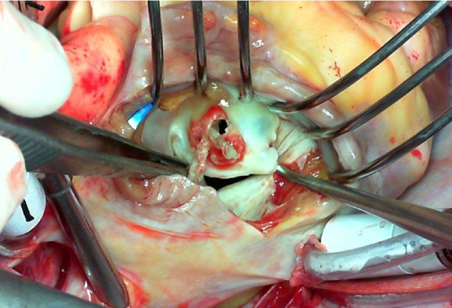
Three-Dimensional Transesophageal Echocardiography in Native Valve Endocarditis: A Case Report and Similarities Between Echocardiography and Surgical Findings

Giulia Bevilacqua Schmitz et al. Amadeu Antonio Bertuol Filho Solano Vinicius Berger Rodrigo Petersen Saadi … Álvaro Albrecht Angela Barreto Santiago Santos

Abstract

Native valve bacterial endocarditis is an uncommon, complex, and highly morbid disease that requires prolonged clinical treatment and challenging surgical interventions. Transthoracic and transesophageal echocardiography are paramount assessment tools whose findings are included in the diagnostic criteria. Three-dimensional echocardiography shows further realistic imaging details. Here we present a case demonstrating the role of echocardiography in the diagnosis of endocarditis and the identification of its complications to show how advanced imaging techniques may have a remarkable resemblance with in vivo surgical findings.
  • Echocardiography
  • Endocarditis
  • Mitral Valve
  • Three-Dimensional
Read Full Article
Creative Commons Attribution (CC BY) Creative Commons license
07/07/2022 DOI: 10.47593/2675-312X/20223503eabc279 Views 1
Figure 1
ABCIC
My Approach To

My Approach to Imaging the Atrial Septal Defect by Three-Dimensional Echocardiography

Daniela Lago Kreuzig

Pre-procedural assessment

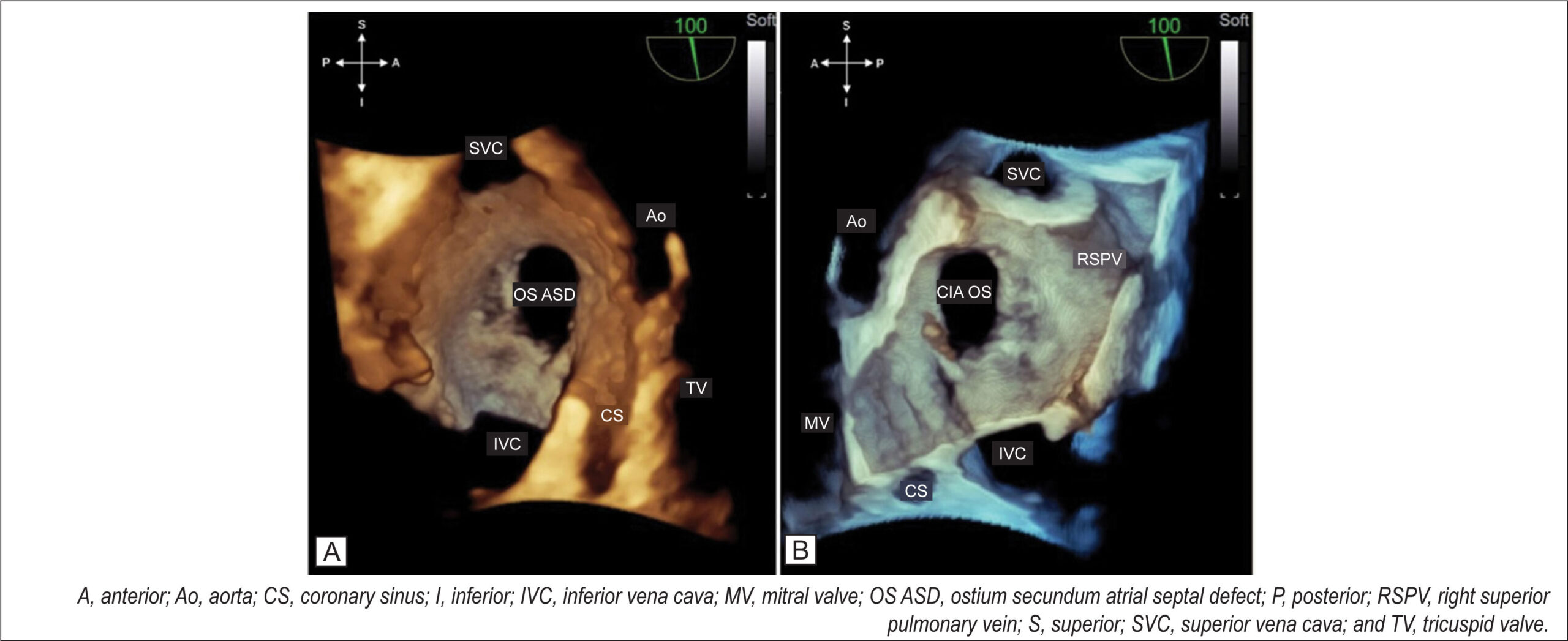
At the end of a detailed 2D TEE with a full septal analysis from the four-chamber view to bicaval section and color mapping, we commence the 3D image acquisition. The en-face projection of the ASD and the defect’s positioning in the anatomic position provide valuable information from a single image: size, shape, location in the interatrial septum, characteristics of the defect’s borders, and the presence of additional fenestration.

I usually start the 3D evaluation from the perspective of the right atrium to facilitate the identification of anatomical markers. I acquire a medium angle block (3D or 4D zoom) or a large block (full-volume using the two-section tool) starting from an initial bicaval section and proceeding with approximate 90° rotation in the z-plane so that the superior and inferior vena cava are aligned in the vertical plane (i.e., superior vena cava at the 11 o’clock position) and the aorta in the anterior and superior region. I prefer not to use the narrow-angle mode for acquisition (live 3D or bird’s eye view) or very high magnification due to a loss of anatomical references adjacent to the defect, which can interfere with this fundamental initial analysis. From this reconstruction (), I can characterize the defect as unique, the OS type, having an oval shape, being central, being medium-sized, and having adequate borders. shows the same ASD and allows assessment of the occurrence of dynamic variation in its dimension with the cardiac cycle.

[…]

  • atrial
  • Congenital
  • Echocardiography
  • Heart Defects
  • Heart septal defects
  • Three-Dimensional
Read Full Article
Creative Commons Attribution (CC BY) Creative Commons license
13/06/2022 DOI: 10.47593/2675-312X/20223502ecom32 Views 3
ABCIC
Editorial

Left Atrium: Novel Insights and Future Directions

Cristiane de Carvalho Singulane et al. Karima Addetia Roberto Miguel Lang

[…]

  • Atrial Function
  • Echocardiography, Left Atrium
  • Strains
  • Three-Dimensional
Read Full Article
Creative Commons Attribution (CC BY) Creative Commons license
17/12/2021 DOI: 10.47593/2675-312X/20203304eabc157
Left ventricular ejection fraction and volumes as measured by 3d echocardiography and ultrafast computed tomography
ABC
Artigo Original

Left ventricular ejection fraction and volumes as measured by 3d echocardiography and ultrafast computed tomography

Marcelo Luiz Campos Vieira et al. César H. Nomura Bernardino Tranchesi Junior Wercules A. de Oliveira … Gustavo Naccarato Bruna S. Serpa Rodrigo B. D. Passos Marcelo B. G. Funari Cláudio H. Fischer Samira S. Morhy

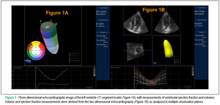
BACKGROUND: Real-time three-dimensional echocargiography (RT-3D-Echo) and ultrafast computed tomography (CT) are two novel methods for the analysis of LV ejection fraction and volumes. OBJECTIVE: To compare LVEF and volume measurements as obtained using RT-3D-Echo and ultrafast CT. METHODS: Thirty nine consecutive patients (27 men, mean age of 57±12 years) were studied using RT-3D-Echo and 64-slice ultrafast CT. LVEF and LV volumes were analyzed. Statistical analysis: coefficient of correlation (r: Pearson), Bland-Altman analysis, linear regression analysis, 95% CI, p<0.05. RESULTS: RT-3D-Echo measurements: LVEF ranged from 56.1 to 78.6 (65.5±5.58)%; end-diastolic volume ranged from 49.6 to 178.2 (87±27.8) ml; end-systolic volume ranged from 11.4 to 78 (33.1±13.6) ml. CT scan measurements: LVEF ranged from 53 to 86% (67.8±7.78); end-diastolic volume ranged from 51 to 186 (106.5±30.3) ml; end-systolic volume ranged from 7 to 72 (35.5±13.4)ml. Correlations between RT-3D-Echo and CT were: LVEF (r: 0.7888, p<0.0001, 95% CI 0.6301 to 0.8843); end-diastolic volume (r: 0.7695, p<0.0001, 95% CI 0.5995 to 0.8730); end-systolic volume (r: 0.8119, p<0.0001, 95% CI 0.6673 to 0.8975). CONCLUSION: Good correlation between LVEF and ventricular volume parameters as measured by RT-3D-Echo and 64-slice ultrafast CT was found in the present case series.

  • Echocardiography
  • emission-computed
  • Heart
  • Stroke Volume
  • Three-Dimensional
  • Tomography
Read Full Article
Creative Commons Attribution (CC BY) Creative Commons license
23/06/2009 DOI: 10.1590/S0066-782X2009000400008 Views 1

How to cite this article

EndNote RIS

Electronic Document Format (Vancouver)

Schmitz GB, Bertuol Filho AA, Berger SV, Saadi RP, Albrecht , Santos ABS. Three-Dimensional Transesophageal Echocardiography in Native Valve Endocarditis: A Case Report and Similarities Between Echocardiography and Surgical Findings. ABC Imagem Cardiovascular 2022;35(3):eabc279.

Electronic Document Format (ABNT)

Schmitz, Giulia Bevilacqua; Bertuol Filho, Amadeu Antonio; Berger, Solano Vinicius; Saadi, Rodrigo Petersen; Albrecht, Álvaro; Santos, Angela Barreto Santiago. Three-Dimensional Transesophageal Echocardiography in Native Valve Endocarditis: A Case Report and Similarities Between Echocardiography and Surgical Findings. ABC Imagem Cardiovascular, v. 35, n. 3, eabc279, Jul. 2022.

Electronic Document Format (APA)

Schmitz, G. B., Bertuol Filho, A. A., Berger, S. V., Saadi, R. P., Albrecht, , & Santos, A. B. S. (2022). Three-Dimensional Transesophageal Echocardiography in Native Valve Endocarditis: A Case Report and Similarities Between Echocardiography and Surgical Findings. ABC Imagem Cardiovascular, 35(3), eabc279.

Electronic Document Format (ISO)

Schmitz, Giulia Bevilacqua and Bertuol Filho, Amadeu Antonio and Berger, Solano Vinicius and Saadi, Rodrigo Petersen and Albrecht, Álvaro and Santos, Angela Barreto Santiago. Three-Dimensional Transesophageal Echocardiography in Native Valve Endocarditis: A Case Report and Similarities Between Echocardiography and Surgical Findings. ABC Imagem Cardiovascular [online]. 2022, vol. 35, n. 3, [cited 2026-10-14], eabc279. Available from: <https://journals.cardiol.br/en/article/three-dimensional-transesophageal-echocardiography-in-native-valve-endocarditis-a-case-report-and-similarities-between-echocardiography-and-surgical-findings/>. ISSN 2318-8219

EndNote RIS

Electronic Document Format (Vancouver)

Kreuzig DL. My Approach to Imaging the Atrial Septal Defect by Three-Dimensional Echocardiography. ABC Imagem Cardiovascular 2022;35(2):ecom32.

Electronic Document Format (ABNT)

Kreuzig, Daniela Lago. My Approach to Imaging the Atrial Septal Defect by Three-Dimensional Echocardiography. ABC Imagem Cardiovascular, v. 35, n. 2, ecom32, Jun. 2022.

Electronic Document Format (APA)

Kreuzig, D. L. (2022). My Approach to Imaging the Atrial Septal Defect by Three-Dimensional Echocardiography. ABC Imagem Cardiovascular, 35(2), ecom32.

Electronic Document Format (ISO)

Kreuzig, Daniela Lago. My Approach to Imaging the Atrial Septal Defect by Three-Dimensional Echocardiography. ABC Imagem Cardiovascular [online]. 2022, vol. 35, n. 2, [cited 2026-10-14], ecom32. Available from: <https://journals.cardiol.br/en/article/my-approach-to-imaging-the-atrial-septal-defect-by-three-dimensional-echocardiography/>. ISSN 2318-8219

EndNote RIS

Electronic Document Format (Vancouver)

Singulane CC, Addetia K, Lang RM. Left Atrium: Novel Insights and Future Directions. Arquivos Brasileiros de Cardiologia Imagem cardiovascular 2021;33(4):1-5.

Electronic Document Format (ABNT)

Singulane, Cristiane de Carvalho; Addetia, Karima; Lang, Roberto Miguel. Left Atrium: Novel Insights and Future Directions. Arquivos Brasileiros de Cardiologia Imagem cardiovascular, v. 33, n. 4, p. 1-5, Dec. 2021.

Electronic Document Format (APA)

Singulane, C. C., Addetia, K., & Lang, R. M. (2021). Left Atrium: Novel Insights and Future Directions. Arquivos Brasileiros de Cardiologia Imagem cardiovascular, 33(4), 1-5.

Electronic Document Format (ISO)

Singulane, Cristiane de Carvalho and Addetia, Karima and Lang, Roberto Miguel. Left Atrium: Novel Insights and Future Directions. Arquivos Brasileiros de Cardiologia Imagem cardiovascular [online]. 2021, vol. 33, n. 4, [cited 2026-10-14], pp. 1-5. Available from: <https://journals.cardiol.br/en/article/left-atrium-novel-insights-and-future-directions/>. ISSN 2318-8219

EndNote RIS

Electronic Document Format (Vancouver)

Vieira MLC, Nomura CH, Tranchesi Junior B, Oliveira WA, Naccarato G, Serpa BS, Passos RBD, et al. Left ventricular ejection fraction and volumes as measured by 3d echocardiography and ultrafast computed tomography. Arquivos Brasileiros de Cardiologia 2009;92(4):294-301.

Electronic Document Format (ABNT)

Vieira, Marcelo Luiz Campos; Nomura, César H.; Tranchesi Junior, Bernardino; Oliveira, Wercules A. de; Naccarato, Gustavo; Serpa, Bruna S.; Passos, Rodrigo B. D.; Funari, Marcelo B. G.; Fischer, Cláudio H.; Morhy, Samira S.. Left ventricular ejection fraction and volumes as measured by 3d echocardiography and ultrafast computed tomography. Arquivos Brasileiros de Cardiologia, v. 92, n. 4, p. 294-301, Jun. 2009.

Electronic Document Format (APA)

Vieira, M. L. C., Nomura, C. H., Tranchesi Junior, B., Oliveira, W. A. , Naccarato, G., Serpa, B. S., Passos, R. B. D., Funari, M. B. G., Fischer, C. H., & Morhy, S. S. (2009). Left ventricular ejection fraction and volumes as measured by 3d echocardiography and ultrafast computed tomography. Arquivos Brasileiros de Cardiologia, 92(4), 294-301.

Electronic Document Format (ISO)

Vieira, Marcelo Luiz Campos and Nomura, César H. and Tranchesi Junior, Bernardino and Oliveira, Wercules A. de and Naccarato, Gustavo and Serpa, Bruna S. and Passos, Rodrigo B. D. and Funari, Marcelo B. G. and Fischer, Cláudio H. and Morhy, Samira S.. Left ventricular ejection fraction and volumes as measured by 3d echocardiography and ultrafast computed tomography. Arquivos Brasileiros de Cardiologia [online]. 2009, vol. 92, n. 4, [cited 2026-10-14], pp. 294-301. Available from: <https://journals.cardiol.br/en/article/left-ventricular-ejection-fraction-and-volumes-as-measured-by-3d-echocardiography-and-ultrafast-computed-tomography/>. ISSN 0066-782X

  • About ABC Family
  • Internal Regulation
  • Editorial Board
  • Brazilian Society of Cardiology (SBC)

Issues

SBC Guidelines

  • Videos
  • Podcasts
  • News