| ABCIC
ABC Family Arquivos Brasileiros de Cardiologia International Journal of Cardiovascular Sciences ABC Heart Failure & Cardiomyopathy ABC Imagem Cardiovascular
Português English
ABC Family | ABCIC
Português English
  • About
    • About the Journal
    • Internal Regulation
    • Editors Section
    • Editorial Board
    • SBC Award
  • Instructions to Authors
    • Guidelines for Publication
    • Editorial Policy
    • Ethics Policy
    • Policy on the Use of Artificial Intelligence
    • Mandatory Publication Form
  • Issues
  • SBC Guidelines
  • About
    • About the Journal
    • Internal Regulation
    • Editors Section
    • Editorial Board
    • SBC Award
  • Instructions to Authors
    • Guidelines for Publication
    • Editorial Policy
    • Ethics Policy
    • Policy on the Use of Artificial Intelligence
    • Mandatory Publication Form
  • Issues
  • SBC Guidelines

ABC Family

  • ABC Family
  • Arquivos Brasileiros de Cardiologia
  • International Journal of Cardiovascular Sciences
  • ABC Heart Failure & Cardiomyopathy
  • ABC Imagem Cardiovascular
Search in

No content found.

Journals
Ordering
Article types
Others

  1. Home
  2. ABCIC
  3. Issues
  4. 2024, Volume 37, Number 1
What do Cardiologists Expect Towards Myocardial Viability Assessment
Review Article

What do Cardiologists Expect Towards Myocardial Viability Assessment

Eduardo Gomes Lima et al. Eduardo Bello Martins Leticia Neves Solon Carvalho Diogo Freitas Cardoso de Azevedo

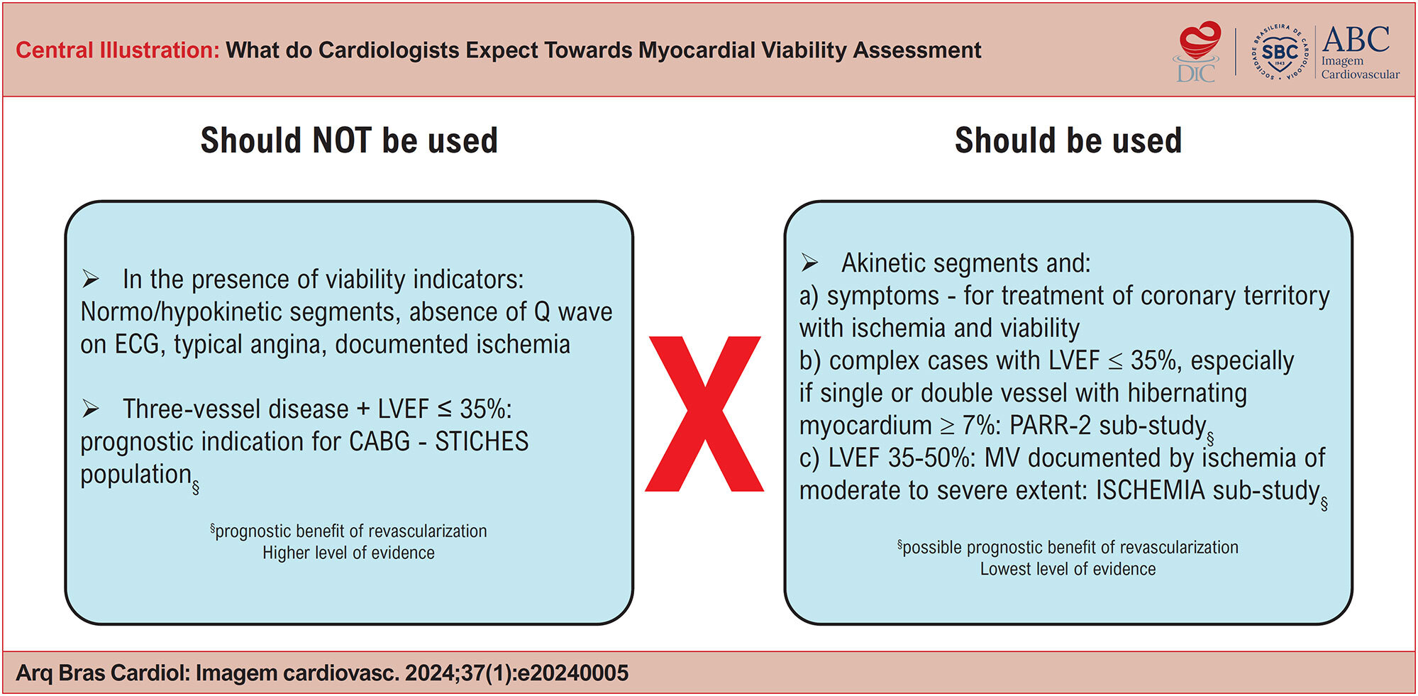
Within the clinical spectrum of chronic coronary syndrome (CCS), the presence of coronary artery disease (CAD) alongside ventricular dysfunction has long been a focal point due to the lower survival rates seen in this population. Improving survival in these patients, whether through medications or interventions, is a key concern for clinical cardiologists. Previous observational studies suggested that myocardial viability (MV) could influence the effectiveness of revascularization strategies in patients with ventricular dysfunction. Thus, viability assessment became a standard practice in cardiology. Different imaging methods assess viability through different parameters (such as cell membrane integrity, mitochondrial function, glycolytic metabolism, contractile reserve, or evidence of fibrosis), leading to differing sensitivities and specificities. However, recent evidence from large randomized studies does not support the notion that the presence of MV, as determined by various methods, is linked to the prognostic benefit of revascularization. Furthermore, the reduction in mortality observed in these studies was not attributed to the recovery of contractile function but rather to a decrease in fatal heart attacks. As a result, the current role of MV as a decision-making tool for revascularization in patients with CCS and left ventricular dysfunction has diminished. Nonetheless, there are still situations in specialist practice where its use may be warranted, particularly in the context of constructing a comprehensive functional revascularization strategy.
  • Left Ventricular Function
  • Myocardial Ischemia
  • Myocardial Stunning
Read Full Article
Creative Commons Attribution (CC BY) Creative Commons license
28/03/2024 DOI: 10.36660/abcimg.20240005i Views 2
Association of Sympathetic Denervation, Myocardial Hypoperfusion, and Fibrosis with Ventricular Arrhythmias in Chronic Chagas Cardiomyopathy
Original Article

Association of Sympathetic Denervation, Myocardial Hypoperfusion, and Fibrosis with Ventricular Arrhythmias in Chronic Chagas Cardiomyopathy

Adriana Soares Xavier de Brito et al. Renata Junqueira Moll-Bernardes Martha Valéria Tavares Pinheiro Gabriel Camargo … Fabio Paiva Rossini Siqueira Adriana Pereira Glavam Sergio Altino Almeida Fernanda Souza Nogueira Sardinha Mendes Paulo Henrique Rosado-de-Castro Andrea Silvestre Sousa

Abstract

Background:

Chronic Chagas cardiomyopathy (CCC) manifests as heart failure, thromboembolic events, and sudden cardiac death (SCD). Although SCD may be the presenting event, there is still no recommendation for early cardioverter/defibrillator implantation in current guidelines.

Objective:

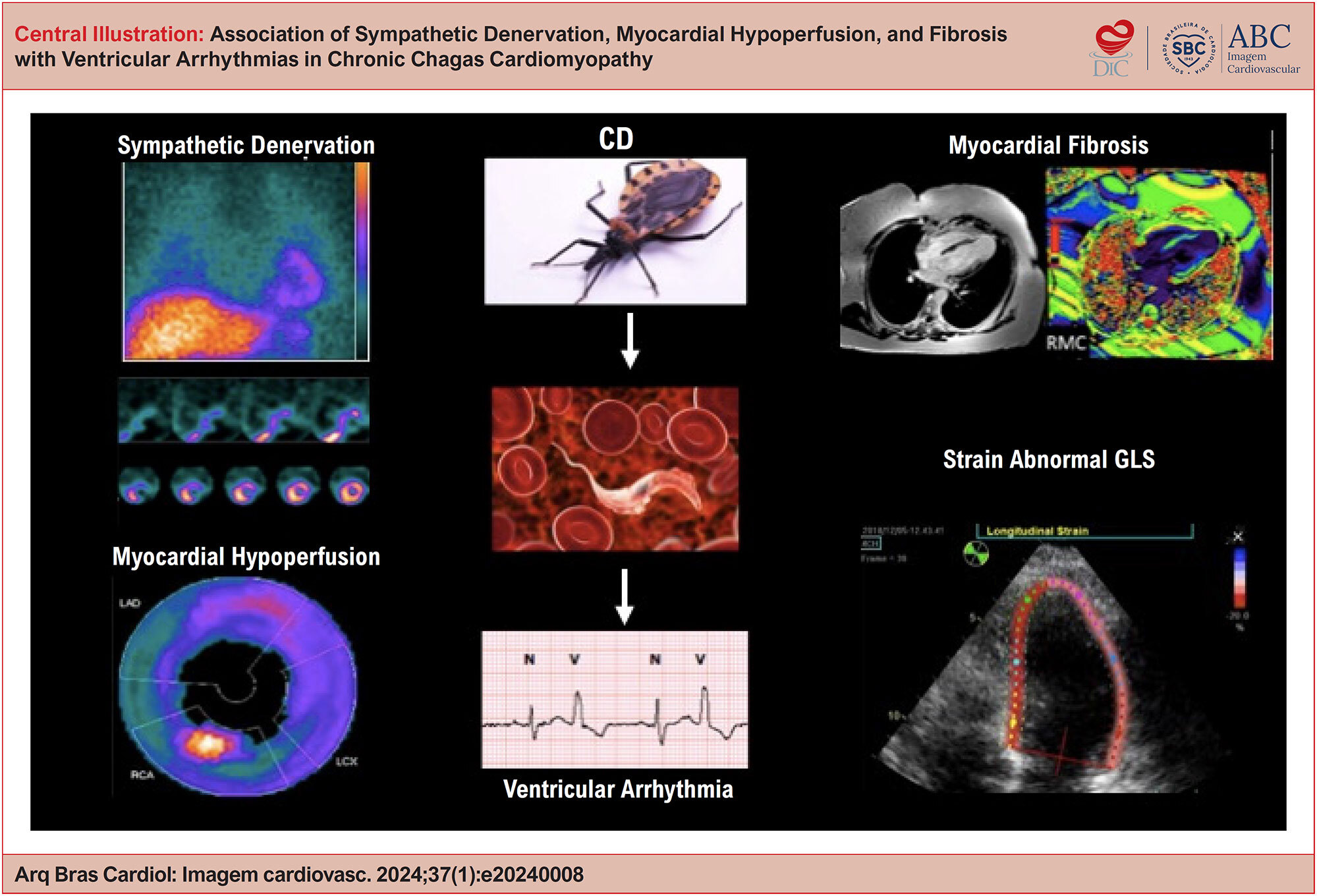
To evaluate the correlation between autonomic denervation, myocardial hypoperfusion, fibrosis, and ventricular arrhythmias in patients in the early stages of CCC.

Methods:

Cross-sectional study of 29 patients with CCC and preserved left ventricular function who underwent SPECT with iodine-123-meta-iodobenzylguanidine (123I-mIBG), myocardial perfusion SPECT with technetium-99m sestamibi (99mTc-MIBI), and cardiac magnetic resonance (CMR) with gadolinium, divided into two groups according to on 24h Holter findings: arrhythmia (> 6 ventricular premature complexes/hour and/or nonsustained ventricular tachycardia; n = 15) or no-arrhythmia (< 6 ventricular premature complexes/hour and no ventricular tachycardia; n = 14).

Results:

Significant correlations were observed between parameters of the three cardiovascular imaging modalities and the presence of ventricular arrhythmia. Denervation on mIBG correlated moderately with diffuse fibrosis, represented by ECV on CMR (r = 0.55, P = 0.002). Hypoperfusion by MIBI-SPECT correlated with fibrosis by both LGE (r = 0.66, P = 0.005) and extracelluar volume (ECV) (r = 0.56, P = 0.002). We also observed a moderate correlation between the extent of myocardial areas with denervation and hypoperfusion (r = 0.48, P = 0.007).

Conclusion:

The presence of autonomic denervation, myocardial hypoperfusion, and fibrosis was associated with ventricular arrhythmia in the early stages of CCC. A combination of these parameters can improve stratification of SCD risk in these patients.

  • Cardiac Arrhythmias
  • Chagas disease
  • Sympathetic Denervation
Read Full Article
Creative Commons Attribution (CC BY) Creative Commons license
28/03/2024 DOI: 10.36660/abcimg.20240008i Views 2
Left Atrial Strain: Clinical Applications and Prognostic Implications
Review Article

Left Atrial Strain: Clinical Applications and Prognostic Implications

Fernanda de Azevedo Figueiredo et al. Admilson Lemos da Costa Filho Flávio de Azevedo Figueiredo Luz Marina Tacuri Chavez … Marcia Fabrícia Almeida Teixeira William Silva Barbosa Pedro Henrique Bronzatto Priscila Rabelo Cintra Maria Carmo Pereira Nunes

Abstract

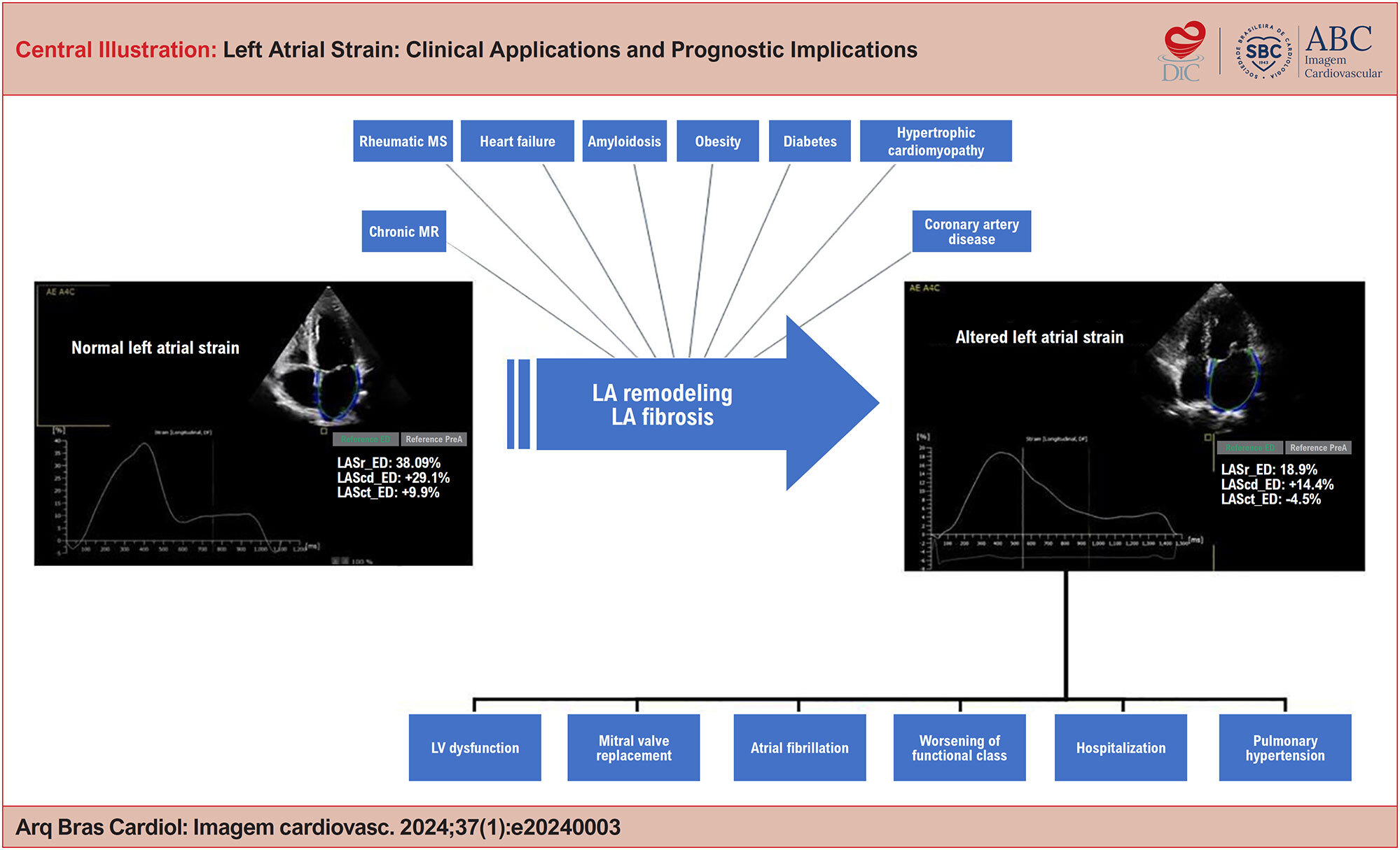
Left atrial (LA) strain, obtained using two-dimensional speckle tracking echocardiography (2DSTE), has emerged as an accurate method for evaluating LA function. Recently, it has gained prominence in clinical practice due to its significant prognostic value in various cardiovascular diseases, standing out due to its greater sensitivity compared to traditional methods of volumetric analysis and Doppler parameters.

This review article addresses the complex function of the LA and its close interdependence with left ventricular (LV) function, highlighting its relevance in cardiac performance. The LA performs three distinct functions, acting as a reservoir during ventricular systole, as a conduit in early diastole, and as a contractile chamber during the atrial contraction phase.

This review analyzes the normal patterns of LA strain and its application in various clinical conditions, such as atrial fibrillation (AF), heart failure, coronary artery disease, obesity, diabetes mellitus, hypertrophic cardiomyopathy, and cardiac amyloidosis. The crucial role of atrial strain in rheumatic mitral stenosis (MS) is highlighted due to its capacity to predict clinical outcomes.

  • Atrial Fibrillation
  • Echocardiography
  • Global Longitudinal Strain
  • Left Atrial Function
Read Full Article
Creative Commons Attribution (CC BY) Creative Commons license
28/03/2024 DOI: 10.36660/abcimg.20240003i Views 2
Figure 1
Case Report

Ultrasound in the Diagnosis of Carotid Web: Report of Two Cases and Review

Ana Claudia Gomes Pereira Petisco et al. Paulo Magno Martins Dourado Larissa Almeida Dourado João Paulo Almeida Dourado Ricardo Thomaz Tebaldi

Introduction

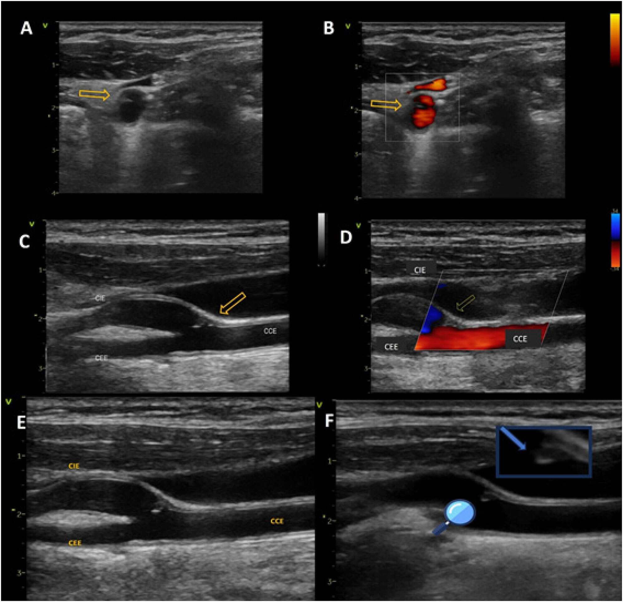
Carotid web (CW) is a nonatherosclerotic disease of the extracranial carotid artery, described as an atypical, still little known, and poorly diagnosed focal fibromuscular dysplasia (FMD). CW has been associated with the occurrence of cerebral vascular accidents (CVAs) and transient ischemic attacks (TIAs), affecting young patients, with no other known causes.

[…]

  • Carotid Arteries Ultrasonography
  • Carotid Artery Diseases
  • Fibromuscular Dysplasia
Read Full Article
Creative Commons Attribution (CC BY) Creative Commons license
28/03/2024 DOI: 10.36660/abcimg.20230117i Views 2
My Approach to Nuclear Medicine in the Assessment of Microvascular Disease in Women
Review Article

My Approach to Nuclear Medicine in the Assessment of Microvascular Disease in Women

Lara Cristiane Terra Ferreira Carreira et al. Lívia Carreira Adriana Soares Xavier de Brito

Abstract

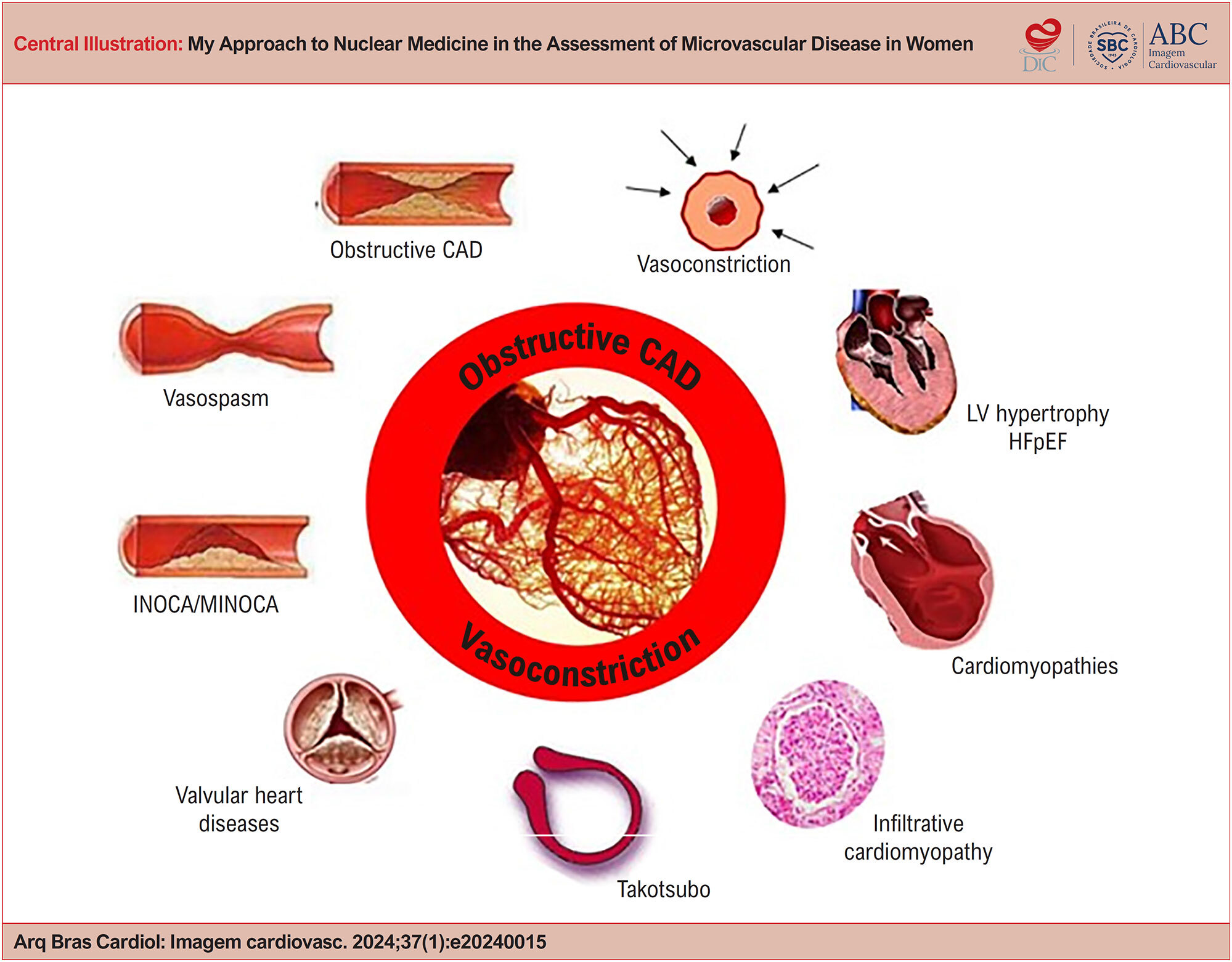
Coronary microvascular dysfunction (CMD) is a condition that has been increasingly recognized as a cause of angina, with prognostic importance in multiple cardiovascular processes, especially in women. It results from abnormalities in the structure and/or function of the coronary microcirculation. Even in the absence of obstructive coronary artery disease (CAD), CMD is associated with worse prognosis, greater morbidity, impaired quality of life, and recurrent hospitalizations due to angina and heart failure, posing a challenge for diagnosis and treatment. In this article, we briefly review CMD and how nuclear medicine can assist in its assessment.

  • Microvascular Angina
  • Myocardial Ischemia
  • Nuclear Medicine
Read Full Article
Creative Commons Attribution (CC BY) Creative Commons license
28/03/2024 DOI: 10.36660/abcimg.20240015i
Figure 1
Case Report

Ischemic Stroke in a Patient with Challenging Pseudoaneurysm in the Left Ventricular Outflow Tract

Saulo Rodrigo Cunha et al. Fernanda Pandolfo Fernando Colares Barros Eduardo Gatti Pianca Pedro Tregnago Barcellos

Introduction

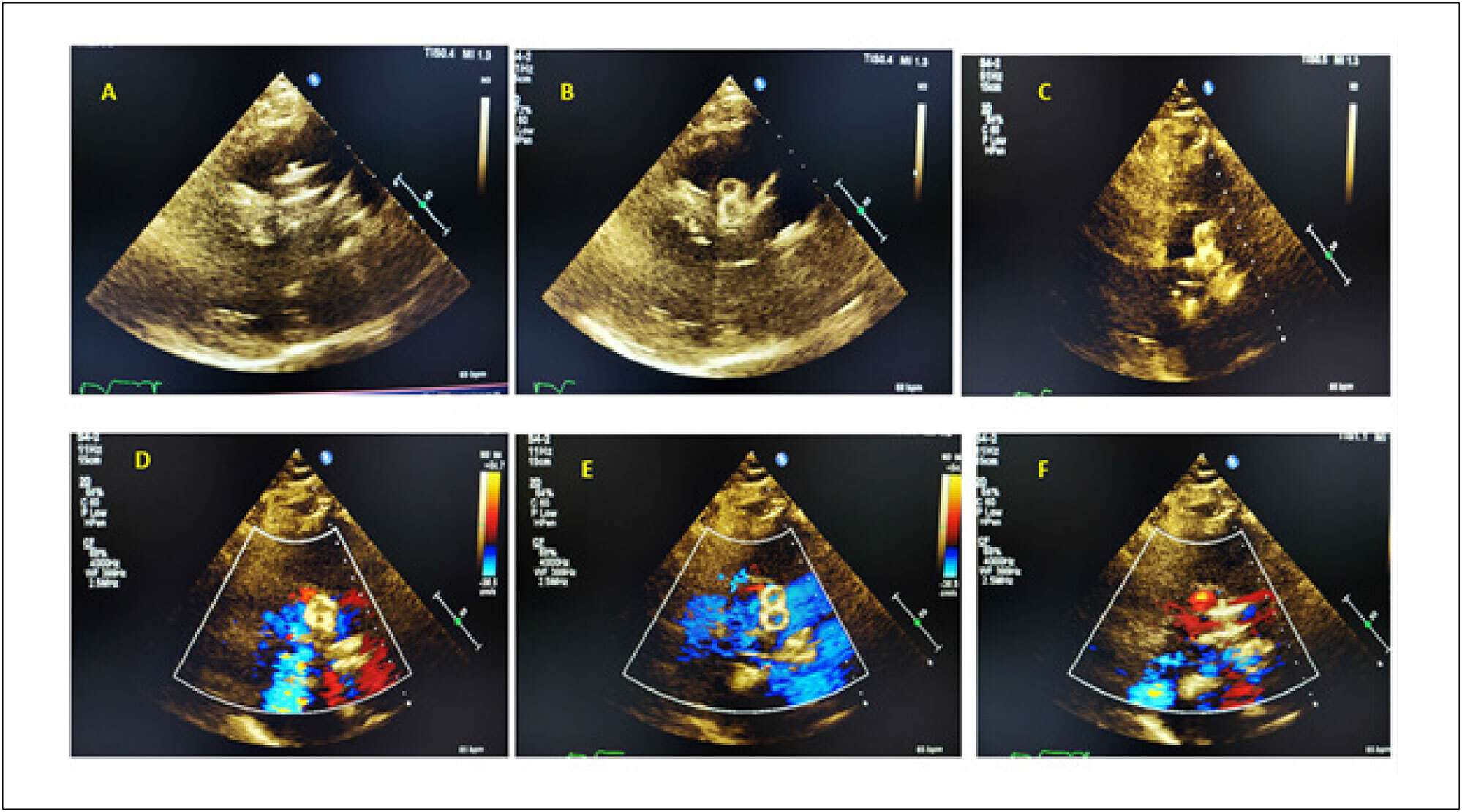
Literature shows cases of cardiac neocavities, often associated with pseudoaneurysms resulting from traumatic injuries or fistulized periprosthetic abscesses involving the left ventricle (LV).

Cardiac pseudoaneurysms are typically complications of acute myocardial infarction but can also occur in the late postoperative period of valve surgery. Certain areas are particularly prone to the development of ascending aortic pseudoaneurysms. Generally, these events occur in sites where aortic cannulation and clamping take place, as well as in the areas of graft sutures.

[…]

  • False Aneurysm
  • Heart Valve Prosthesis
  • Stroke
Read Full Article
Creative Commons Attribution (CC BY) Creative Commons license
27/03/2024 DOI: 10.36660/abcimg.20240001i Views 2
My Approach to Assess Cardiac Sympathetic Activity
Review Article

My Approach to Assess Cardiac Sympathetic Activity

Adriana Soares Xavier de Brito et al. Jessica Costa Leite Simone Cristina Soares Brandão

Abstract

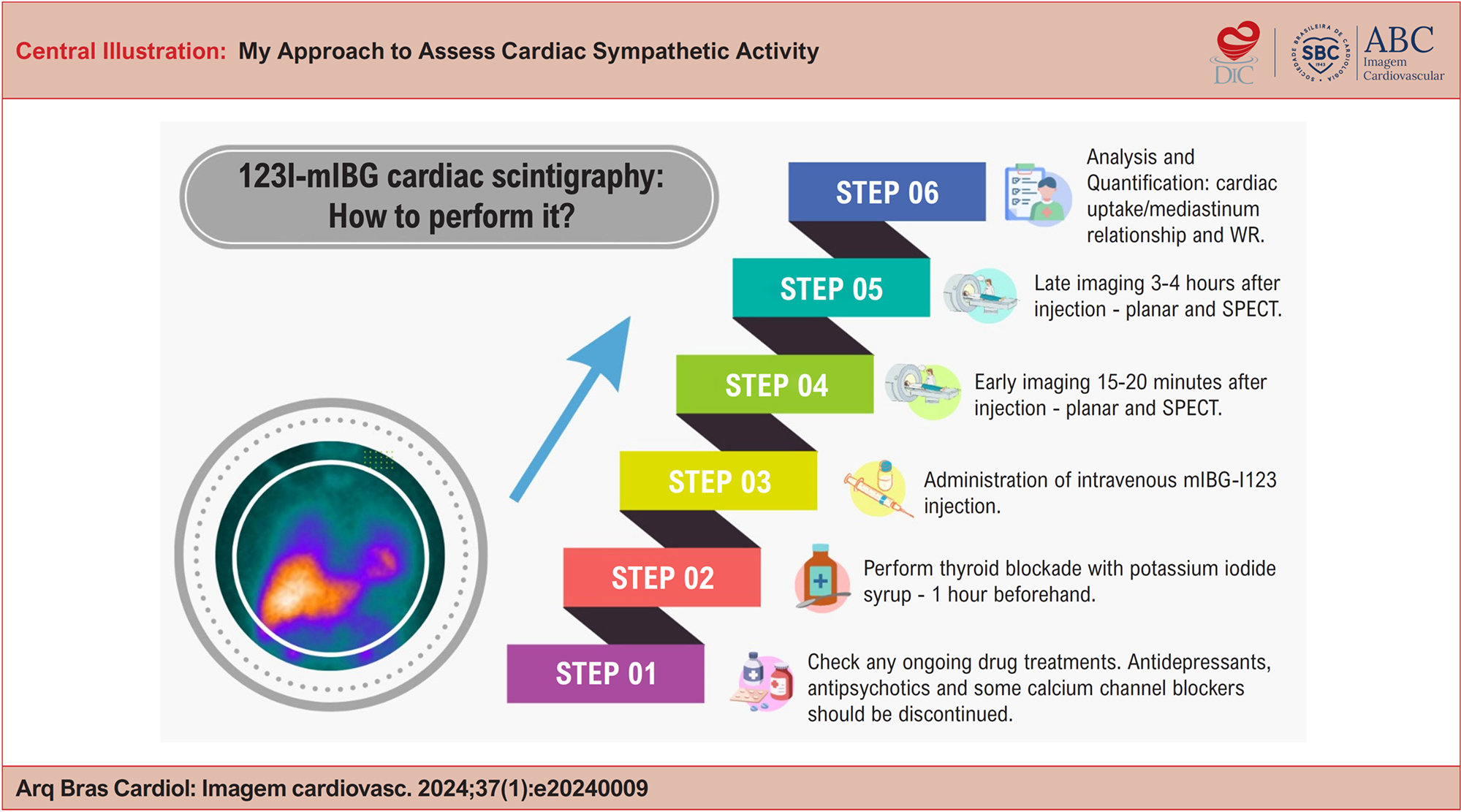
Sympathetic innervation plays a crucial role in regulating blood flow, chronotropism, dromotropism, lusitropism, and myocardial inotropism. In several cardiac and neurodegenerative disorders, the autonomic nervous system (ANS) undergoes alterations. Cardiac neuronal imaging can aid in understanding the pathophysiology, diagnosis, and prognosis of these conditions. This article provides a clear and objective overview of the step-by-step procedure for mIBG-I123 scintigraphy, which assesses cardiac sympathetic activity in primary clinical practice settings.

  • 123-metaiodobenzylguanidine
  • Heart Failure
  • Heart Rate
  • Parkinson Disease
Read Full Article
Creative Commons Attribution (CC BY) Creative Commons license
27/03/2024 DOI: 10.36660/abcimg.20240009i Views 1
Cardiac Sarcoidosis: The Chameleon of Cardiology
Review Article

Cardiac Sarcoidosis: The Chameleon of Cardiology

Diego Moraes De Moura et al. Aluísio José De Oliveira Monteiro Neto Marcelo Dantas Tavares de Melo Fábio Fernandes

Abstract

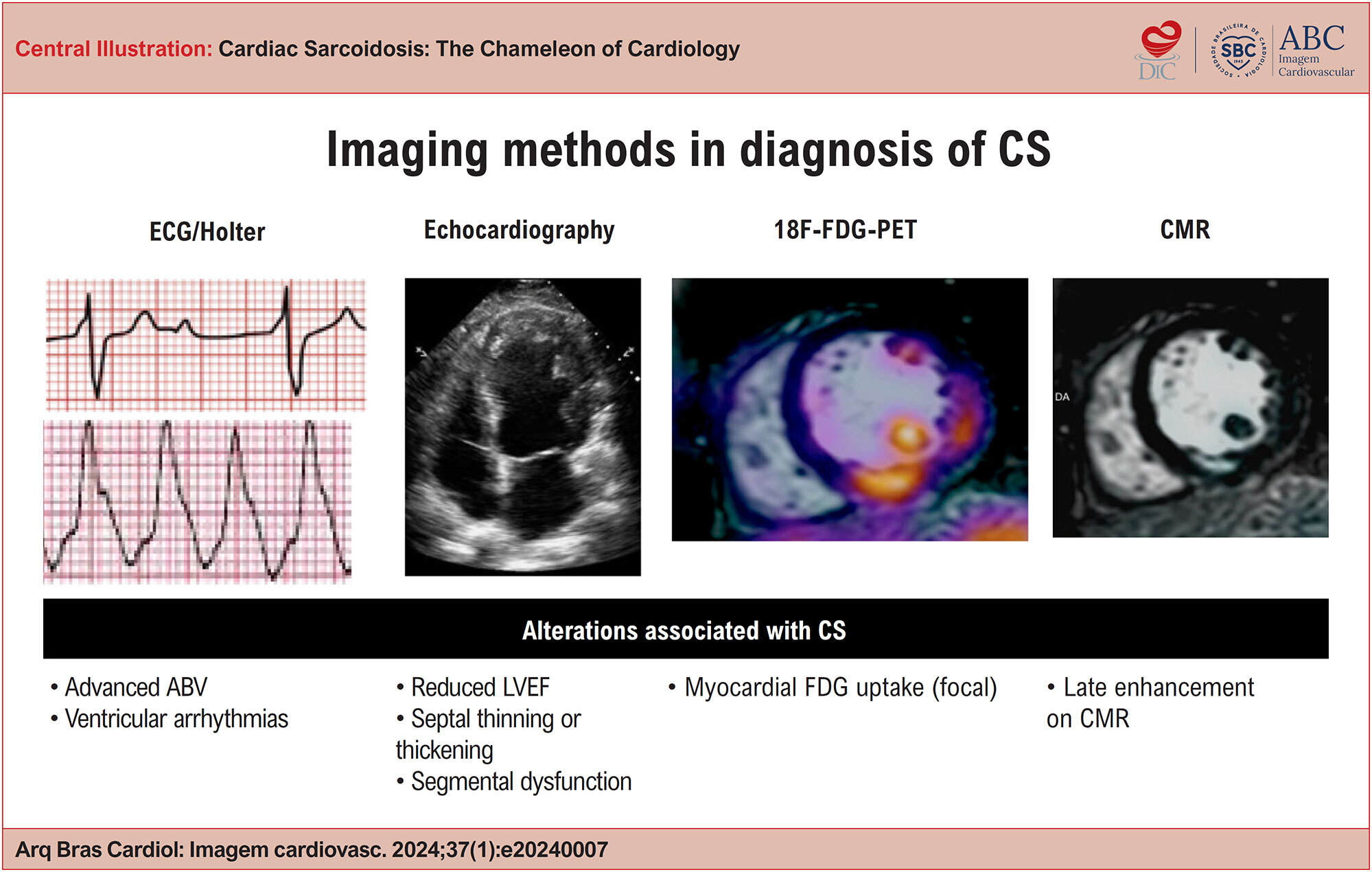
Sarcoidosis is a multisystemic granulomatous disease of unknown etiology, characterized by the formation of non-caseating granulomas in multiple organs. Cardiac involvement, an important cause of morbidity and mortality in these patients, has been generating interest in cardiology, because it is a cause of heart failure, atrioventricular blocks, and ventricular arrhythmias with unfavorable prognosis; however, there are specific treatments with the potential to change the natural history of this condition. The main challenge of cardiac sarcoidosis (CS) is diagnosis, given that the gold standard method of endomyocardial biopsy has limited sensitivity due to the focal nature of the pathology.

Accordingly, cardiovascular imaging methods play the role of guiding most diagnoses of CS. In this scenario, knowledge about these methods, their main findings, and their rational use are essential to the diagnosis of this disease with such diverse presentations.

Electrocardiogram and echocardiography are practical and widely available exams; however, they provide greater diagnostic capacity in patients with clinically manifest disease. On the other hand, to identify incipient forms, which are often silent, it is necessary to use advanced imaging methods, such as positron emission tomography with 18F-fluorodeoxyglucose and cardiac magnetic resonance, which primarily identify signs of active inflammatory activity and fibrosis, respectively. Despite the advances in these imaging methods, due to the lack of studies comparing them with the gold standard (endomyocardial biopsy), the diagnosis of CS currently remains a major challenge.

  • Echocardiography
  • Electrocardiography
  • Positron Emission Tomography-Computed Tomography
  • Sarcoidosis
  • X-Ray Computed Tomography
Read Full Article
Creative Commons Attribution (CC BY) Creative Commons license
27/03/2024 DOI: 10.36660/abcimg.20240007i Views 1
Echocardiographic Evaluation of Right Atrial Function in Patients with Precapillary Pulmonary Hypertension
Original Article

Echocardiographic Evaluation of Right Atrial Function in Patients with Precapillary Pulmonary Hypertension

Wanessa Alves de Carvalho et al. Andressa Alves de Carvalho Eliauria Rosa Martins Agostinho Hermes de Medeiros Neto … Fernando Bacal Marcelo Dantas Tavares de Melo

Abstract

Background:

Considering that the effect of right ventricular (RV) failure on the right atrium (RA) is an essential part of the pathophysiology of pulmonary hypertension (PH), the assessment of RA function seems to have prognostic value in the course of the disease, but there are still few studies on this subject.

Objective:

To evaluate myocardial strain of the RA in patients with precapillary PH.

Methods:

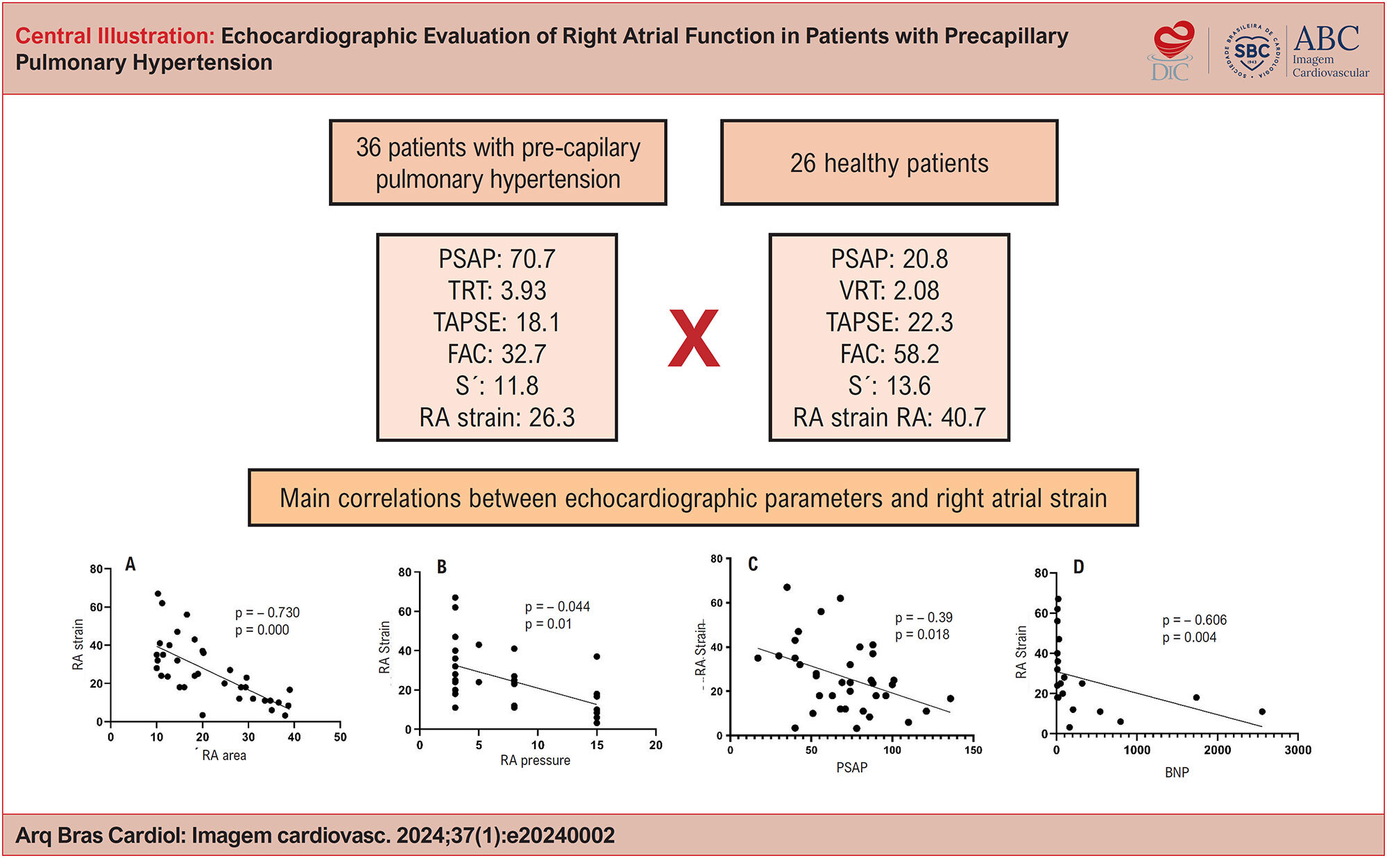
This is an observational, longitudinal, prospective study involving 36 patients with precapillary PH, in whom two-dimensional Doppler echocardiography was performed. A control group consisting of 26 healthy individuals was used.

Results:

There was a statistically significant difference in mean RA strain between the group with PH (26.3%) and the control group (40.7%). Regarding RA strain of the patients, there was a negative and moderate correlation with RA pressure, RV diameter, and RV hypokinesia; negative and weak correlation with pulmonary artery systolic pressure (PASP); negative and strong correlation with RA area; and positive and moderate correlation with, tricuspid annular plane systolic excursion (TAPSE), TAPSE/PASP, and RV strain. Patients with RA strain ≤ 23 had significantly reduced overall survival compared with patients with RA strain > 23, but this value was not a predictor of mortality.

Conclusion:

It was possible to demonstrate that the analysis of myocardial strain of the RA provides more accurate information about RA function and can be used as an additional parameter in the follow-up of patients with PH, given the early evaluation of atrial dysfunction.

  • Doppler Echocardiography
  • Pulmonary Hypertension
  • Right Atrial Function
Read Full Article
Creative Commons Attribution (CC BY) Creative Commons license
27/03/2024 DOI: 10.36660/abcimg.20240002i Views 1
Review Article

The Indiscriminate Use Of Androgenic Anabolic Steroids: The Contribution of Cardiovascular Imaging

Irving Gabriel Araöşjo Bispo et al. Isabella Montanher Zago

Abstract

Anabolic androgenic steroids (AAS) are natural or synthetic compounds similar to the male hormone testosterone. The literature also describes cases of AAS abuse, such as in bodybuilding, cosmetics, recreational use, and aesthetic enhancement. It was discovered that AAS alone directly induce myocardial injury, with the main pathological finding in autopsied hearts being left ventricle (LV) hypertrophy in frequent association with myocyte hypertrophy, increased collagen deposition in the matrix, increased activity of the cardiac angiotensin, and myocardial fibrosis.

Patients who used illicit AAS present decreased LV systolic function. In athletes who self-administer AAS, LV hypertrophy and elevated sympathetic modulation were observed, as well as elevated blood pressure (BP). High doses of AAS can cause the user to have ventricular arrhythmias and sudden death. Furthermore, AAS users demonstrated greater coronary artery plaque volume than non-users. The objective of this review is to revisit the main effects of the use of AAS on cardiac changes through cardiovascular imaging as well as to establish a difference in relation to athletic heart syndrome.

  • Doppler Echocardiography
  • Global Longitudinal Strain
  • Steroids
  • Tomography
Read Full Article
Creative Commons Attribution (CC BY) Creative Commons license
21/03/2024 DOI: 10.36660/abcimg.20230113i
Figure 1
Original Article

Relationship Between Echocardiographic Parameters and The Systemic Immune-Inflammation Index in Patients With Pulmonary Arterial Hypertension

Sefa Erdi Ömür et al. Gulsen Genc Tapar Mustafa Yilmaz Abdullah Emre Bektaş Osman Demir

Abstract

Background:

Pulmonary arterial hypertension (PAH) is a chronic disease with high morbidity and mortality. The diagnosis of PAH is mainly made based on echocardiographic parameters and natriuretic peptide levels. However, given the low incidence of PAH worldwide, the diagnosis of PAH can be challenging.

Objectives:

To evaluate the relationship between systemic immune-inflammation (SII) index and PAH.

Methods:

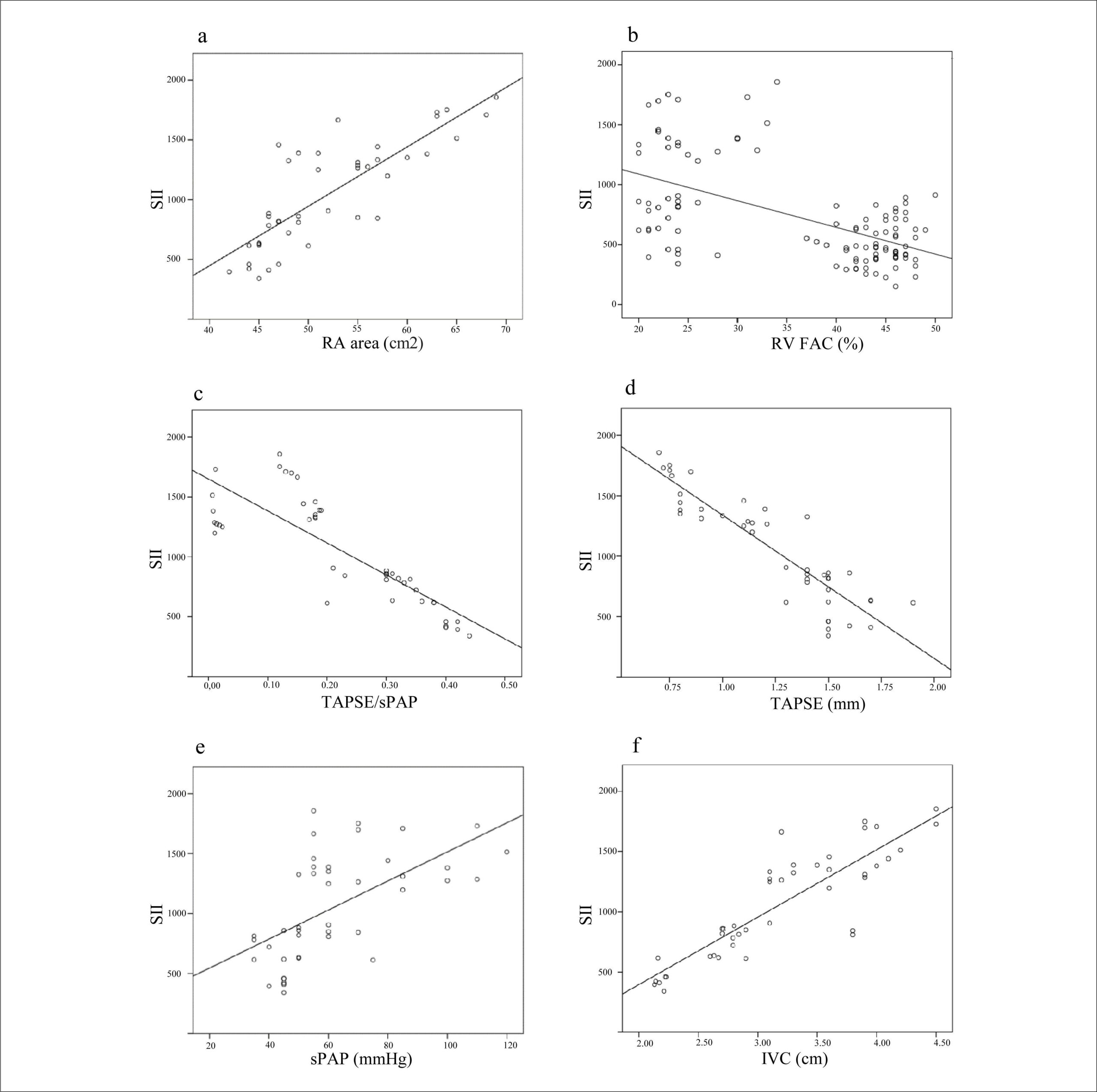
This was a retrospective, cross-sectional study of 110 patients (43 PAH patients and 67 controls). The SII index was compared between the PAH and control groups. A probability(p) <0.05 were deemed to indicate statistical significance.

Results:

The findings of this study indicated that SII index was significantly higher in the PAH group than in the control group (1054.15±439.99 vs. 506.7±180.55, p<0.001). The correlation analysis between SII index and echocardiographic parameters revealed that SII index was moderately correlated with right ventricular fractional area change (FAC) (r:-0.567,p<0.001), systolic pulmonary artery pressure (sPAP) (r:0.593, p<0.001), and tricuspid regurgitation velocity (TRV) (r:0.662,p<0.001). Additionally, the SII index was strongly correlated with right atrial (RA) area (r:0.822,p<0.001), pulmonary artery (PA) diameter (r:0.819, p<0.001), left atrium (LA) diameter (r:0.937,p<0.001), inferior vena cava diameter (r:0.869,p<0.001), tricuspid annular plane systolic excursion (TAPSE) (r:-0.902,p<0.001), TAPSE/sPAP (r:-0.831,p<0.001). In addition, the SII index significantly increased as patients' functional capacity (FC) decreased.

Conclusion:

The SII index is as a simple, inexpensive, noninvasive and easily accessible biochemical parameter that may be useful in the diagnosis and follow-up of PAH patients, especially in centers where echocardiography (ECHO) is not available.

  • Echocardiography
  • Inflammation
  • Pulmonary Arterial Hypertension
Read Full Article
Creative Commons Attribution (CC BY) Creative Commons license
21/03/2024 DOI: 10.36660/abcimg.20230086i Views 1
Figure 1
Brief Communication

Correlation of Telemedicine-guided POCUS Echocardiography and In Situ Data: Pilot Study in a Remote Area in Brazil

Juliane Rompkoski et al. Tarso Augusto Duenhas Accorsi Bruna Dayanne Reges Amaral Christian Barbosa de Freitas … Flavio Tocci Moreira Karen Francine Kohler Karine De Amicis Lima Renata Albaladejo Morbeck Carlos Henrique Sartorato Pedrotti

Abstract

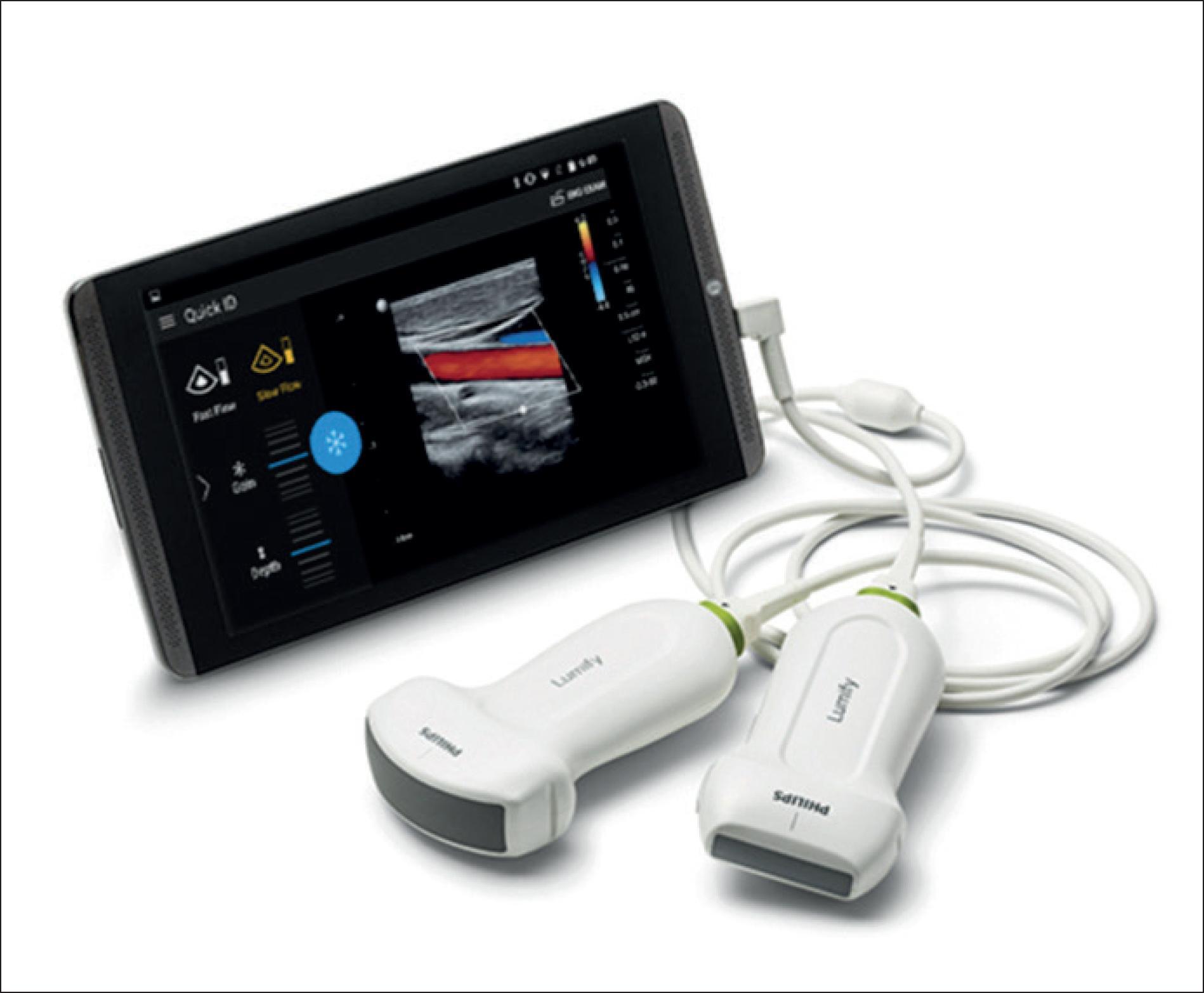
Cardiovascular diseases remain the leading cause of mortality in Brazil. Nevertheless, a significant portion of the Brazilian population still faces challenges in accessing specialized care and undergoing complementary exams, especially in remote areas. Currently, data transmission and remote specialized assessment are possible, but obtaining echocardiographic images still relies on a trained professional on-site. This pilot study aimed to assess the feasibility of Point-of-Care Ultrasound (POCUS) tele-echocardiography administered by a general physician with specialist guidance to improve accessibility to cardiovascular exams. Ten patients (mean age was 58.6+8 years, 50% female) from the North region participated, undergoing remote-guided tests using Philips Lumify™ ultrasound and Facetime for transmission. These patients had recent official echocardiographic exams. As part of the POCUS examination, seven echocardiographic views were obtained — parasternal long axis, parasternal short axis (PSAX), apical four chambers (A4C), apical five chambers (A5C), apical two chambers (A2C), subcostal (SC), and suprasternal (SSN). The procedure included linear measurements of cardiac chambers, and valve analysis was performed using color flow. The on-site examinations were carried out by a general physician who underwent brief training prior to the official exams. Despite encountering technical challenges, the study demonstrated the viability of acquiring echocardiographic images. Overall agreement in examination results was observed, except for left ventricular segmental contractility in two cases and the systolic diameter of the left ventricle. The approach, though performed by untrained professionals, showed promise in screening cardiovascular diseases in remote locations, focusing on ventricular function and valvular diseases. Nevertheless, further research is necessary to improve the quality of this process.

  • Echocardiography
  • Telecardiology
  • Telediagnostics
  • Telemedicine
  • Ultrasonography
Read Full Article
Creative Commons Attribution (CC BY) Creative Commons license
12/03/2024 DOI: 10.36660/abcimg.20230110i Views 1
Figure 1
Case Report

Rare Presentation of Pseudoaneurysm Obstructing the Left Ventricular Inflow Tract in a Patient With Acute Myocardial Infarction

Janine Daiana Stürmer et al. Raphael dos Santos Silva Willer Cesar Bica Tiago Hansel Basile Vigil … Gabriel Soder Renata Pibernat de Moraes Mathias Silvestre de Brida Rodrigo Moraes Reis

Introduction

Left ventricular (LV) pseudoaneurysm is nowadays a rare but still potentially fatal complication of myocardial infarction (MI). Recent studies have estimated that following ST-elevation MI, 0.27 to 0.91% of patients develop mechanical complications. LV free wall rupture (VFWR) is estimated to occur in 0.01 to 0.52% of patients, showing a clear decrease in incidence associated with the widespread adoption of reperfusion therapy. Nonetheless, there has been no significant decrease in associated mortality rates and this complication remains an important determinant of outcomes after MI.

A pseudoaneurysm is formed when a cardiac rupture is contained by adherent pericardium or scar tissue, with no myocardial tissue. Transmural MI is the most common cause, usually following occlusion of the right coronary artery or the circumflex artery, therefore more often occurring on the posterior or lateral wall.

[…]

  • Echocardiography
  • Myocardial Infarction
  • Pseudoaneurysm
Read Full Article
Creative Commons Attribution (CC BY) Creative Commons license
12/03/2024 DOI: 10.36660/abcimg.20230106i Views 1
Figure 1
Case Report

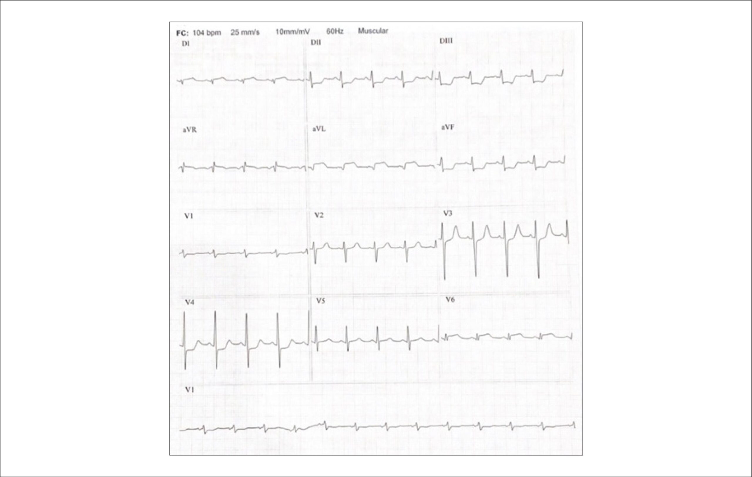
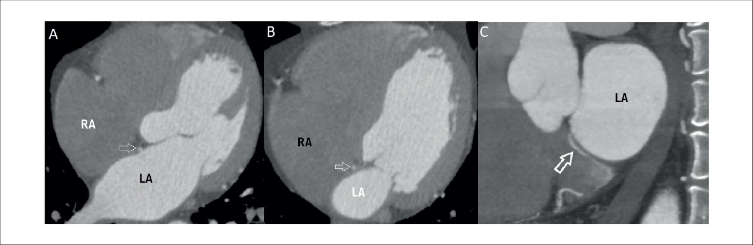
A Bridging Vein Connecting Left Atrium and Coronary Sinus: Coronary Computed Tomography Angiography Findings

Sercan Şahin et al. Nilgün Işıksalan Özbülbül

Introduction

Until recent years, coronary venous system imaging was overshadowed by coronary artery imaging. However, in recent times, the significance of coronary venous system imaging has risen markedly, particularly due to its pivotal role in diverse interventional cardiology procedures. These interventions encompass a spectrum of tasks, including left ventricular pacing, mapping and ablation of arrhythmias, and retrograde cardioplegia. These procedures are facilitated by utilizing the potential of the coronary sinus (CS). As a result, comprehending the anatomy and anomalies of the CS holds paramount importance, and coronary computed tomography (CT) angiography has proven invaluable for achieving this objective.

In this case report, we present an extraordinary cardiac venous anomaly: a bridging vein connecting the left atrium to the CS with non-specific chest pain. We believe that this case serves as an illustrative example for understanding rarely observed cardiac venous vascular anomalies, while also making a valuable contribution to the existing literature through the utilization of clear and direct coronary CT angiography images.

[…]

  • Computed Tomography Angiography
  • Coronary sinus
  • Heart Atria
Read Full Article
Creative Commons Attribution (CC BY) Creative Commons license
12/03/2024 DOI: 10.36660/abcimg.20230111i Views 1
Central Illustration
Review Article

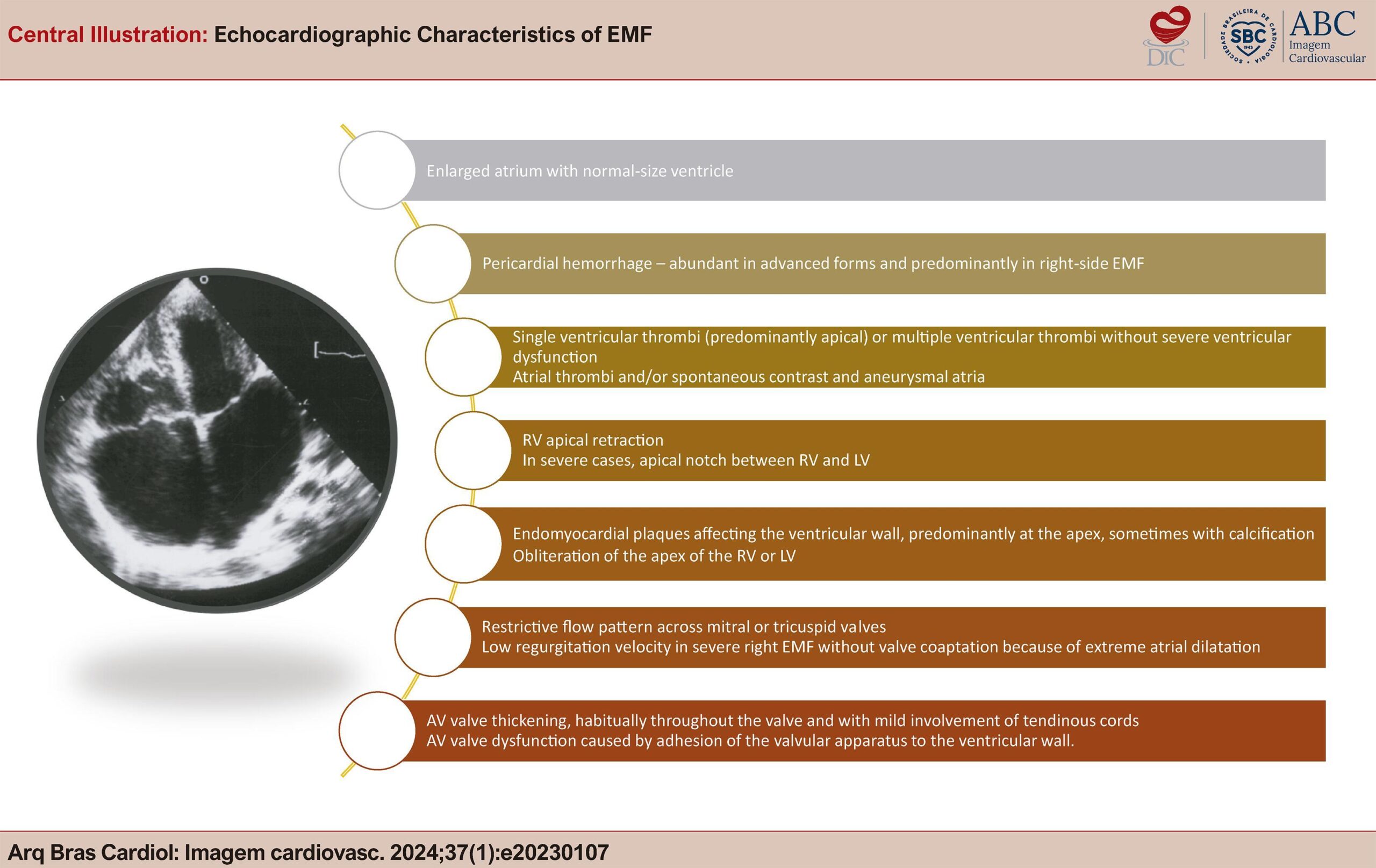
How Do I Diagnose and Classify Endomyocardial Fibrosis in Under-Resourced Settings

Ana Mocumbi

Abstract

Endomyocardial fibrosis (EMF) is a disease of unclear etiology and pathogenesis that is characterized by fibrosis in the ventricular cavities and mainly affects children and adolescents from tropical regions of Africa, South America, and Asia. It is classified as a restrictive cardiomyopathy, with pathophysiology of restriction to diastolic filling associated with valve abnormalities, both caused by endocardial fibrosis. Typical echocardiographic features of this condition are endocardial thickening, severe atrioventricular (AV) valve regurgitation, aneurysmal atria, and heart distortion.

In endemic areas of Africa, in the absence of advanced imaging techniques, EMF diagnosis and management relies on transthoracic echocardiography for careful assessment of structural and hemodynamic abnormalities, aiming at planning medical or surgical management. This review highlights the key echocardiographic aspects of diagnosis and classification of EMF, as used in under-resourced settings in Africa. This is a standardized approach to screening populations with known occurrence of the disease and for informing medical and surgical management.

  • Diagnosis
  • Echocardiography
  • Endomyocardial Fibrosis
Read Full Article
Creative Commons Attribution (CC BY) Creative Commons license
28/02/2024 DOI: 10.36660/abcimg.20230107i Views 2
Review Article

My Approach to Patent Foramen Ovale Closure

Fernando Melo Netto et al. Maria Estefânia Bosco Otto

Abstract

The patent foramen ovale (PFO), a relatively common and generally benign finding, has been associated with paradoxical embolisms. The closure of PFO, performed in patients with cryptogenic stroke, has been shown to reduce the recurrence rates of ischemic events, as evidenced by recent clinical trials. In this article, the authors synthesize the crucial role played by echocardiography in this context, from diagnosis and risk stratification to planning and monitoring of percutaneous intervention.

  • Brain Ischemia
  • Foramen Ovale
Read Full Article
Creative Commons Attribution (CC BY) Creative Commons license
28/02/2024 DOI: 10.36660/abcimg.20230103i
Central Illustration
Review Article

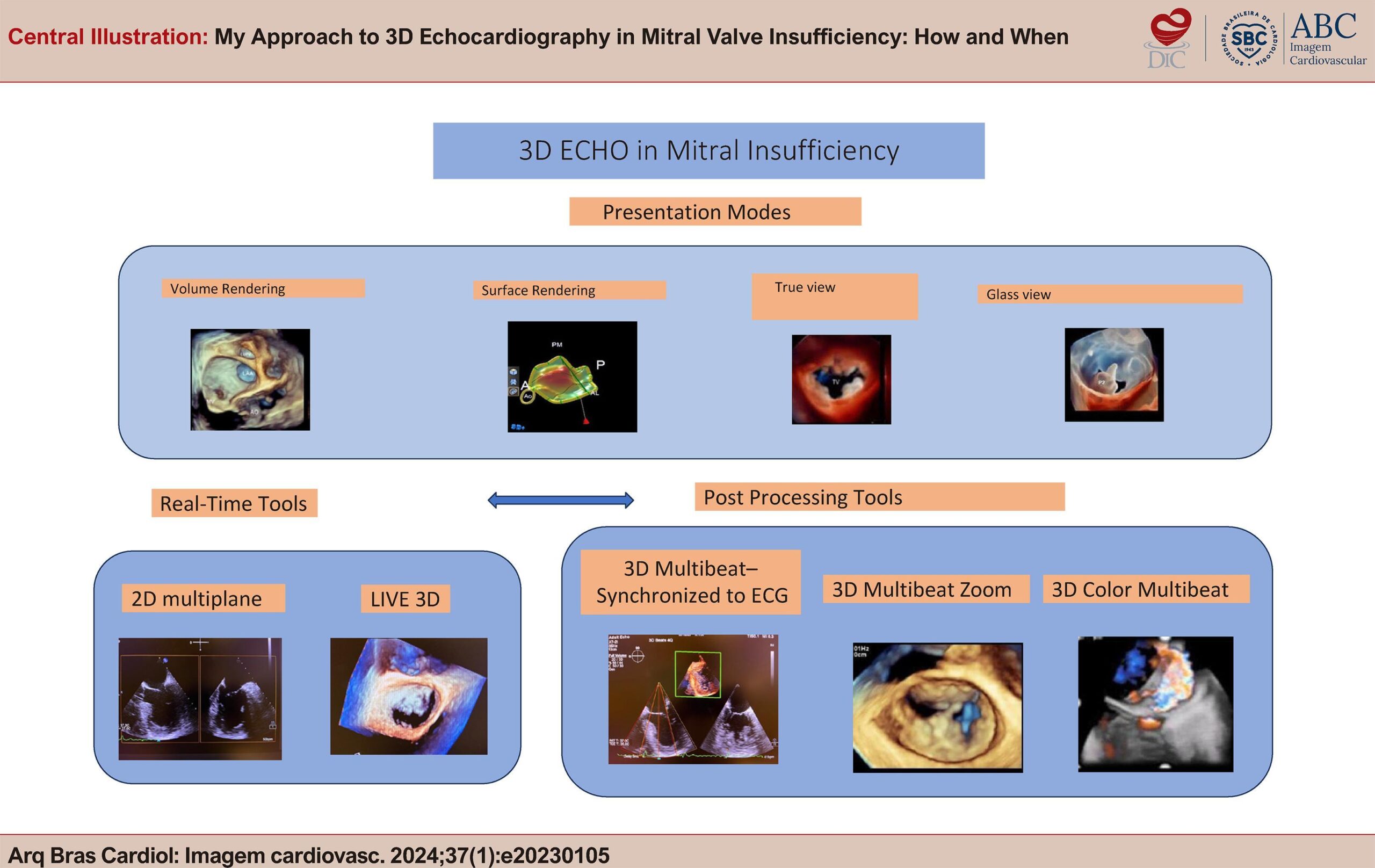
My Approach to 3D Echocardiography in Mitral Valve Insufficiency: How and When

Fábio Cañellas

Abstract

3D Echocardiogram is an indispensable tool in all stages of mitral valve disease, from its diagnosis to the moment of treatment, playing a fundamental role mainly in the decision for surgical plasty or percutaneous correction. In this article, we sought to present very important basic concepts in the use of the technique, from obtaining a 2D image, through the main tools for obtaining 3D images, to new solutions brought by manufacturers for presenting images and for post-processing software. This knowledge is fundamental and contributes to the demystification of 3D Echo, often labeled as difficult to perform and still little incorporated into our clinical practice.

  • Mitral Valve Insufficiency Diagnosis
  • Three-Dimensional Echocardiography
Read Full Article
Creative Commons Attribution (CC BY) Creative Commons license
28/02/2024 DOI: 10.36660/abcimg.20230105i
Figure 1
Review Article

Atrial Functional Mitral Regurgitation

Alexsander da Silva Pretto

Abstract

Secondary mitral insufficiency (MI) in the setting of ventricular disease has always been a topic at conferences and has achieved extensive pathophysiological knowledge over the years. More recently, with the increasing incidence of atrial fibrillation (AF) and heart failure with preserved ejection fraction (HFpEF), a new phenotype that had been little discussed has come to light: atrial functional mitral regurgitation. In this entity, special attention should be addressed to the left atrium and the mitral annulus because, in early stages, they present normal left ventricular dimensions and function.

  • Atrial Fibrillation
  • Echocardiography
  • Heart Failure
  • Mitral Valve Insufficiency
Read Full Article
Creative Commons Attribution (CC BY) Creative Commons license
29/01/2024 DOI: 10.36660/abcimg.20230097i Views 1
Figure 1
Case Report

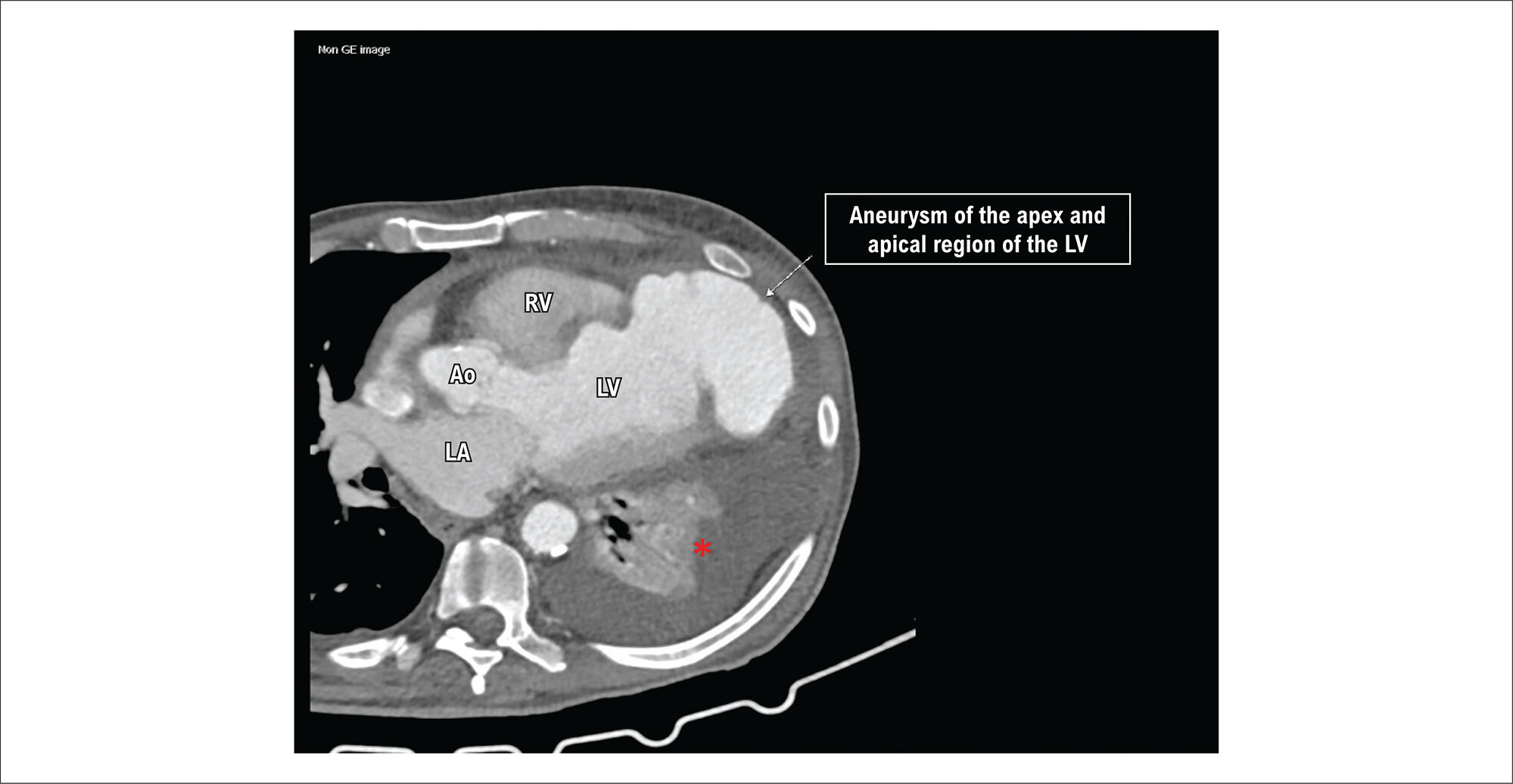
Left Ventricular Aneurysm With Contained Rupture

Priscila Nasser de Carvalho et al. Mariane Higa Shinzato Roberto Tadeu Magro Kroll Andrea Cunha Cortellazzi Aloysio Abdo Silva Campos

Introduction

Left ventricular aneurysms (LVA) and left ventricular pseudoaneurysms (LVPA) are serious complications resulting from transmural myocardial infarction. Their incidence has decreased considerably with the advancements of interventional cardiology, using early revascularization of the compromised coronary artery, preventing ventricular remodeling. Differential diagnosis is often not easy; however, it must be made early.

An LVA occurs months or years after acute myocardial infarction. It is formed by an area of scar tissue and contains the three layers of the ventricular wall (endocardium, myocardium, and pericardium). Its wall is preserved and may contain thrombi. An LVPA is a rare complication that can occur days or months after the infarction. It is formed by the weakened wall that ruptures after the infarction, in a way that this rupture is contained by the pericardium, resulting in discontinuity of the endocardial border. Usually, an LVA is located in the apical and anterior regions, while an LVPA is located in the posterolateral region of the left ventricle.

[…]

  • Heart Aneurysm
  • Myocardial Infarction
  • Thoracic Surgery
Read Full Article
Creative Commons Attribution (CC BY) Creative Commons license
29/01/2024 DOI: 10.36660/abcimg.20230072i Views 1

How to cite this article

EndNote RIS

Electronic Document Format (Vancouver)

Lima EG, Martins EB, Carvalho LNS, Azevedo DFC. What do Cardiologists Expect Towards Myocardial Viability Assessment. ABC Imagem Cardiovascular 2024;37(1):e20240005.

Electronic Document Format (ABNT)

Lima, Eduardo Gomes; Martins, Eduardo Bello; Carvalho, Leticia Neves Solon; Azevedo, Diogo Freitas Cardoso de. What do Cardiologists Expect Towards Myocardial Viability Assessment. ABC Imagem Cardiovascular, v. 37, n. 1, e20240005, Mar. 2024.

Electronic Document Format (APA)

Lima, E. G., Martins, E. B., Carvalho, L. N. S., & Azevedo, D. F. C. (2024). What do Cardiologists Expect Towards Myocardial Viability Assessment. ABC Imagem Cardiovascular, 37(1), e20240005.

Electronic Document Format (ISO)

Lima, Eduardo Gomes and Martins, Eduardo Bello and Carvalho, Leticia Neves Solon and Azevedo, Diogo Freitas Cardoso de. What do Cardiologists Expect Towards Myocardial Viability Assessment. ABC Imagem Cardiovascular [online]. 2024, vol. 37, n. 1, [cited 2026-10-14], e20240005. Available from: <https://journals.cardiol.br/en/article/what-do-cardiologists-expect-towards-myocardial-viability-assessment/>. ISSN 2318-8219

EndNote RIS

Electronic Document Format (Vancouver)

Brito ASX, Moll-Bernardes RJ, Pinheiro MVT, Camargo G, Siqueira FPR, Glavam AP, Almeida SA, et al. Association of Sympathetic Denervation, Myocardial Hypoperfusion, and Fibrosis with Ventricular Arrhythmias in Chronic Chagas Cardiomyopathy. ABC Imagem Cardiovascular 2024;37(1):e20240008.

Electronic Document Format (ABNT)

Brito, Adriana Soares Xavier de; Moll-Bernardes, Renata Junqueira; Pinheiro, Martha Valéria Tavares; Camargo, Gabriel; Siqueira, Fabio Paiva Rossini; Glavam, Adriana Pereira; Almeida, Sergio Altino; Mendes, Fernanda Souza Nogueira Sardinha; Rosado-de-Castro, Paulo Henrique; Sousa, Andrea Silvestre. Association of Sympathetic Denervation, Myocardial Hypoperfusion, and Fibrosis with Ventricular Arrhythmias in Chronic Chagas Cardiomyopathy. ABC Imagem Cardiovascular, v. 37, n. 1, e20240008, Mar. 2024.

Electronic Document Format (APA)

Brito, A. S. X. , Moll-Bernardes, R. J., Pinheiro, M. V. T., Camargo, G., Siqueira, F. P. R., Glavam, A. P., Almeida, S. A., Mendes, F. S. N. S., Rosado-de-Castro, P. H., & Sousa, A. S. (2024). Association of Sympathetic Denervation, Myocardial Hypoperfusion, and Fibrosis with Ventricular Arrhythmias in Chronic Chagas Cardiomyopathy. ABC Imagem Cardiovascular, 37(1), e20240008.

Electronic Document Format (ISO)

Brito, Adriana Soares Xavier de and Moll-Bernardes, Renata Junqueira and Pinheiro, Martha Valéria Tavares and Camargo, Gabriel and Siqueira, Fabio Paiva Rossini and Glavam, Adriana Pereira and Almeida, Sergio Altino and Mendes, Fernanda Souza Nogueira Sardinha and Rosado-de-Castro, Paulo Henrique and Sousa, Andrea Silvestre. Association of Sympathetic Denervation, Myocardial Hypoperfusion, and Fibrosis with Ventricular Arrhythmias in Chronic Chagas Cardiomyopathy. ABC Imagem Cardiovascular [online]. 2024, vol. 37, n. 1, [cited 2026-10-14], e20240008. Available from: <https://journals.cardiol.br/en/article/association-of-sympathetic-denervation-myocardial-hypoperfusion-and-fibrosis-with-ventricular-arrhythmias-in-chronic-chagas-cardiomyopathy/>. ISSN 2318-8219

EndNote RIS

Electronic Document Format (Vancouver)

Figueiredo FA, Costa Filho AL, Figueiredo FA, Chavez LMT, Teixeira MFA, Barbosa WS, Bronzatto PH, et al. Left Atrial Strain: Clinical Applications and Prognostic Implications. ABC Imagem Cardiovascular 2024;37(1):e20240003.

Electronic Document Format (ABNT)

Figueiredo, Fernanda de Azevedo; Costa Filho, Admilson Lemos da; Figueiredo, Flávio de Azevedo; Chavez, Luz Marina Tacuri; Teixeira, Marcia Fabrícia Almeida; Barbosa, William Silva; Bronzatto, Pedro Henrique; Cintra, Priscila Rabelo; Nunes, Maria Carmo Pereira. Left Atrial Strain: Clinical Applications and Prognostic Implications. ABC Imagem Cardiovascular, v. 37, n. 1, e20240003, Mar. 2024.

Electronic Document Format (APA)

Figueiredo, F. A., Costa Filho, A. L. , Figueiredo, F. A., Chavez, L. M. T., Teixeira, M. F. A., Barbosa, W. S., Bronzatto, P. H., Cintra, P. R., & Nunes, M. C. P. (2024). Left Atrial Strain: Clinical Applications and Prognostic Implications. ABC Imagem Cardiovascular, 37(1), e20240003.

Electronic Document Format (ISO)

Figueiredo, Fernanda de Azevedo and Costa Filho, Admilson Lemos da and Figueiredo, Flávio de Azevedo and Chavez, Luz Marina Tacuri and Teixeira, Marcia Fabrícia Almeida and Barbosa, William Silva and Bronzatto, Pedro Henrique and Cintra, Priscila Rabelo and Nunes, Maria Carmo Pereira. Left Atrial Strain: Clinical Applications and Prognostic Implications. ABC Imagem Cardiovascular [online]. 2024, vol. 37, n. 1, [cited 2026-10-14], e20240003. Available from: <https://journals.cardiol.br/en/article/left-atrial-strain-clinical-applications-and-prognostic-implications/>. ISSN 2318-8219

EndNote RIS

Electronic Document Format (Vancouver)

Petisco ACGP, Dourado PMM, Dourado LA, Dourado JPA, Tebaldi RT. Ultrasound in the Diagnosis of Carotid Web: Report of Two Cases and Review. ABC Imagem Cardiovascular 2024;37(1):e20230117.

Electronic Document Format (ABNT)

Petisco, Ana Claudia Gomes Pereira; Dourado, Paulo Magno Martins; Dourado, Larissa Almeida; Dourado, João Paulo Almeida; Tebaldi, Ricardo Thomaz. Ultrasound in the Diagnosis of Carotid Web: Report of Two Cases and Review. ABC Imagem Cardiovascular, v. 37, n. 1, e20230117, Mar. 2024.

Electronic Document Format (APA)

Petisco, A. C. G. P., Dourado, P. M. M., Dourado, L. A., Dourado, J. P. A., & Tebaldi, R. T. (2024). Ultrasound in the Diagnosis of Carotid Web: Report of Two Cases and Review. ABC Imagem Cardiovascular, 37(1), e20230117.

Electronic Document Format (ISO)

Petisco, Ana Claudia Gomes Pereira and Dourado, Paulo Magno Martins and Dourado, Larissa Almeida and Dourado, João Paulo Almeida and Tebaldi, Ricardo Thomaz. Ultrasound in the Diagnosis of Carotid Web: Report of Two Cases and Review. ABC Imagem Cardiovascular [online]. 2024, vol. 37, n. 1, [cited 2026-10-14], e20230117. Available from: <https://journals.cardiol.br/en/article/ultrasound-in-the-diagnosis-of-carotid-web-report-of-two-cases-and-review/>. ISSN 2318-8219

EndNote RIS

Electronic Document Format (Vancouver)

Carreira LCTF, Carreira L, Brito ASX. My Approach to Nuclear Medicine in the Assessment of Microvascular Disease in Women. ABC Imagem Cardiovascular 2024;37(1):e20240015.

Electronic Document Format (ABNT)

Carreira, Lara Cristiane Terra Ferreira; Carreira, Lívia; Brito, Adriana Soares Xavier de. My Approach to Nuclear Medicine in the Assessment of Microvascular Disease in Women. ABC Imagem Cardiovascular, v. 37, n. 1, e20240015, Mar. 2024.

Electronic Document Format (APA)

Carreira, L. C. T. F., Carreira, L., & Brito, A. S. X. (2024). My Approach to Nuclear Medicine in the Assessment of Microvascular Disease in Women. ABC Imagem Cardiovascular, 37(1), e20240015.

Electronic Document Format (ISO)

Carreira, Lara Cristiane Terra Ferreira and Carreira, Lívia and Brito, Adriana Soares Xavier de. My Approach to Nuclear Medicine in the Assessment of Microvascular Disease in Women. ABC Imagem Cardiovascular [online]. 2024, vol. 37, n. 1, [cited 2026-10-14], e20240015. Available from: <https://journals.cardiol.br/en/article/my-approach-to-nuclear-medicine-in-the-assessment-of-microvascular-disease-in-women/>. ISSN 2318-8219

EndNote RIS

Electronic Document Format (Vancouver)

Cunha SR, Pandolfo F, Barros FC, Pianca EG, Barcellos PT. Ischemic Stroke in a Patient with Challenging Pseudoaneurysm in the Left Ventricular Outflow Tract. ABC Imagem Cardiovascular 2024;37(1):e20240001.

Electronic Document Format (ABNT)

Cunha, Saulo Rodrigo; Pandolfo, Fernanda; Barros, Fernando Colares; Pianca, Eduardo Gatti; Barcellos, Pedro Tregnago. Ischemic Stroke in a Patient with Challenging Pseudoaneurysm in the Left Ventricular Outflow Tract. ABC Imagem Cardiovascular, v. 37, n. 1, e20240001, Mar. 2024.

Electronic Document Format (APA)

Cunha, S. R., Pandolfo, F., Barros, F. C., Pianca, E. G., & Barcellos, P. T. (2024). Ischemic Stroke in a Patient with Challenging Pseudoaneurysm in the Left Ventricular Outflow Tract. ABC Imagem Cardiovascular, 37(1), e20240001.

Electronic Document Format (ISO)

Cunha, Saulo Rodrigo and Pandolfo, Fernanda and Barros, Fernando Colares and Pianca, Eduardo Gatti and Barcellos, Pedro Tregnago. Ischemic Stroke in a Patient with Challenging Pseudoaneurysm in the Left Ventricular Outflow Tract. ABC Imagem Cardiovascular [online]. 2024, vol. 37, n. 1, [cited 2026-10-14], e20240001. Available from: <https://journals.cardiol.br/en/article/ischemic-stroke-in-a-patient-with-challenging-pseudoaneurysm-in-the-left-ventricular-outflow-tract/>. ISSN 2318-8219

EndNote RIS

Electronic Document Format (Vancouver)

Brito ASX, Leite JC, Brandão SCS. My Approach to Assess Cardiac Sympathetic Activity. ABC Imagem Cardiovascular 2024;37(1):e20240009.

Electronic Document Format (ABNT)

Brito, Adriana Soares Xavier de; Leite, Jessica Costa; Brandão, Simone Cristina Soares. My Approach to Assess Cardiac Sympathetic Activity. ABC Imagem Cardiovascular, v. 37, n. 1, e20240009, Mar. 2024.

Electronic Document Format (APA)

Brito, A. S. X. , Leite, J. C., & Brandão, S. C. S. (2024). My Approach to Assess Cardiac Sympathetic Activity. ABC Imagem Cardiovascular, 37(1), e20240009.

Electronic Document Format (ISO)

Brito, Adriana Soares Xavier de and Leite, Jessica Costa and Brandão, Simone Cristina Soares. My Approach to Assess Cardiac Sympathetic Activity. ABC Imagem Cardiovascular [online]. 2024, vol. 37, n. 1, [cited 2026-10-14], e20240009. Available from: <https://journals.cardiol.br/en/article/my-approach-to-assess-cardiac-sympathetic-activity/>. ISSN 2318-8219

EndNote RIS

Electronic Document Format (Vancouver)

Moura DMD, Monteiro Neto AJDO, Melo MDT, Fernandes F. Cardiac Sarcoidosis: The Chameleon of Cardiology. ABC Imagem Cardiovascular 2024;37(1):e20240007.

Electronic Document Format (ABNT)

Moura, Diego Moraes De; Monteiro Neto, Aluísio José De Oliveira; Melo, Marcelo Dantas Tavares de; Fernandes, Fábio. Cardiac Sarcoidosis: The Chameleon of Cardiology. ABC Imagem Cardiovascular, v. 37, n. 1, e20240007, Mar. 2024.

Electronic Document Format (APA)

Moura, D. M. D., Monteiro Neto, A. J. D. O., Melo, M. D. T. , & Fernandes, F. (2024). Cardiac Sarcoidosis: The Chameleon of Cardiology. ABC Imagem Cardiovascular, 37(1), e20240007.

Electronic Document Format (ISO)

Moura, Diego Moraes De and Monteiro Neto, Aluísio José De Oliveira and Melo, Marcelo Dantas Tavares de and Fernandes, Fábio. Cardiac Sarcoidosis: The Chameleon of Cardiology. ABC Imagem Cardiovascular [online]. 2024, vol. 37, n. 1, [cited 2026-10-14], e20240007. Available from: <https://journals.cardiol.br/en/article/cardiac-sarcoidosis-the-chameleon-of-cardiology/>. ISSN 2318-8219

EndNote RIS

Electronic Document Format (Vancouver)

Carvalho WA, Carvalho AA, Martins ER, Medeiros Neto AH, Bacal F, Melo MDT. Echocardiographic Evaluation of Right Atrial Function in Patients with Precapillary Pulmonary Hypertension. ABC Imagem Cardiovascular 2024;37(1):e20240002.

Electronic Document Format (ABNT)

Carvalho, Wanessa Alves de; Carvalho, Andressa Alves de; Martins, Eliauria Rosa; Medeiros Neto, Agostinho Hermes de; Bacal, Fernando; Melo, Marcelo Dantas Tavares de. Echocardiographic Evaluation of Right Atrial Function in Patients with Precapillary Pulmonary Hypertension. ABC Imagem Cardiovascular, v. 37, n. 1, e20240002, Mar. 2024.

Electronic Document Format (APA)

Carvalho, W. A. , Carvalho, A. A. , Martins, E. R., Medeiros Neto, A. H. , Bacal, F., & Melo, M. D. T. (2024). Echocardiographic Evaluation of Right Atrial Function in Patients with Precapillary Pulmonary Hypertension. ABC Imagem Cardiovascular, 37(1), e20240002.

Electronic Document Format (ISO)

Carvalho, Wanessa Alves de and Carvalho, Andressa Alves de and Martins, Eliauria Rosa and Medeiros Neto, Agostinho Hermes de and Bacal, Fernando and Melo, Marcelo Dantas Tavares de. Echocardiographic Evaluation of Right Atrial Function in Patients with Precapillary Pulmonary Hypertension. ABC Imagem Cardiovascular [online]. 2024, vol. 37, n. 1, [cited 2026-10-14], e20240002. Available from: <https://journals.cardiol.br/en/article/echocardiographic-evaluation-of-right-atrial-function-in-patients-with-precapillary-pulmonary-hypertension/>. ISSN 2318-8219

EndNote RIS

Electronic Document Format (Vancouver)

Bispo IGA, Zago IM. The Indiscriminate Use Of Androgenic Anabolic Steroids: The Contribution of Cardiovascular Imaging. ABC Imagem Cardiovascular 2024;37(1):e20230113.

Electronic Document Format (ABNT)

Bispo, Irving Gabriel Araöşjo; Zago, Isabella Montanher. The Indiscriminate Use Of Androgenic Anabolic Steroids: The Contribution of Cardiovascular Imaging. ABC Imagem Cardiovascular, v. 37, n. 1, e20230113, Mar. 2024.

Electronic Document Format (APA)

Bispo, I. G. A., & Zago, I. M. (2024). The Indiscriminate Use Of Androgenic Anabolic Steroids: The Contribution of Cardiovascular Imaging. ABC Imagem Cardiovascular, 37(1), e20230113.

Electronic Document Format (ISO)

Bispo, Irving Gabriel Araöşjo and Zago, Isabella Montanher. The Indiscriminate Use Of Androgenic Anabolic Steroids: The Contribution of Cardiovascular Imaging. ABC Imagem Cardiovascular [online]. 2024, vol. 37, n. 1, [cited 2026-10-14], e20230113. Available from: <https://journals.cardiol.br/en/article/the-indiscriminate-use-of-androgenic-anabolic-steroids-the-contribution-of-cardiovascular-imaging/>. ISSN 2318-8219

EndNote RIS

Electronic Document Format (Vancouver)

Ömür SE, Tapar GG, Yilmaz M, Bektaş AE, Demir O. Relationship Between Echocardiographic Parameters and The Systemic Immune-Inflammation Index in Patients With Pulmonary Arterial Hypertension. ABC Imagem Cardiovascular 2024;37(1):e20230086.

Electronic Document Format (ABNT)

Ömür, Sefa Erdi; Tapar, Gulsen Genc; Yilmaz, Mustafa; Bektaş, Abdullah Emre; Demir, Osman. Relationship Between Echocardiographic Parameters and The Systemic Immune-Inflammation Index in Patients With Pulmonary Arterial Hypertension. ABC Imagem Cardiovascular, v. 37, n. 1, e20230086, Mar. 2024.

Electronic Document Format (APA)

Ömür, S. E., Tapar, G. G., Yilmaz, M., Bektaş, A. E., & Demir, O. (2024). Relationship Between Echocardiographic Parameters and The Systemic Immune-Inflammation Index in Patients With Pulmonary Arterial Hypertension. ABC Imagem Cardiovascular, 37(1), e20230086.

Electronic Document Format (ISO)

Ömür, Sefa Erdi and Tapar, Gulsen Genc and Yilmaz, Mustafa and Bektaş, Abdullah Emre and Demir, Osman. Relationship Between Echocardiographic Parameters and The Systemic Immune-Inflammation Index in Patients With Pulmonary Arterial Hypertension. ABC Imagem Cardiovascular [online]. 2024, vol. 37, n. 1, [cited 2026-10-14], e20230086. Available from: <https://journals.cardiol.br/en/article/relationship-between-echocardiographic-parameters-and-the-systemic-immune-inflammation-index-in-patients-with-pulmonary-arterial-hypertension/>. ISSN 2318-8219

EndNote RIS

Electronic Document Format (Vancouver)

Rompkoski J, Accorsi TAD, Amaral BDR, Freitas CB, Moreira FT, Kohler KF, Lima KDA, et al. Correlation of Telemedicine-guided POCUS Echocardiography and In Situ Data: Pilot Study in a Remote Area in Brazil. ABC Imagem Cardiovascular 2024;37(1):e20230110.

Electronic Document Format (ABNT)

Rompkoski, Juliane; Accorsi, Tarso Augusto Duenhas; Amaral, Bruna Dayanne Reges; Freitas, Christian Barbosa de; Moreira, Flavio Tocci; Kohler, Karen Francine; Lima, Karine De Amicis; Morbeck, Renata Albaladejo; Pedrotti, Carlos Henrique Sartorato. Correlation of Telemedicine-guided POCUS Echocardiography and In Situ Data: Pilot Study in a Remote Area in Brazil. ABC Imagem Cardiovascular, v. 37, n. 1, e20230110, Mar. 2024.

Electronic Document Format (APA)

Rompkoski, J., Accorsi, T. A. D., Amaral, B. D. R., Freitas, C. B. , Moreira, F. T., Kohler, K. F., Lima, K. D. A., Morbeck, R. A., & Pedrotti, C. H. S. (2024). Correlation of Telemedicine-guided POCUS Echocardiography and In Situ Data: Pilot Study in a Remote Area in Brazil. ABC Imagem Cardiovascular, 37(1), e20230110.

Electronic Document Format (ISO)

Rompkoski, Juliane and Accorsi, Tarso Augusto Duenhas and Amaral, Bruna Dayanne Reges and Freitas, Christian Barbosa de and Moreira, Flavio Tocci and Kohler, Karen Francine and Lima, Karine De Amicis and Morbeck, Renata Albaladejo and Pedrotti, Carlos Henrique Sartorato. Correlation of Telemedicine-guided POCUS Echocardiography and In Situ Data: Pilot Study in a Remote Area in Brazil. ABC Imagem Cardiovascular [online]. 2024, vol. 37, n. 1, [cited 2026-10-14], e20230110. Available from: <https://journals.cardiol.br/en/article/correlation-of-telemedicine-guided-pocus-echocardiography-and-in-situ-data-pilot-study-in-a-remote-area-in-brazil/>. ISSN 2318-8219

EndNote RIS

Electronic Document Format (Vancouver)

Stürmer JD, Silva RS, Bica WC, Vigil THB, Soder G, Moraes RP, Brida MS, et al. Rare Presentation of Pseudoaneurysm Obstructing the Left Ventricular Inflow Tract in a Patient With Acute Myocardial Infarction. ABC Imagem Cardiovascular 2024;37(1):e20230106.

Electronic Document Format (ABNT)

Stürmer, Janine Daiana; Silva, Raphael dos Santos; Bica, Willer Cesar; Vigil, Tiago Hansel Basile; Soder, Gabriel; Moraes, Renata Pibernat de; Brida, Mathias Silvestre de; Reis, Rodrigo Moraes. Rare Presentation of Pseudoaneurysm Obstructing the Left Ventricular Inflow Tract in a Patient With Acute Myocardial Infarction. ABC Imagem Cardiovascular, v. 37, n. 1, e20230106, Mar. 2024.

Electronic Document Format (APA)

Stürmer, J. D., Silva, R. S., Bica, W. C., Vigil, T. H. B., Soder, G., Moraes, R. P. , Brida, M. S. , & Reis, R. M. (2024). Rare Presentation of Pseudoaneurysm Obstructing the Left Ventricular Inflow Tract in a Patient With Acute Myocardial Infarction. ABC Imagem Cardiovascular, 37(1), e20230106.

Electronic Document Format (ISO)

Stürmer, Janine Daiana and Silva, Raphael dos Santos and Bica, Willer Cesar and Vigil, Tiago Hansel Basile and Soder, Gabriel and Moraes, Renata Pibernat de and Brida, Mathias Silvestre de and Reis, Rodrigo Moraes. Rare Presentation of Pseudoaneurysm Obstructing the Left Ventricular Inflow Tract in a Patient With Acute Myocardial Infarction. ABC Imagem Cardiovascular [online]. 2024, vol. 37, n. 1, [cited 2026-10-14], e20230106. Available from: <https://journals.cardiol.br/en/article/rare-presentation-of-pseudoaneurysm-obstructing-the-left-ventricular-inflow-tract-in-a-patient-with-acute-myocardial-infarction/>. ISSN 2318-8219

EndNote RIS

Electronic Document Format (Vancouver)

Şahin S, Özbülbül NI. A Bridging Vein Connecting Left Atrium and Coronary Sinus: Coronary Computed Tomography Angiography Findings. ABC Imagem Cardiovascular 2024;37(1):e20230111.

Electronic Document Format (ABNT)

Şahin, Sercan; Özbülbül, Nilgün Işıksalan. A Bridging Vein Connecting Left Atrium and Coronary Sinus: Coronary Computed Tomography Angiography Findings. ABC Imagem Cardiovascular, v. 37, n. 1, e20230111, Mar. 2024.

Electronic Document Format (APA)

Şahin, S., & Özbülbül, N. I. (2024). A Bridging Vein Connecting Left Atrium and Coronary Sinus: Coronary Computed Tomography Angiography Findings. ABC Imagem Cardiovascular, 37(1), e20230111.

Electronic Document Format (ISO)

Şahin, Sercan and Özbülbül, Nilgün Işıksalan. A Bridging Vein Connecting Left Atrium and Coronary Sinus: Coronary Computed Tomography Angiography Findings. ABC Imagem Cardiovascular [online]. 2024, vol. 37, n. 1, [cited 2026-10-14], e20230111. Available from: <https://journals.cardiol.br/en/article/a-bridging-vein-connecting-left-atrium-and-coronary-sinus-coronary-computed-tomography-angiography-findings/>. ISSN 2318-8219

EndNote RIS

Electronic Document Format (Vancouver)

Mocumbi A. How Do I Diagnose and Classify Endomyocardial Fibrosis in Under-Resourced Settings. ABC Imagem Cardiovascular 2024;37(1):e20230107.

Electronic Document Format (ABNT)

Mocumbi, Ana. How Do I Diagnose and Classify Endomyocardial Fibrosis in Under-Resourced Settings. ABC Imagem Cardiovascular, v. 37, n. 1, e20230107, Feb. 2024.

Electronic Document Format (APA)

Mocumbi, A. (2024). How Do I Diagnose and Classify Endomyocardial Fibrosis in Under-Resourced Settings. ABC Imagem Cardiovascular, 37(1), e20230107.

Electronic Document Format (ISO)

Mocumbi, Ana. How Do I Diagnose and Classify Endomyocardial Fibrosis in Under-Resourced Settings. ABC Imagem Cardiovascular [online]. 2024, vol. 37, n. 1, [cited 2026-10-14], e20230107. Available from: <https://journals.cardiol.br/en/article/how-do-i-diagnose-and-classify-endomyocardial-fibrosis-in-under-resourced-settings/>. ISSN 2318-8219

EndNote RIS

Electronic Document Format (Vancouver)

Netto FM, Otto MEB. My Approach to Patent Foramen Ovale Closure. ABC Imagem Cardiovascular 2024;37(1):e20230103.

Electronic Document Format (ABNT)

Netto, Fernando Melo; Otto, Maria Estefânia Bosco. My Approach to Patent Foramen Ovale Closure. ABC Imagem Cardiovascular, v. 37, n. 1, e20230103, Feb. 2024.

Electronic Document Format (APA)

Netto, F. M., & Otto, M. E. B. (2024). My Approach to Patent Foramen Ovale Closure. ABC Imagem Cardiovascular, 37(1), e20230103.

Electronic Document Format (ISO)

Netto, Fernando Melo and Otto, Maria Estefânia Bosco. My Approach to Patent Foramen Ovale Closure. ABC Imagem Cardiovascular [online]. 2024, vol. 37, n. 1, [cited 2026-10-14], e20230103. Available from: <https://journals.cardiol.br/en/article/my-approach-to-patent-foramen-ovale-closure/>. ISSN 2318-8219

EndNote RIS

Electronic Document Format (Vancouver)

Cañellas F. My Approach to 3D Echocardiography in Mitral Valve Insufficiency: How and When. ABC Imagem Cardiovascular 2024;37(1):e20230105.

Electronic Document Format (ABNT)

Cañellas, Fábio. My Approach to 3D Echocardiography in Mitral Valve Insufficiency: How and When. ABC Imagem Cardiovascular, v. 37, n. 1, e20230105, Feb. 2024.

Electronic Document Format (APA)

Cañellas, F. (2024). My Approach to 3D Echocardiography in Mitral Valve Insufficiency: How and When. ABC Imagem Cardiovascular, 37(1), e20230105.

Electronic Document Format (ISO)

Cañellas, Fábio. My Approach to 3D Echocardiography in Mitral Valve Insufficiency: How and When. ABC Imagem Cardiovascular [online]. 2024, vol. 37, n. 1, [cited 2026-10-14], e20230105. Available from: <https://journals.cardiol.br/en/article/my-approach-to-3d-echocardiography-in-mitral-valve-insufficiency-how-and-when/>. ISSN 2318-8219

EndNote RIS

Electronic Document Format (Vancouver)

Pretto AS. Atrial Functional Mitral Regurgitation. ABC Imagem Cardiovascular 2024;37(1):e20230097.

Electronic Document Format (ABNT)

Pretto, Alexsander da Silva. Atrial Functional Mitral Regurgitation. ABC Imagem Cardiovascular, v. 37, n. 1, e20230097, Jan. 2024.

Electronic Document Format (APA)

Pretto, A. S. (2024). Atrial Functional Mitral Regurgitation. ABC Imagem Cardiovascular, 37(1), e20230097.

Electronic Document Format (ISO)

Pretto, Alexsander da Silva. Atrial Functional Mitral Regurgitation. ABC Imagem Cardiovascular [online]. 2024, vol. 37, n. 1, [cited 2026-10-14], e20230097. Available from: <https://journals.cardiol.br/en/article/atrial-functional-mitral-regurgitation/>. ISSN 2318-8219

EndNote RIS

Electronic Document Format (Vancouver)

Carvalho PN, Shinzato MH, Kroll RTM, Cortellazzi AC, Campos AAS. Left Ventricular Aneurysm With Contained Rupture. ABC Imagem Cardiovascular 2024;37(1):e20230072.

Electronic Document Format (ABNT)

Carvalho, Priscila Nasser de; Shinzato, Mariane Higa; Kroll, Roberto Tadeu Magro; Cortellazzi, Andrea Cunha; Campos, Aloysio Abdo Silva. Left Ventricular Aneurysm With Contained Rupture. ABC Imagem Cardiovascular, v. 37, n. 1, e20230072, Jan. 2024.

Electronic Document Format (APA)

Carvalho, P. N. , Shinzato, M. H., Kroll, R. T. M., Cortellazzi, A. C., & Campos, A. A. S. (2024). Left Ventricular Aneurysm With Contained Rupture. ABC Imagem Cardiovascular, 37(1), e20230072.

Electronic Document Format (ISO)

Carvalho, Priscila Nasser de and Shinzato, Mariane Higa and Kroll, Roberto Tadeu Magro and Cortellazzi, Andrea Cunha and Campos, Aloysio Abdo Silva. Left Ventricular Aneurysm With Contained Rupture. ABC Imagem Cardiovascular [online]. 2024, vol. 37, n. 1, [cited 2026-10-14], e20230072. Available from: <https://journals.cardiol.br/en/article/left-ventricular-aneurysm-with-contained-rupture/>. ISSN 2318-8219

Sede – São Paulo
Rua Barata Ribeiro, 380 cj 54 Bela Vista
São Paulo, Brasil – CEP: 01308-000
Tel.: +55 (11) 3120-3363
Telefax: +55 (11) 3259-2988
E-mail: depeco@cardiol.br

Sede – Rio de Janeiro
Av. Marechal Câmara, 160
3º andar – Sala: 330 – Centro
Rio de Janeiro – CEP: 20020-907
Tel.: +55 (21) 3478-2700
ou: +55 (21) 99386-3961
E-mail: abcimaging@cardiol.br

  • About the Journal
  • Internal Regulation
  • Editors Section
  • Editorial Board
  • SBC Award

  • Guidelines for Publication
  • Editorial Policy
  • Ethics Policy
  • Policy on the Use of Artificial Intelligence
  • Mandatory Publication Form

Issues

SBC Guidelines