Matheus Rodrigues Barbosa et al. Henrique Turin Moreira Gustavo Jardim Volpe Danilo Tadao Wada … Minna Moreira Dias Romano Andre Schmidt
Discussion
Pericardial cyst is an uncommon clinical entity, with an estimated incidence of 1/100,000 to 1/10,000 autopsies. Its etiology is not yet completely understood, but it is believed to be due to an abnormal development of the pericardium during embryogenesis. It is necessary to differentiate the cyst from the pericardial diverticulum, which possibly have the same origin but, in the latter, there is communication with the pericardial cavity. Most cases are asymptomatic, even with large dimensions, and the lesion is often discovered incidentally during imaging exams or heart surgery. When symptomatic, the clinical presentation is variable and may include cardiorespiratory symptoms, such as chest pain, dyspnea, cough, sudden cardiac death or signs of cardiac tamponade.
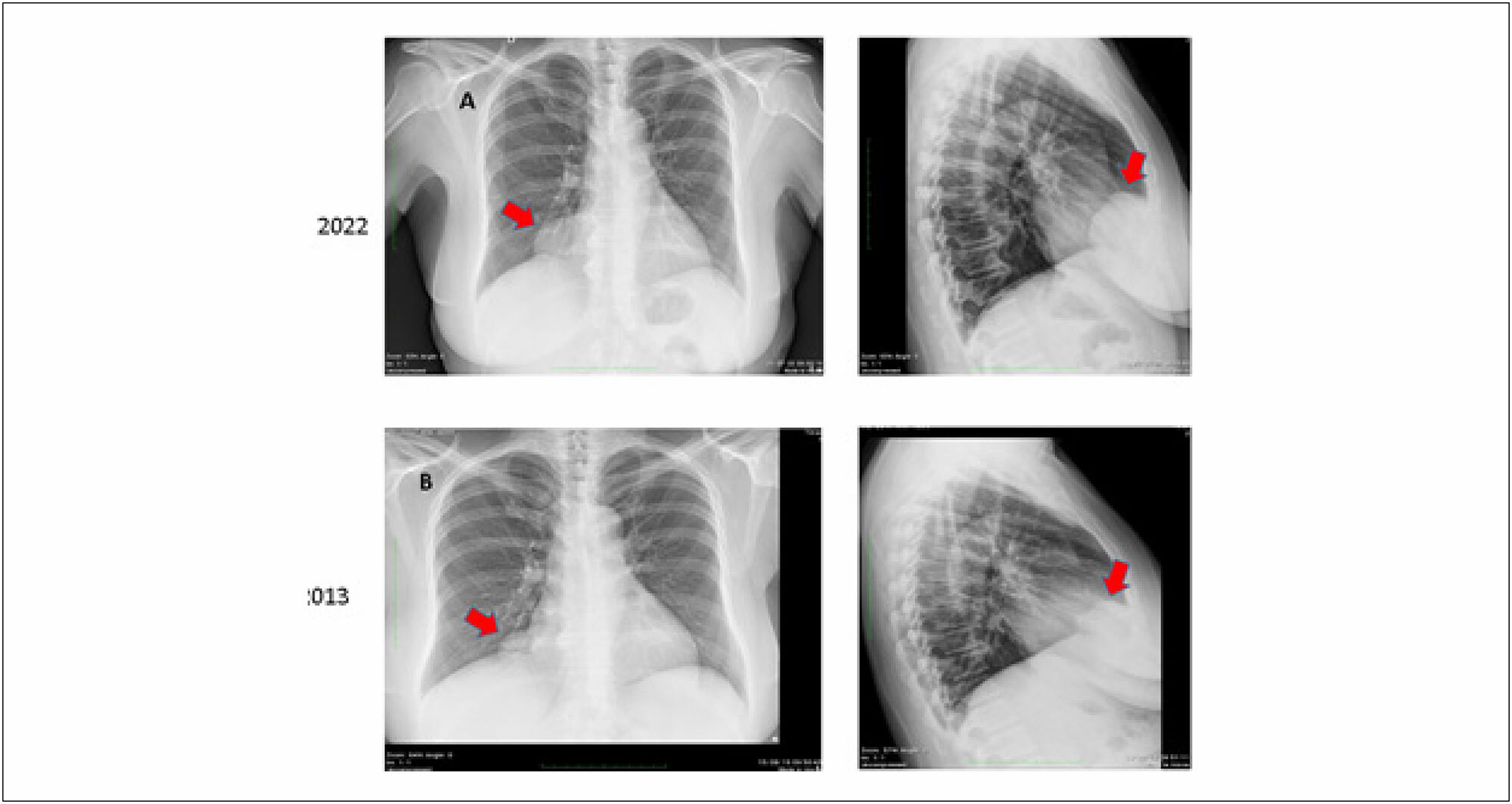
The diagnosis of pericardial cyst is initially suspected by the incidental performance of a chest x-ray, which shows the mass attached to the cardiac silhouette. Doppler echocardiography, computed tomography and cardiac MRI allow the diagnosis to be confirmed, notably the last two. Chest x-ray may reveal a mediastinal mass or an area of increased density. Computed tomography (CT) scans and cardiac MRIs provide more detailed information about the location, size, and characteristics of the cyst. Transthoracic echocardiography can be useful in evaluating cardiac function and the presence of cyst-related complications, such as cardiac compression. Differential diagnosis should include neoplasms and, more rarely, migratory hydatid cysts, which may be single.
[…]