ABC Family Arquivos Brasileiros de Cardiologia International Journal of Cardiovascular Sciences ABC Heart Failure & Cardiomyopathy ABC Imagem Cardiovascular
Português English
ABC Family
Português English
  • About
    • About ABC Family
    • Internal Regulation
    • Editorial Board
    • Brazilian Society of Cardiology (SBC)
  • Issues
  • SBC Guidelines
  • Content
    • Videos
    • Podcasts
    • News
  • About
    • About ABC Family
    • Internal Regulation
    • Editorial Board
    • Brazilian Society of Cardiology (SBC)
  • Issues
  • SBC Guidelines
  • Content
    • Videos
    • Podcasts
    • News

ABC Family

  • ABC Family
  • Arquivos Brasileiros de Cardiologia
  • International Journal of Cardiovascular Sciences
  • ABC Heart Failure & Cardiomyopathy
  • ABC Imagem Cardiovascular
Search in

No content found.

Journals
Ordering
Article types
Others

  1. Home
  2. Keyword: Cardiac Ouput
ABC
Editorial

Continuous Non-Invasive Cardiac Output: Myth or Reality

João Manoel Rossi Neto

Cardiac output (CO) is an important cardiovascular system function parameter. Changes in cardiac function are commonly observed as a response to physical training and pharmacological interventions. Unfortunately, the methods for assessing CO are invasive, leading to well-known complications and considered inconvenient in daily practice. For this reason, the search for new noninvasive methods that can accurately detect CO at rest, at physical exertion or as a response to a clinical intervention has become desirable in academic and non-academic circles. The ideal method for measuring CO at rest and during exercise should be noninvasive, safe, reproducible and inexpensive.

The Cardiopulmonary Exercise Testing (CPT) is recommended in the evaluation of cardiorespiratory fitness and exercise tolerance in athletes, the general population and in patients. Briefly, CO and systolic volume can be estimated during CPT through measured VO2. In 2001, Williams et al. were the first ones to integrate CPT with non-invasive measures of CO using rebreathing (RB) of carbon dioxide, but the technique was quickly abandoned due to its difficulty and inaccuracy. Another non-invasive method is thoracic electrical bioimpedance (TEB), first described in 1966 by Kubicek et al., which measures thoracic resistance as a result of changes in blood velocity during the cardiac cycle and uses an algorithm to calculate the CO.

[…]

  • Breathing Exercise
  • Cardiac Ouput
  • Electric
  • Impedance
  • Interventions
  • Pharmacological
Read Full Article
Creative Commons Attribution (CC BY) Creative Commons license
02/09/2019 DOI: 10.5935/abc.20190163
Transthoracic impedance compared to magnetic resonance imaging in the assessment of cardiac output
ABC
Artigo Original

Transthoracic impedance compared to magnetic resonance imaging in the assessment of cardiac output

Humberto Villacorta Junior et al. Aline Sterque Villacorta Fernanda Amador Marcelo Hadlich … Denilson Campos de Albuquerque Clerio Francisco Azevedo

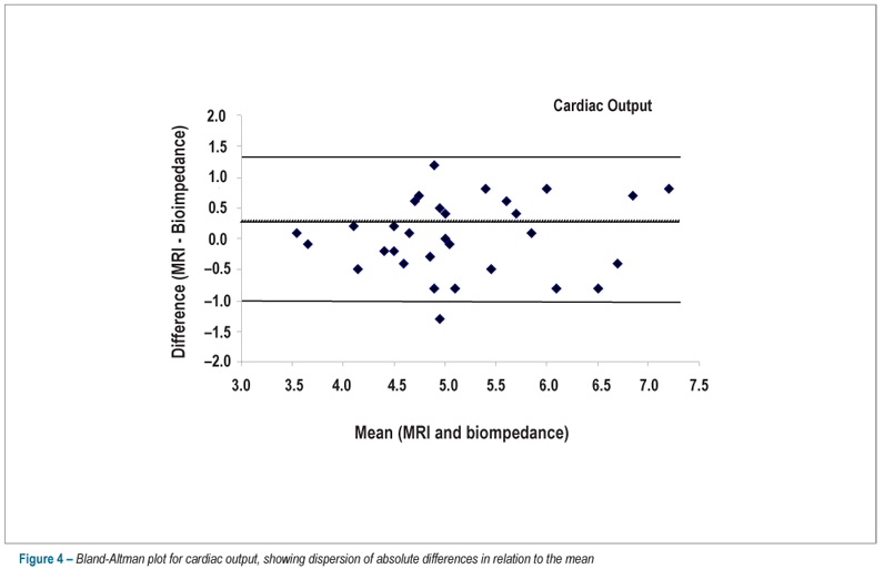
BACKGROUND: Cardiac magnetic resonance imaging is considered the gold-standard method for the calculation of cardiac volumes. Transthoracic impedance cardiography assesses the cardiac output. No studies validating this measurement, in comparison to that obtained by magnetic resonance imaging, are available. OBJECTIVE: To evaluate the performance of transthoracic impedance cardiography in the calculation of the cardiac output, cardiac index and stroke volume using magnetic resonance imaging as the gold-standard. METHODS: 31 patients with a mean age of 56.7 ± 18 years were assessed; of these, 18 (58%) were males. Patients whose indication for magnetic resonance imaging required pharmacologic stress test were excluded. Correlation between methods was assessed using the Pearson’s coefficient, and dispersion of absolute differences in relation to the mean was demonstrated using the Bland-Altman’s method. Agreement between methods was analyzed using the intraclass correlation coefficient. RESULTS: The mean cardiac output by transthoracic impedance cardiography and by magnetic resonance imaging was 5.16 ± 0.9 and 5.13 ± 0.9 L/min, respectively. Good agreement between methods was observed for cardiac output (r = 0.79; p = 0.0001), cardiac index (r = 0.74; p = 0.0001) and stroke volume (r = 0.88; p = 0.0001). The analysis by the Bland-Altman plot showed low dispersion of differences in relation to the mean, with a low amplitude of agreement intervals. Good agreement between the two methods was observed when analyzed by the intraclass correlation coefficient, with coefficients for cardiac output, cardiac index and stroke volume of 0.78, 0.73 and 0.88, respectively (p < 0.0001 for all comparisons). CONCLUSION: Transthoracic impedance cardiography proved accurate in the calculation of the cardiac output in comparison to cardiac magnetic resonance imaging.

  • Cardiac Ouput
  • Electric Impedance
  • Heart Failure
  • Magnetic Resonance Spectroscopy
Read Full Article
Creative Commons Attribution (CC BY) Creative Commons license
16/11/2012 DOI: 10.1590/S0066-782X2012005000104 Views 1

How to cite this article

EndNote RIS

Electronic Document Format (Vancouver)

Rossi Neto JM. Continuous Non-Invasive Cardiac Output: Myth or Reality. Arquivos Brasileiros de Cardiologia 2019;113(2):240-1.

Electronic Document Format (ABNT)

Rossi Neto, João Manoel. Continuous Non-Invasive Cardiac Output: Myth or Reality. Arquivos Brasileiros de Cardiologia, v. 113, n. 2, p. 240-241, Sep. 2019.

Electronic Document Format (APA)

Rossi Neto, J. M. (2019). Continuous Non-Invasive Cardiac Output: Myth or Reality. Arquivos Brasileiros de Cardiologia, 113(2), 240-241.

Electronic Document Format (ISO)

Rossi Neto, João Manoel. Continuous Non-Invasive Cardiac Output: Myth or Reality. Arquivos Brasileiros de Cardiologia [online]. 2019, vol. 113, n. 2, [cited 2026-10-14], pp. 240-241. Available from: <https://journals.cardiol.br/en/article/continuous-non-invasive-cardiac-output-myth-or-reality/>. ISSN 0066-782X

EndNote RIS

Electronic Document Format (Vancouver)

Villacorta Junior H, Villacorta AS, Amador F, Hadlich M, Albuquerque DC, Azevedo CF. Transthoracic impedance compared to magnetic resonance imaging in the assessment of cardiac output. Arquivos Brasileiros de Cardiologia 2012;99(6):1149-55.

Electronic Document Format (ABNT)

Villacorta Junior, Humberto; Villacorta, Aline Sterque; Amador, Fernanda; Hadlich, Marcelo; Albuquerque, Denilson Campos de; Azevedo, Clerio Francisco. Transthoracic impedance compared to magnetic resonance imaging in the assessment of cardiac output. Arquivos Brasileiros de Cardiologia, v. 99, n. 6, p. 1149-1155, Nov. 2012.

Electronic Document Format (APA)

Villacorta Junior, H., Villacorta, A. S., Amador, F., Hadlich, M., Albuquerque, D. C. , & Azevedo, C. F. (2012). Transthoracic impedance compared to magnetic resonance imaging in the assessment of cardiac output. Arquivos Brasileiros de Cardiologia, 99(6), 1149-1155.

Electronic Document Format (ISO)

Villacorta Junior, Humberto and Villacorta, Aline Sterque and Amador, Fernanda and Hadlich, Marcelo and Albuquerque, Denilson Campos de and Azevedo, Clerio Francisco. Transthoracic impedance compared to magnetic resonance imaging in the assessment of cardiac output. Arquivos Brasileiros de Cardiologia [online]. 2012, vol. 99, n. 6, [cited 2026-10-14], pp. 1149-1155. Available from: <https://journals.cardiol.br/en/article/transthoracic-impedance-compared-to-magnetic-resonance-imaging-in-the-assessment-of-cardiac-output/>. ISSN 0066-782X

  • About ABC Family
  • Internal Regulation
  • Editorial Board
  • Brazilian Society of Cardiology (SBC)

Issues

SBC Guidelines

  • Videos
  • Podcasts
  • News