| ABCIC
ABC Family Arquivos Brasileiros de Cardiologia International Journal of Cardiovascular Sciences ABC Heart Failure & Cardiomyopathy ABC Imagem Cardiovascular
Português English
ABC Family | ABCIC
Português English
  • About
    • About the Journal
    • Internal Regulation
    • Editors Section
    • Editorial Board
    • SBC Award
  • Instructions to Authors
    • Guidelines for Publication
    • Editorial Policy
    • Ethics Policy
    • Policy on the Use of Artificial Intelligence
    • Mandatory Publication Form
  • Issues
  • SBC Guidelines
  • About
    • About the Journal
    • Internal Regulation
    • Editors Section
    • Editorial Board
    • SBC Award
  • Instructions to Authors
    • Guidelines for Publication
    • Editorial Policy
    • Ethics Policy
    • Policy on the Use of Artificial Intelligence
    • Mandatory Publication Form
  • Issues
  • SBC Guidelines

ABC Family

  • ABC Family
  • Arquivos Brasileiros de Cardiologia
  • International Journal of Cardiovascular Sciences
  • ABC Heart Failure & Cardiomyopathy
  • ABC Imagem Cardiovascular
Search in

No content found.

Journals
Ordering
Article types
Others

  1. Home
  2. ABCIC
  3. Issues
  4. 2021, Volume 34, Number 3
Figure 1
Case Report

Myocarditis and Cardiogenic in a Covid-19 Positive Patient

Murilo Zomer Frasson et al. Cristiano Pederneiras Jaeger Daniel Souto Silveira Diego Raul Romero Cawen

Case Report

A 38-year-old man was taken to the emergency room complaining of chest pain, dyspnea, myalgia, and a significant decline in his general condition. He had a history of hyperthyroidism controlled with thiamazole 5 mg/day and depressive disorder treated with escitalopram 10 mg/day and bupropion 150 mg/day. He had no previous history of infarction, established cardiovascular diseases, or familial sudden death.

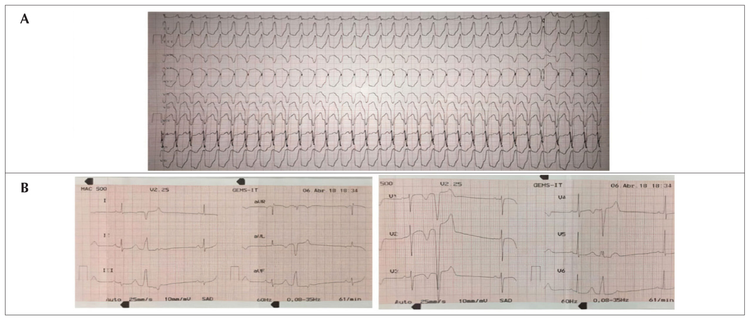
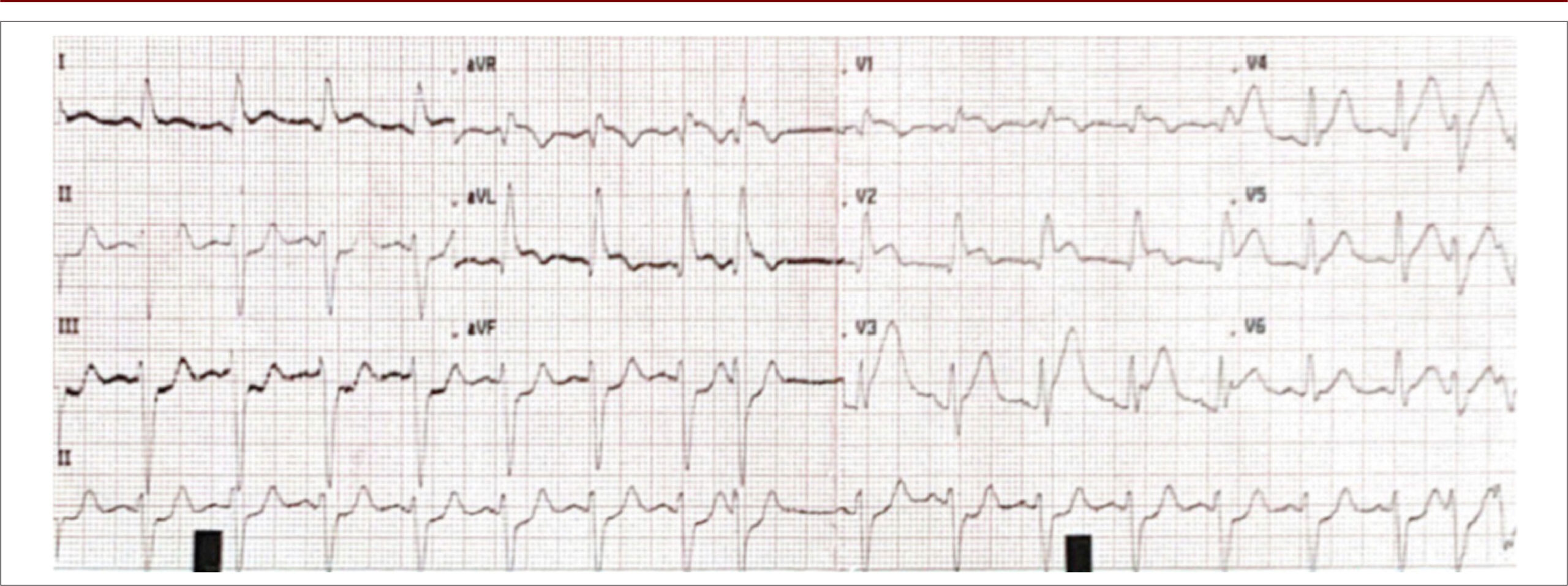
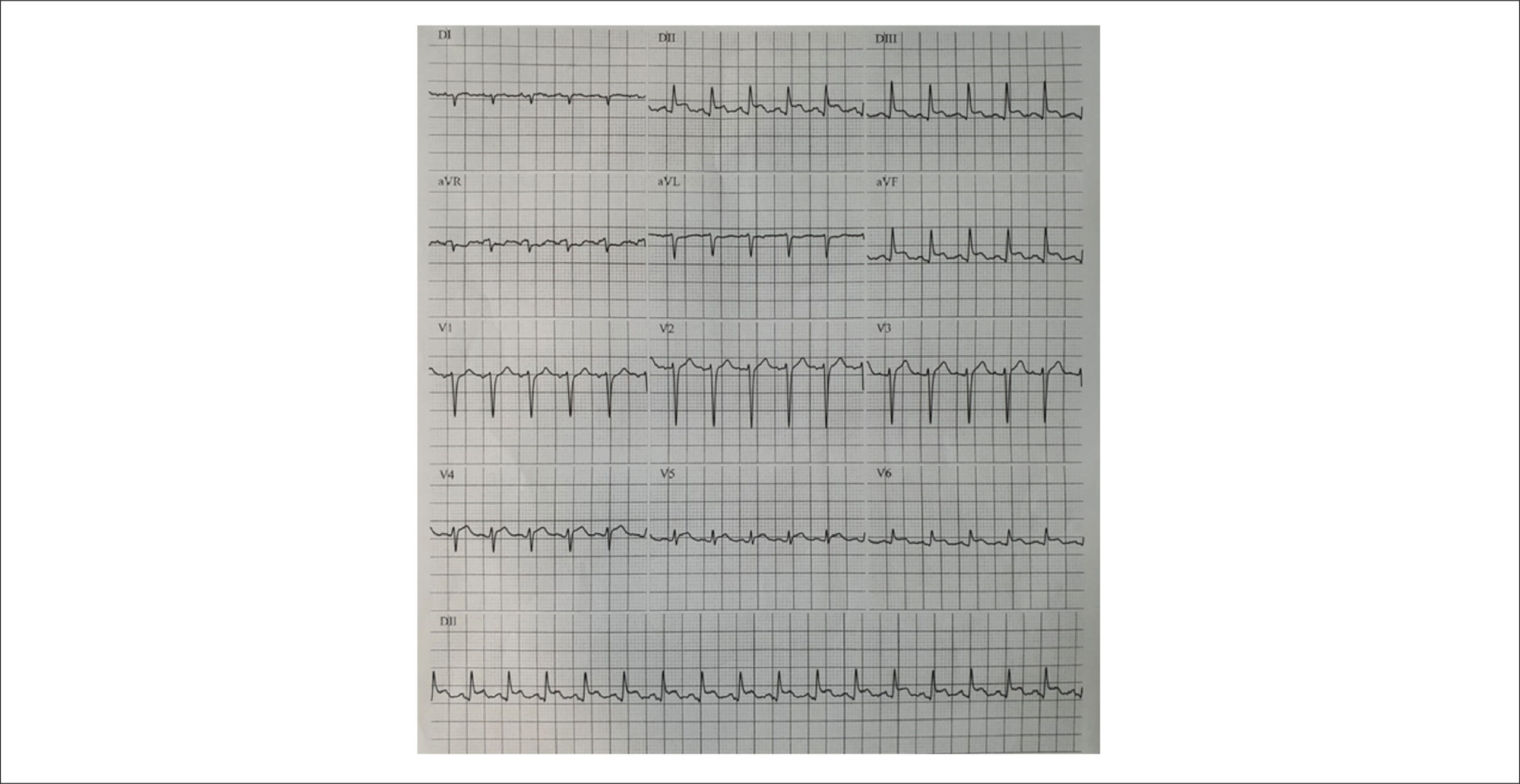
The patient reported symptoms of prostration and a dry cough for a few days for which he was tested for severe acute respiratory syndrome coronavirus 2 (Sars-CoV-2) infection at another institution. On admission, the patient was sweating, had cold extremities, and was anxious. His blood pressure was 128/58 mmHg, heart rate was approximately 140 bpm, and peripheral oxygen saturation was 99% on room air. His initial electrocardiogram () revealed an elevated ST-segment in the inferior and lateral wall but no clear evidence of reciprocal changes.

[…]

  • Cardiogenic shock
  • Coronavirus infections
  • COVID-19
  • Myocarditis
Read Full Article
Creative Commons Attribution (CC BY) Creative Commons license
26/11/2021 DOI: 10.47593/2675-312X/20213403eabc216
Figure 1
Original Article

Myocardial Deformation (Strain) and Myocardial Fibrosis in Mild Cardiac Chagas Disease

Polyana Evangelista Lima et al. Rafael de Castro da Silva André Maurício Souza Fernandes Marta Silva Menezes Edmundo José Nassri Camara

Abstract

Background:

The early identification of myocardial damage seems important in the management of patients with Chagas disease. However, it is unknown whether speckle tracking echocardiography strain and cardiac magnetic resonance imaging are promising methods for assessing myocardial involvement and fibrosis, respectively.

Objective:

To evaluate myocardial involvement in the mild chronic cardiac form of Chagas disease using speckle tracking strain and myocardial fibrosis by cardiac magnetic resonance and assess their correlations.

Method:

This cross-sectional study analyzed patients with the mild chronic cardiac form of Chagas disease (preserved ejection fraction) using speckle tracking echocardiography and cardiac magnetic resonance.

Results:

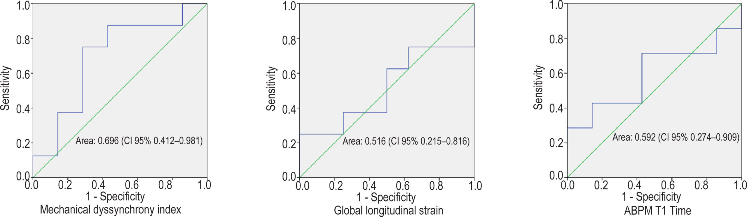
The study included 21 participants (women: 62%; age: 54 ± 5 years). The prevalence of myocardial fibrosis was 50% on delayed myocardial enhancement. The median global longitudinal strain was decreased (14.1%; interquartile range, 12.1–16.3%) in 17 patients (81%). The mean T1 mapping value was high in patients with Chagas disease (993 ± 163 ms). The T1 map was significantly correlated with the global longitudinal strain (r = 0.634; p = 0.015). In addition, the mechanical dispersion index obtained by strain was increased (>55 ms) by 84%, with the largest area under the receiver operating characteristic curve (area under the curve, 0.696; 95% confidence interval, 0.412–0.981) for fibrosis discrimination by delayed myocardial enhancement.

Conclusion:

Myocardial strain and T1 mapping are early markers of myocardial damage in mild chronic Chagas heart disease. The mechanical dispersion index was high and the most closely correlated with delayed myocardial enhancement.

  • Cardiac magnetic resonance
  • Chagas disease
  • Myocardial fibrosis
  • Myocardial strain
  • Speckle tracking echocardiography
Read Full Article
Creative Commons Attribution (CC BY) Creative Commons license
26/11/2021 DOI: 10.47593/2675-312X/20213403eabc173
Figure 1
Case Report

Arrhythmogenic Right Ventricular Cardiomyopathy in Marathon Runners: Case Report

Isabelle Adjanine Borges de Lima et al. Diego Vital Campos Ana Karolina Bento da Silva Evellyn Pereira de Melo

Introduction

Arrhythmogenic right ventricular cardiomyopathy (ARVC) is an inherited disease of the cardiac muscle characterized by right ventricular dysfunction (global or regional) with or without left ventricular (LV) involvement in the presence of histological evidence of the disease and/or electrocardiographic abnormalities according to published criteria.

ARVC has age-dependent penetrance and is mainly characterized by episodes of ventricular arrhythmia that can progress to sudden cardiac death (SCD). The estimated prevalence ranges from 1:2,000 to 1:5,000, with a predominance in the Caucasian population and in people who practice strenuous exercise or competitive sports. Despite its low prevalence, ARVC represents approximately 5–20% of CSM cases in young people.

[…]

  • Arrhythmogenic right ventricular dysplasia;
  • Echocardiography
  • Ventricular tachycardia
Read Full Article
Creative Commons Attribution (CC BY) Creative Commons license
26/11/2021 DOI: 10.47593/2675-312X/20213403eabc188 Views 1
Figure 1
Image

Left Ventricular Diverticulum

Márcio Silva Miguel Lima

A

[…]

  • Cardiomyopathies
  • cardiomyopathy
  • hypertrophic
  • left
  • Ventricular Dysfunction
Read Full Article
Creative Commons Attribution (CC BY) Creative Commons license
26/11/2021 DOI: 10.47593/2675-312X/20213403eabc234
Figure 1
Case Report

Left Internal Thoracic Artery Fistula to the Bronchial Artery: a Rare Cause of Coronary Flow Theft

Marcela Gomes de Souza et al. Alice Mirane Malta Carrijo Vinicius Ferreira Aratani Flávia Bittar Britto Arantes

Introduction

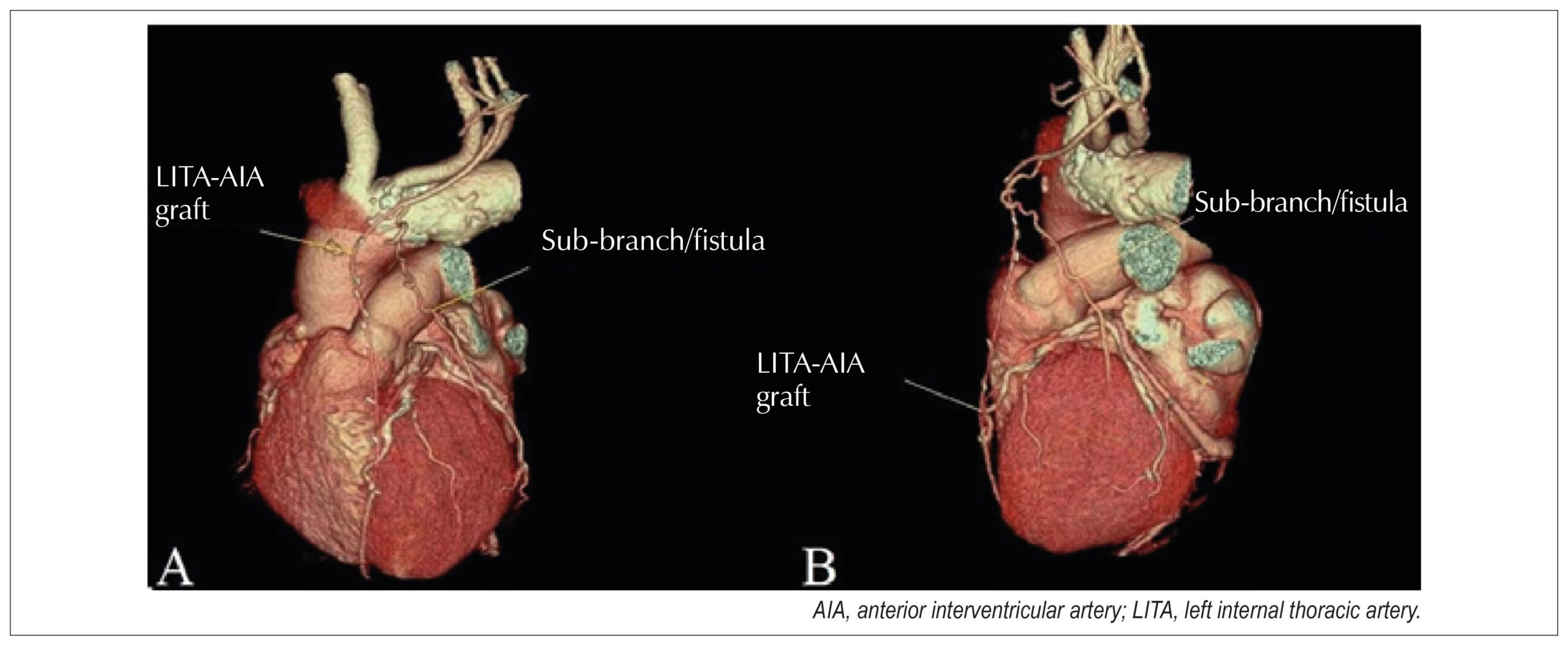
The left internal thoracic artery (LITA) fistula to the pulmonary artery (PA) and/or its branches is a rare condition that was first described in 1947 by Burchell and Clagett and can be acquired or congenital. Its etiologies include, trauma, infection, neoplasms, and mainly myocardial revascularization surgery (MRS)., The surgical manipulation of the intrathoracic vessels during MRS may predispose a patient to postoperative LITA fistulization to the PA, causing coronary flow leakage and myocardial ischemia. LITA should be considered an etiological diagnosis in cases of persistent angina after MRS., Its diagnosis requires coronary cineangiography,– and its treatment options include conservative drug therapy, surgical ligation, or endovascular intervention.

[…]

  • Arteries
  • Case report
  • Coronary Vessel Anomalies
  • Coronary Vessels
  • Diagnostic Imaging
  • Fistula
Read Full Article
Creative Commons Attribution (CC BY) Creative Commons license
26/11/2021 DOI: 10.47593/2675-312X/20213403eabc203
Figure 1
Case Report

Spontaneous Coronary Artery Dissection and Diagnosis by Computed Tomography Angiography: a Case Report

Mathias Silvestre de Brida et al. Georgia Pergher Postingher Fábio Vieira Caovilla Juarez Neuhaus Barbisan

Introduction

Spontaneous coronary artery dissection (SCAD), defined as separation of the layers of the coronary wall unrelated to trauma, iatrogenesis, or atherosclerosis, often occurs in young women without cardiovascular risk factors. First described almost a century ago, it was poorly understood for some time, known only to be associated with the peripartum and pregnancy periods. SCAD is a rare cause of acute coronary syndrome (ACS), and despite technological advances in invasive or non-invasive imaging tests increasing our understanding of its pathophysiology and treatment, it remains underdiagnosed. This report describes the case of an ACS patient without obstructive lesions on coronary angiography in which coronary computed tomography angiography (CCTA) was an indispensable tool in the diagnosis of SCAD with an unusual location, highlighting its growing importance based on the current scientific literature.

[…]

  • Acute coronary syndrome
  • Chest Pain
  • Computed Tomography Angiography
  • Dissection
Read Full Article
Creative Commons Attribution (CC BY) Creative Commons license
26/11/2021 DOI: 10.47593/2675-312X/20213403eabc191
Figure 1
Original Article

Functional Classification versus Ejection Fraction in Patients with Heart Failure and Chagas Disease

Joseylton Gonssçalves Santana et al. Marcela Gordilho Aras Luca Nascimento Roque Aras Júnior

Abstract

Introduction:

Chagas disease, an infection caused by the protozoan Trypanosoma cruzi, is an important health problem worldwide that causes cardiac dilation, arrhythmias, and death. Heart failure is a complex syndrome with high morbidity and mortality rates that progresses with similar complications. The New York Heart Association functional classification is used to categorize heart failure severity and stratify heart disease risks and therapies. A reduced left ventricular ejection fraction measured by echocardiography is directly related to a poor prognosis.

Objective:

To compare the relationship between New York Heart Association functional classification and left ventricular ejection fraction in Chagas versus no Chagas disease outpatients.

Methods:

Cross-sectional study in a cohort of patients followed at a heart failure clinic. Medical records, clinical interviews, functional classification, and left ventricular ejection fraction by echocardiography were analyzed. The data were filed in a database and analyzed using SPSS software.

Results:

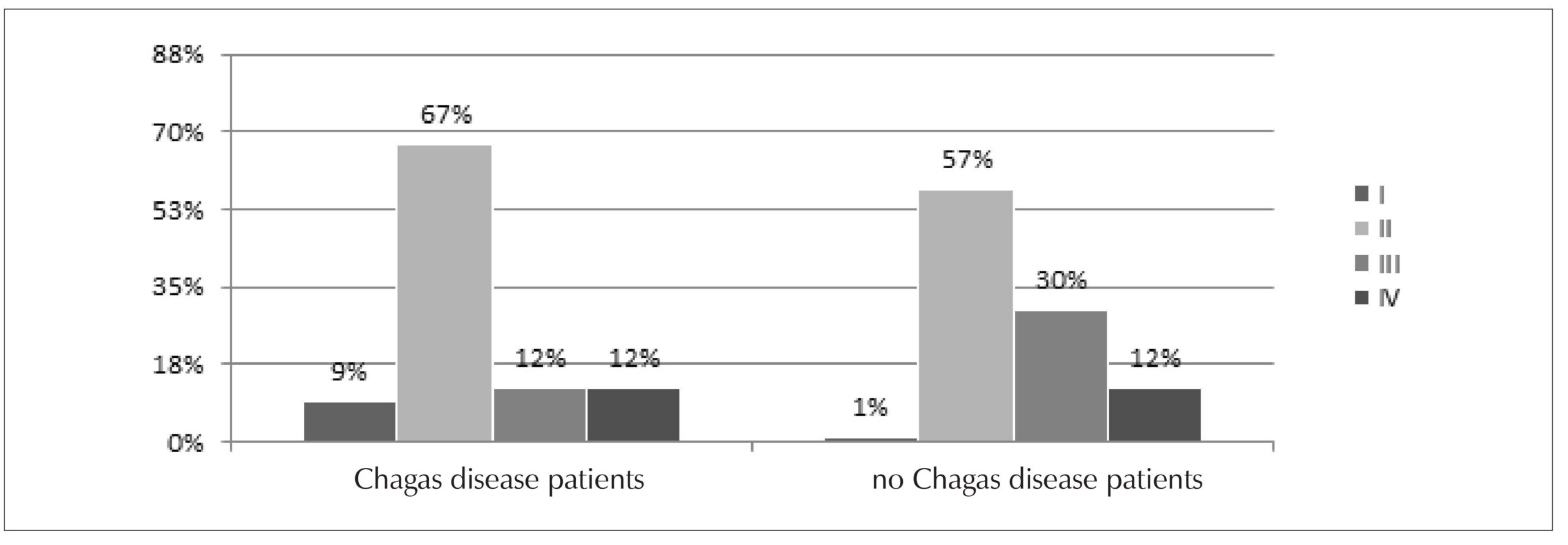
A total of 127 patients with heart failure were selected from August 2018 to July 2019. Of them, 34 (26.8%) had Chagas disease and 93 (73.3%) had no Chagas disease. There was a predominance of men (53.5%) and patients aged > 60 years (61.4%). There was also a predominance of functional class II. Of the Chagas and no Chagas disease patients, 71% versus 93% had a reduced ejection fraction, 21% versus 6% had a mid-range ejection fraction, and 8% versus 1% had a preserved ejection fraction, respectively.

Conclusion:

There was an association between advanced functional class and reduced left ventricular ejection fraction, especially in Chagas patients, information that can be used for outpatient follow-up.

  • Chagas disease
  • Heart Failure
  • Stroke Volume
Read Full Article
Creative Commons Attribution (CC BY) Creative Commons license
26/11/2021 DOI: 10.47593/2675-312X/20213403eabc137 Views 1
Figure 1
Case Report

Subacute Myocarditis with Malignant Progression in a Young Patient: Contribution of Cardiac Magnetic Resonance

Roberto Ramos Barbosa et al. Guilherme Rodrigues Moreira Gabriella Martins Curcio Osmar Araujo Calil … Renato Giestas Serpa Luiz Fernando Machado Barbosa

Introduction

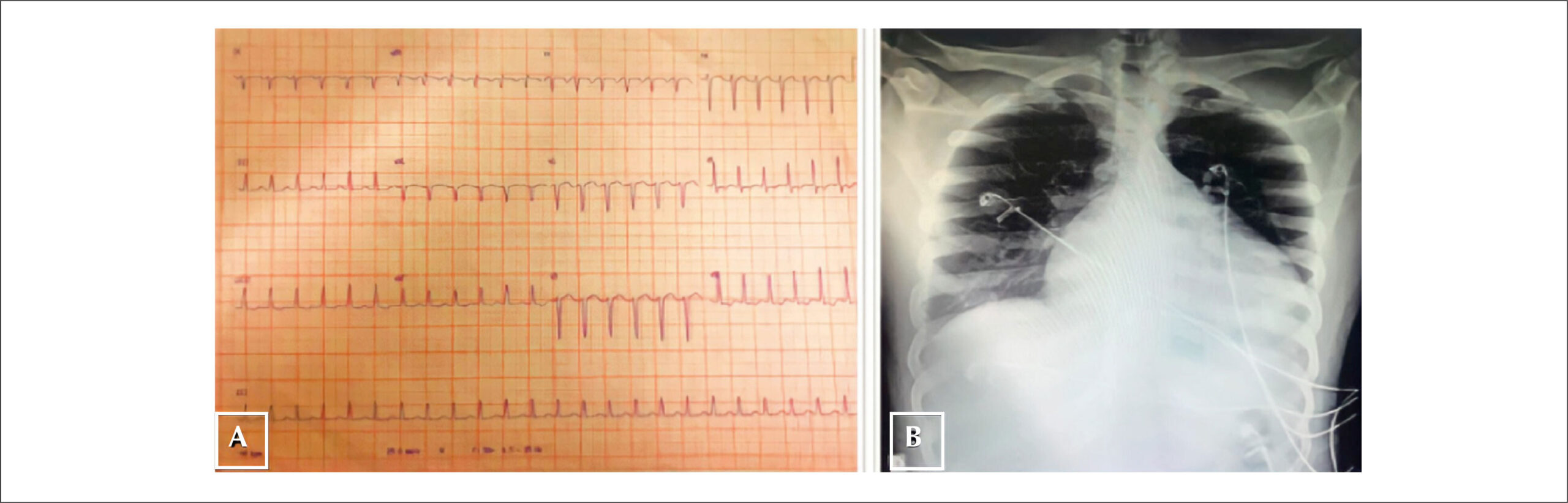
Myocarditis can have a varied clinical presentation ranging from chest pain and mild dyspnea to cardiogenic shock and sudden death. It is defined as myocardial inflammation with myocyte necrosis or degeneration without ischemia and may result from infectious and non-infectious causes, with viral myocarditis being the most prevalent etiology. Myocarditis most commonly affects young male adults, being detected in up to 12% of patients aged under 40 years who had sudden death. Its diagnosis is fundamentally based on clinical suspicion due to recent viral infection associated with a current myocardial lesion, the absence of previous heart disease, and heart failure (HF) of no other apparent cause. Cardiac magnetic resonance (CMR) imaging findings can currently help confirm the diagnosis.

The low availability of endomyocardial biopsy, the gold standard diagnostic method, brings major challenges to the management of myocarditis. On the other hand, the increased use of CMR provides important diagnostic and prognostic data. This report describes the clinical issues and CMR contribution in a case of myocarditis in a young patient.

[…]

  • Death
  • Magnetic Resonance Imaging
  • Myocarditis
  • sudden
Read Full Article
Creative Commons Attribution (CC BY) Creative Commons license
26/11/2021 DOI: 10.47593/2675-312X/20213403eabc218 Views 2
Figure 1
Image

Malignancy of a Benign Tumor

Andre L Antunes et al. David Prieto Pedro E Antunes

Background

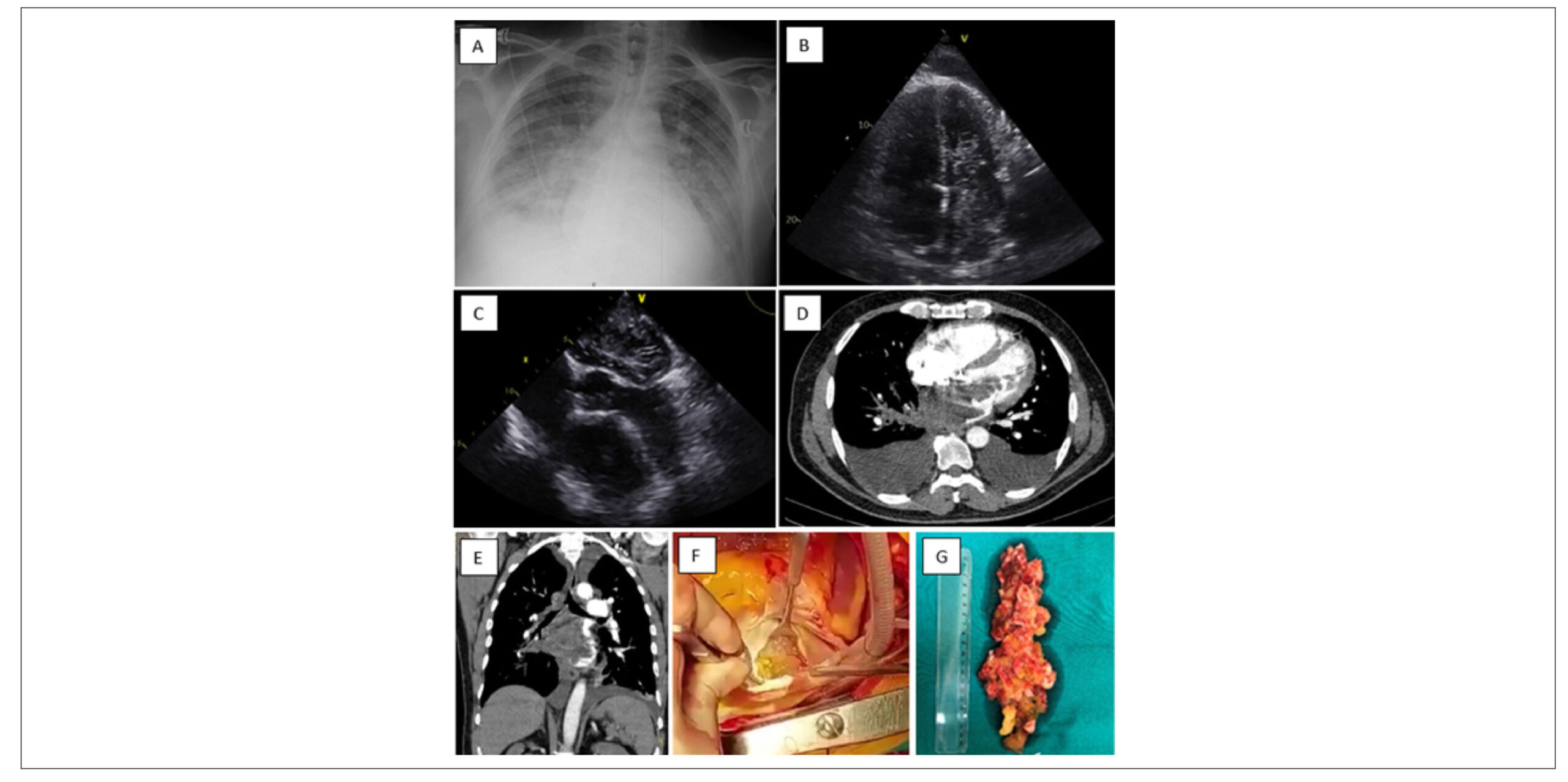
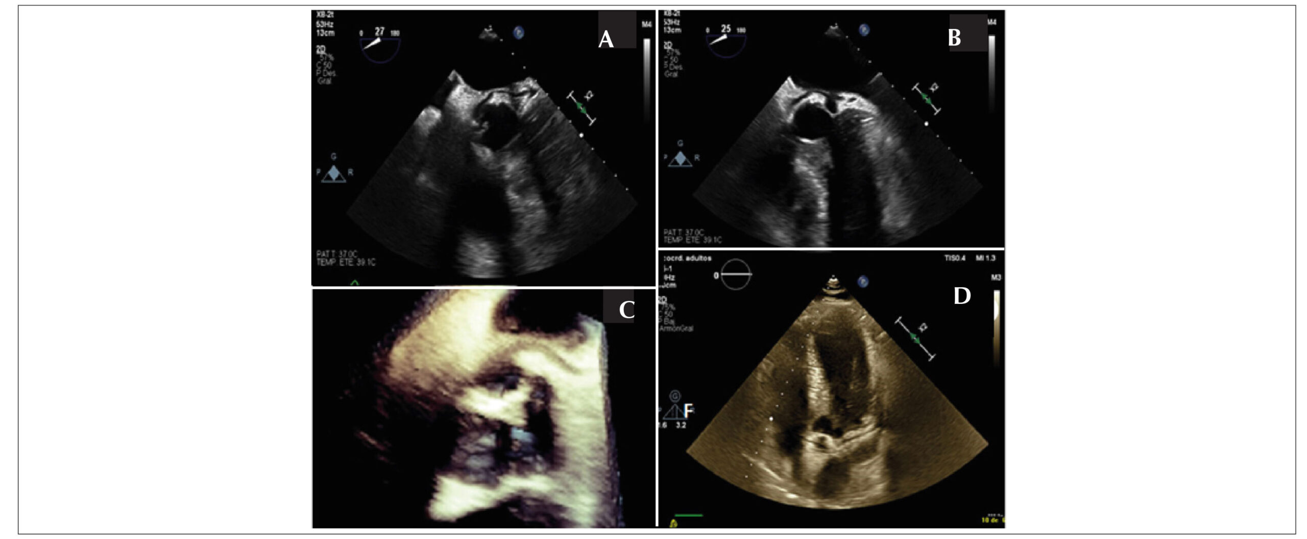
Cardiac tumors can be classified into primary and secondary types. Primary tumors are more common, and the majority of them are benign (70%), with myxomas being the most frequent. However, the overall incidence is rare, ranging from 0.15% to 0.2% in autopsy series. Here we present an image of malignancy of a benign tumor.

[…]

  • Atrial Myxoma
  • Cardiac Primary Tumor
  • Pulmonary Vein Invasion
Read Full Article
Creative Commons Attribution (CC BY) Creative Commons license
26/11/2021 DOI: 10.47593/2675-312X/20213403eabc197 Views 1
Figure 1
Original Article

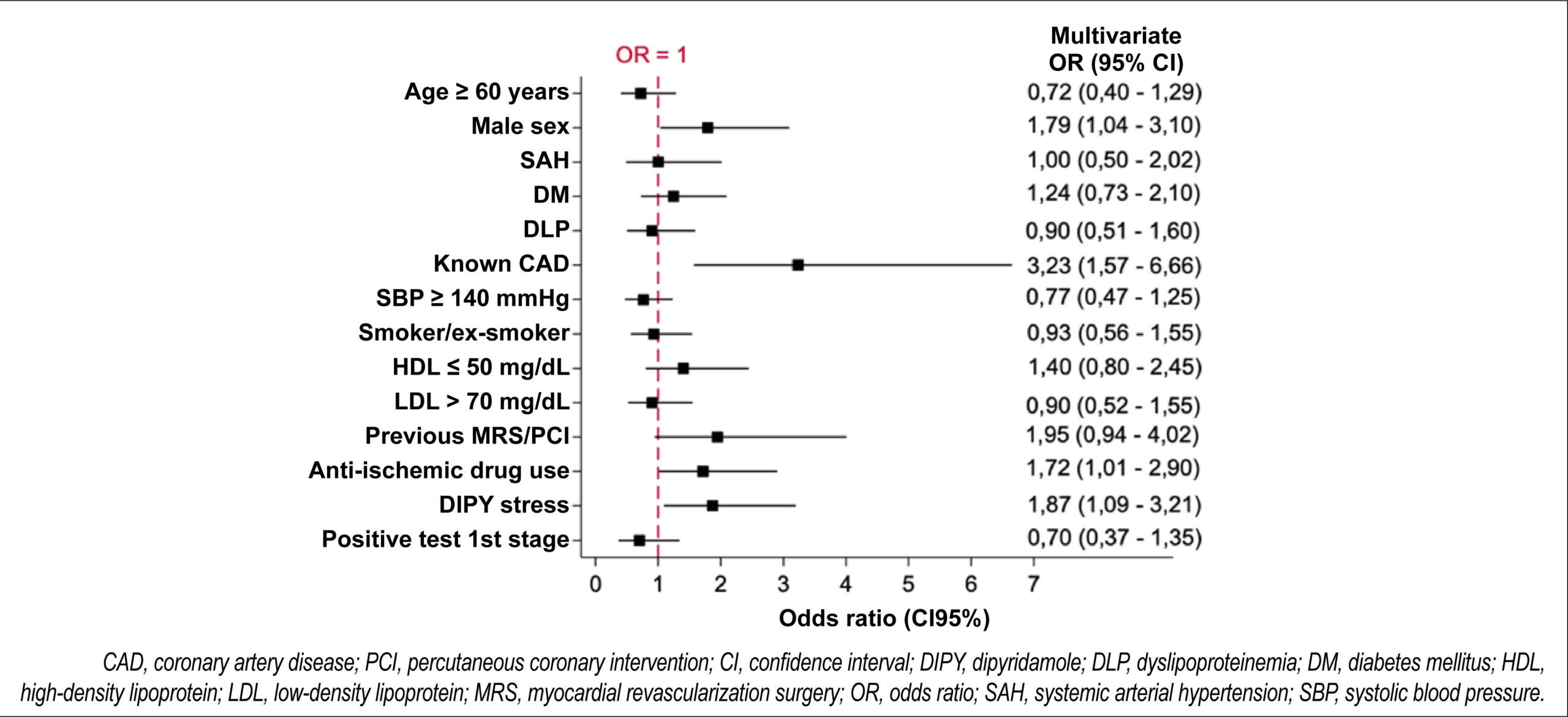
Appropriate Use of Myocardial Perfusion Scintigraphy Criteria for Assessing Asymptomatic Patients with High Cardiovascular Risk or Known Coronary Disease

Vinicius Silva Freire Alvarenga et al. Paola Emanuela Poggio Smanio

Abstract

Background:

The use of myocardial perfusion scintigraphy in asymptomatic patients remains restricted to very specific clinical situations, many of which are addressed in the appropriate use criteria (AUC) for myocardial perfusion scintigraphy.

Objective:

To critically analyze the application of these criteria in indicating the tests performed in asymptomatic patients of the Dante Pazzanese Institute of Cardiology, a population that is notably at high cardiovascular risk.

Methods:

The study selected asymptomatic patients undergoing myocardial perfusion scintigraphy to investigate ischemia. Indications for tests were classified as appropriate, inappropriate, or uncertain. A fixed low uptake, transient low uptake, or transient ischemic dilation were considered changed results. The statistical analysis assessed the correlation between the degree of recommendation of indications and the presence of changed test results.

Results:

From an initial selection of 2,999 medical records, 490 were considered asymptomatic and included according to the previously established inclusion criteria. Only 9.8% of the indications were inappropriate, while 61.4% were appropriate and 28.8% were uncertain. Fixed low uptake of the radiopharmaceutical occurred in 43.5% of cases versus transient low uptake in 16.1%. An appropriate or uncertain test request was a predictor of a changed test result in this population.

Conclusion:

The use of AUC for myocardial perfusion scintigraphy proved effective at predicting abnormal test results in an asymptomatic population at high cardiovascular risk, especially patients with uncertain indications, which may mean that some of the indications considered uncertain may be appropriate for a population at high cardiovascular risk.

  • Cardiovascular risk
  • Diagnosis
  • Nuclear Medicine
Read Full Article
Creative Commons Attribution (CC BY) Creative Commons license
26/11/2021 DOI: 10.47593/2675-312X/20213403eabc189
Figure 1
Review Article

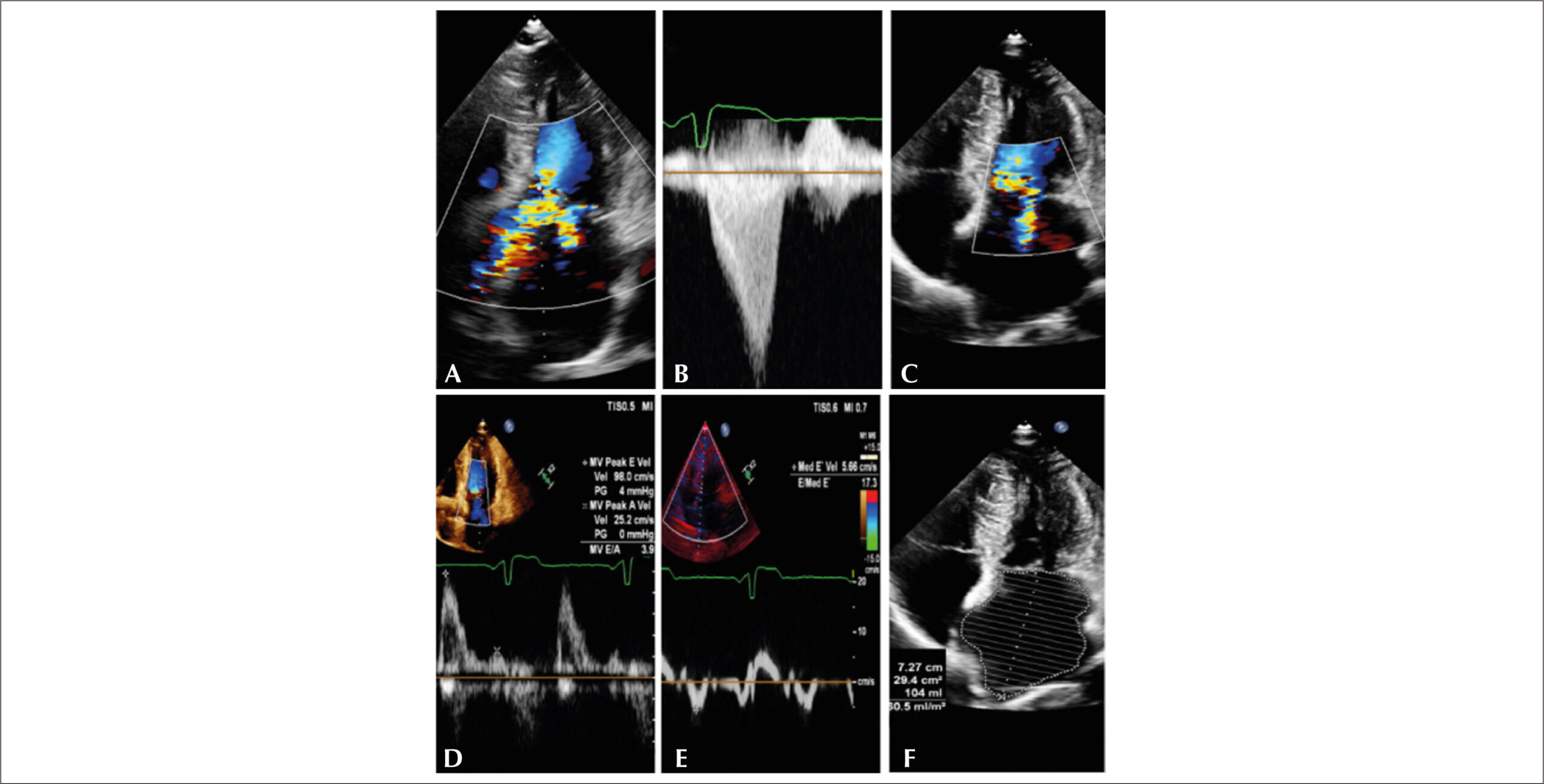
Exercise Stress Echocardiography in Hypertrophic Cardiomyopathy: How, When and Why?

Joselina Luzia Menezes Oliveira et al. Katia Oliveira Nunes Leal Larissa Rebeca da Silva Tavares Irlaneide da Silva Tavares … Luiz Flávio Galvāo Gonçalves Daniel Pio de Oliveira Ursula Maria Moreira Costa Burgos Eduardo José Ferreira José Oliveira Belem Marilia Marques Aquino Antonio Carlos Sobral Sousa

Abstract

Historically, the role of exercise stress echocardiography in the management of hypertrophic cardiomyopathy has been neglected in clinical practice according to the analysis of the 2002 Guidelines of the American College of Cardiology/ American Heart Association, which recommended caution use of this methodology in patients with hypertrophic cardiomyopathy due to the risk of both cardiac arrhythmia and hemodynamic collapse on exertion. Exercise stress testing in patients with hypertrophic cardiomyopathy is currently part of the routine assessment of symptomatic patients with or without a resting left ventricular outflow tract gradient < 50 mmHg. For this group, it is a safe and reliable method for measuring the left ventricular outflow tract gradient during exertion and a solid differentiator of patients with nonobstructive hypertrophic cardiomyopathy (absent gradient, both at rest and on exertion) from those with labile gradients (absent gradient at rest and present on exertion). Therefore, in the assessment of hypertrophic cardiomyopathy, exercise stress is equally useful for quantifying the degree of mitral regurgitation and left ventricular segmental contractility changes as well as assessing left ventricular diastolic function on exertion and is able to predict the future development of heart failure symptoms. The method is also important for determining different treatment strategies for each patient, from surgical myomectomy or alcohol septal ablation for those with a labile gradient, limiting symptoms, and who refractory to drug treatment to heart transplantation for those without a gradient.

  • Echocardiography, Stress
  • Hypertrophic Cardiomyopathy
  • Ventricular Outflow Obstruction
Read Full Article
Creative Commons Attribution (CC BY) Creative Commons license
26/11/2021 DOI: 10.47593/2675-312X/20213403eabc130 Views 1
Figure 1
Case Report

Cor triatriatum sinister and giant eustachian valve mimicking a tetratrial heart

Daniel García-Fuertes et al. Elena Villanueva-Fernández Manuel Crespín-Crespín Francsico José Castillo-Bernal … Ernesto Martín-Dorado María Teresa Gómez-San Román

Introduction

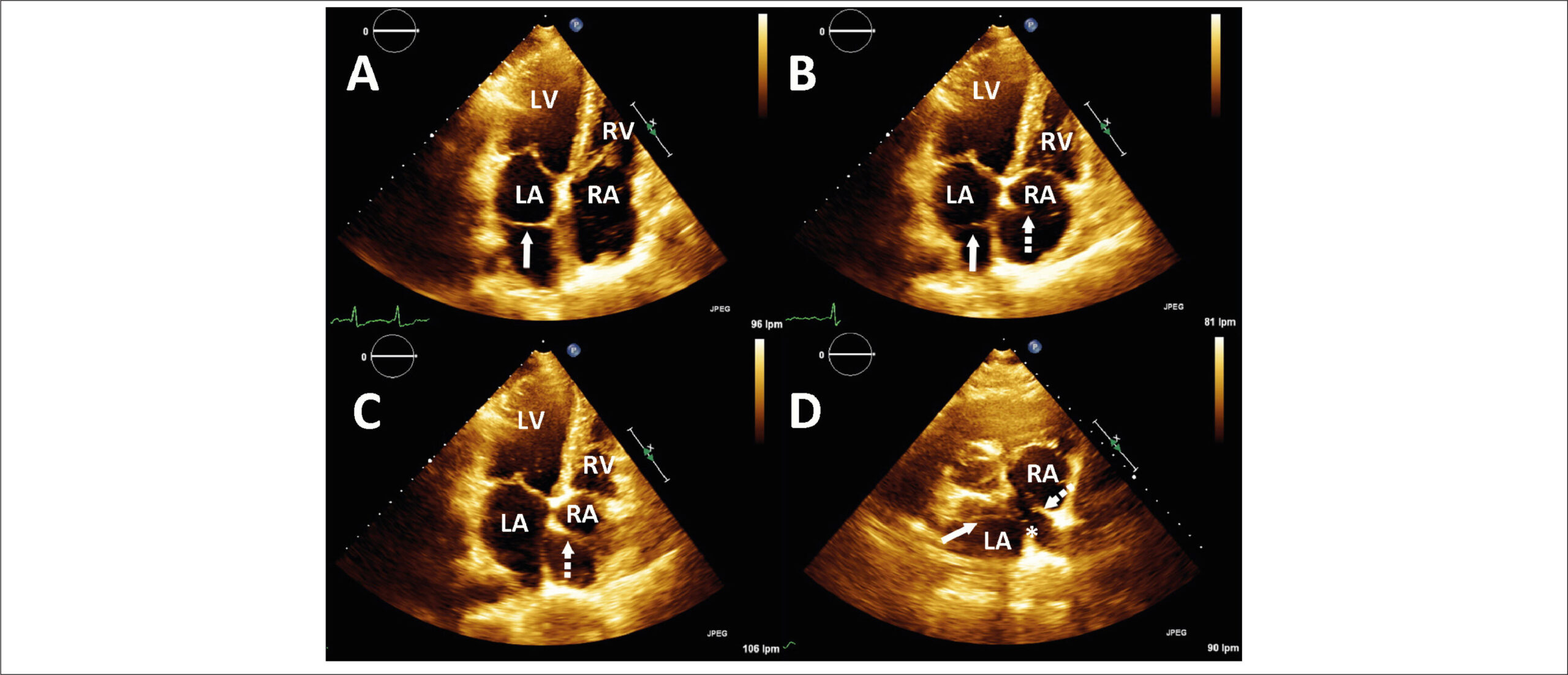
Cor triatriatum is a rare congenital cardiac anomaly that is usually associated with other congenital heart defects such as anomalous pulmonary vein return, atrial septal defect, or patent foramen ovale. Cor triatriatum sinister is found in an approximately 0.1% of necropsy examinations. In this congenital malformation, the left atrium is divided into two chambers, usually by a fibromuscular septum, with the pulmonary veins entering a posterosuperior chamber separated from the anteroinferior distal chamber containing the mitral valve and the left atrial appendage. Although several theories have been hypothesized, its embryologic origin remains not fully understood. An inadequate incorporation of the common pulmonary vein into the left atrium or abnormal growth of the septum primum have been postulated to explain it. On the other hand, cor triatriatum dexter is an even more rare congenital cardiac anomaly in which a membranous structure divides the right atrium into two chambers. In such cases, complete persistence of the right valve of the sinus venosus is responsible for the atrial division. This valve usually regresses as a part of normal embryological development between 9 and 15 weeks’ gestation, leaving behind remnants such as the crista terminalis superiorly and the eustachian valve of the inferior vena cava and the thebesian valve of the coronary sinus inferiorly. The severity of this defect is directly related to the degree of flow obstruction, which in turn is related to the degree of valve persistence.– Although it shares the same embryologic origin and might show a similar echocardiographic appearance, the differential diagnosis includes a prominent eustachian valve, which also results in an apparent division of the right atrium into two chambers.

[…]

  • Cor triatriatum
Read Full Article
Creative Commons Attribution (CC BY) Creative Commons license
26/11/2021 DOI: 10.47593/2675-312X/20213403eabc204 Views 1
Figure 1
Image

Retroaortic Circunflex Coronary Artery Appearance on Transesophageal Echocardiography

Lucia Fernandez Gasso et al. Carmen Jimenez Lopez-Guarch

Abstract

The retroaortic anomalous coronary (RAC) sign is an unknown finding that has been described on transthoracic echocardiography as an echogenic tubular structure located on the atrial side of the atrioventricular sulcus. Here we present a case in which the RAC sign on transesophageal echocardiography (TEE). Knowledge of RAC sign and its evaluation on TEE increases sensitivity and specificity and provides the opportunity to assess high-risk anatomic features that are important in evaluating the risk of sudden death.

  • RAC Sign
  • Retroaortic Circumflex Coronary Artery
  • Transesophageal Echocardiographyt
Read Full Article
Creative Commons Attribution (CC BY) Creative Commons license
26/11/2021 DOI: 10.47593/2675-312X/20213403eabc190
Strain in Monitoring Cancer Therapy-Induced Cardiotoxicity: Where are We After the SUCCOUR Study?
Editorial

Strain in Monitoring Cancer Therapy-Induced Cardiotoxicity: Where are We After the SUCCOUR Study?

Silvio Henrique Barberato et al. Ludhmila Abrão Hajjar

In recent decades, advances in cancer treatment have led to a significantly increased survival rate for several types of cancer. On the other hand, cancer survival increases patients’ vulnerability to cardiovascular complications induced by potentially cardiotoxic cancer therapy., Cardiovascular diseases are currently recognized as a main cause of morbidity and mortality in cancer survivors depending on the type of therapeutic approach used, patient age, and the presence of preexisting cardiovascular risk factors. Due to the high incidence of breast cancer in women, anthracyclines and monoclonal antibodies against human epidermal growth factor receptor type 2 (HER2) are the drugs most well-known as potential cardiotoxicity (CTX) inducers. However, other medications such as vascular endothelial growth factor inhibitors, tyrosine kinase inhibitors, proteasome inhibitors, and immunological checkpoint inhibitors, in addition to mediastinal radiotherapy, are also recognized for having a possible deleterious effect on the cardiovascular system. It is worth mentioning that CTX can express not only as myocardial dysfunction but also as acute coronary syndrome, arrhythmia, valve disease, pericardial disease, systemic arterial hypertension, and pulmonary hypertension. Thus, although left ventricular (LV) systolic dysfunction secondary to anticancer therapy is not the only CTX presentation, its development is among the most worrisome in clinical cardio-oncology practice, and the terms are often used interchangeably.

The diagnosis of LV dysfunction in a CTX setting is traditionally based on the serial evaluation of the LV ejection fraction (LVEF) using the Simpson method on two-dimensional echocardiography. Despite variation between international guidelines and cancer trials, the most commonly used CTX definition is a decrease of at least ten absolute points for a LVEF below the lower limit of normal after cancer treatment (considered below 53% by the cardiovascular imaging consensus of the European Association of Cardiovascular Imaging/American Society of Echocardiography [EACVI/ASE], or below 50% by the European Society of Cardiology [ESC]). However, LVEF measurements have important limitations, which include significant inter- and intra-observer variability, dependence on loading conditions, and low sensitivity to small LV function changes. Therefore, it was postulated that LVEF changes would occur later, when myocardial damage is severe enough to determine an irreversible cardiomyopathy.

[…]
  • Antineoplastic Agents
  • Cardiotoxicity
  • Doppler
  • Echocardiography
Read Full Article
Creative Commons Attribution (CC BY) Creative Commons license
26/11/2021 DOI: 10.47593/2675-312X/20213403eabc247
Figure 1
Systematic Review

Inferior Vena Cava Ultrasound for Assessing Volume Status and Fluid Responsiveness in Critically ill Patients: A Systematic Review

Djalma Gomes Xavier Filho et al. Akeme Laissa Novais Coutinho Romero Henrique de Almeida Barbosa Matheus Rodrigues Lopes Adirlene Pontes de Oliveira Tenório

Abstract

Background:

The assessment of intravascular volume and fluid responsiveness is challenging in the management of critically ill patients. Diagnostic methods of hemodynamic monitoring must be safe, reproducible, and practical.

Objective:

To describe the applicability of ultrasound indices of the inferior vena cava in the assessment of intravascular volume and prediction of fluid responsiveness in critically ill patients.

Method:

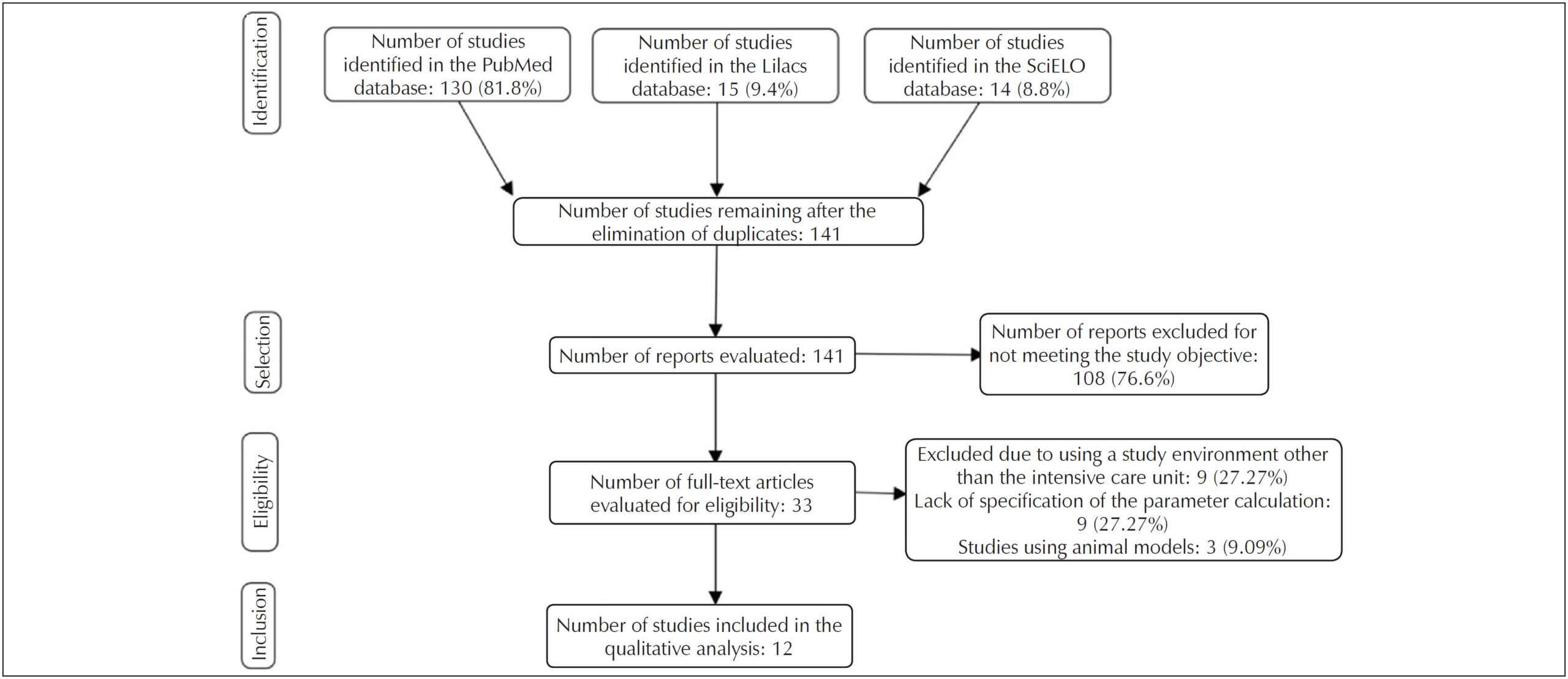
A systematic review performed of the PubMed®, Latin American and Caribbean Health Sciences Literature, and Scientific Electronic Library Online databases for articles published in the previous five years. The descriptors used were “inferior vena cava,” “ultrasonography,” “fluid responsiveness,” and “volume status.”

Results:

The search identified 13 relevant articles. The collapsibility index of the inferior vena cava was 25%–50% as the cutoff point for defining hypovolemia and showed applicability in predicting fluid responsiveness in patients breathing spontaneously with cutoff points of 25%–57%. In mechanical ventilation scenarios, the distensibility index of the inferior vena cava was more effective at predicting fluid responsiveness than the other measurements with variation of 10.2%–20.5%. The inferior vena cava/aorta diameter index was especially useful in the pediatric population for defining intravascular volume; however, in adults, there were many divergences in its applicability.

Conclusion:

The assessment of intravascular volume and fluid responsiveness through ultrasound indices of the inferior vena cava is applicable and safe for diagnosing and monitoring hemodynamic instability. However, studies of value standardization are necessary due to divergences in the cutoff points used in each index.

  • Hemodynamics
  • Inferior vena cava
  • Intensive Care Unit
  • Organism fluid status
  • Ultrasonography
Read Full Article
Creative Commons Attribution (CC BY) Creative Commons license
26/11/2021 DOI: 10.47593/2675-312X/20213403eabc193 Views 2

How to cite this article

EndNote RIS

Electronic Document Format (Vancouver)

Frasson MZ, Jaeger CP, Silveira DS, Cawen DRR. Myocarditis and Cardiogenic in a Covid-19 Positive Patient. ABC Imagem Cardiovascular 2021;34(3):eabc216.

Electronic Document Format (ABNT)

Frasson, Murilo Zomer; Jaeger, Cristiano Pederneiras; Silveira, Daniel Souto; Cawen, Diego Raul Romero. Myocarditis and Cardiogenic in a Covid-19 Positive Patient. ABC Imagem Cardiovascular, v. 34, n. 3, eabc216, Nov. 2021.

Electronic Document Format (APA)

Frasson, M. Z., Jaeger, C. P., Silveira, D. S., & Cawen, D. R. R. (2021). Myocarditis and Cardiogenic in a Covid-19 Positive Patient. ABC Imagem Cardiovascular, 34(3), eabc216.

Electronic Document Format (ISO)

Frasson, Murilo Zomer and Jaeger, Cristiano Pederneiras and Silveira, Daniel Souto and Cawen, Diego Raul Romero. Myocarditis and Cardiogenic in a Covid-19 Positive Patient. ABC Imagem Cardiovascular [online]. 2021, vol. 34, n. 3, [cited 2026-10-14], eabc216. Available from: <https://journals.cardiol.br/en/article/myocarditis-and-cardiogenic-in-a-covid-19-positive-patient/>. ISSN 2318-8219

EndNote RIS

Electronic Document Format (Vancouver)

Lima PE, Silva RC, Fernandes AMS, Menezes MS, Camara EJN. Myocardial Deformation (Strain) and Myocardial Fibrosis in Mild Cardiac Chagas Disease. ABC Imagem Cardiovascular 2021;34(3):eabc173.

Electronic Document Format (ABNT)

Lima, Polyana Evangelista; Silva, Rafael de Castro da; Fernandes, André Maurício Souza; Menezes, Marta Silva; Camara, Edmundo José Nassri. Myocardial Deformation (Strain) and Myocardial Fibrosis in Mild Cardiac Chagas Disease. ABC Imagem Cardiovascular, v. 34, n. 3, eabc173, Nov. 2021.

Electronic Document Format (APA)

Lima, P. E., Silva, R. C. , Fernandes, A. M. S., Menezes, M. S., & Camara, E. J. N. (2021). Myocardial Deformation (Strain) and Myocardial Fibrosis in Mild Cardiac Chagas Disease. ABC Imagem Cardiovascular, 34(3), eabc173.

Electronic Document Format (ISO)

Lima, Polyana Evangelista and Silva, Rafael de Castro da and Fernandes, André Maurício Souza and Menezes, Marta Silva and Camara, Edmundo José Nassri. Myocardial Deformation (Strain) and Myocardial Fibrosis in Mild Cardiac Chagas Disease. ABC Imagem Cardiovascular [online]. 2021, vol. 34, n. 3, [cited 2026-10-14], eabc173. Available from: <https://journals.cardiol.br/en/article/myocardial-deformation-strain-and-myocardial-fibrosis-in-mild-cardiac-chagas-disease/>. ISSN 2318-8219

EndNote RIS

Electronic Document Format (Vancouver)

Lima IAB, Campos DV, Silva AKB, Melo EP. Arrhythmogenic Right Ventricular Cardiomyopathy in Marathon Runners: Case Report. ABC Imagem Cardiovascular 2021;34(3):eabc188.

Electronic Document Format (ABNT)

Lima, Isabelle Adjanine Borges de; Campos, Diego Vital; Silva, Ana Karolina Bento da; Melo, Evellyn Pereira de. Arrhythmogenic Right Ventricular Cardiomyopathy in Marathon Runners: Case Report. ABC Imagem Cardiovascular, v. 34, n. 3, eabc188, Nov. 2021.

Electronic Document Format (APA)

Lima, I. A. B. , Campos, D. V., Silva, A. K. B. , & Melo, E. P. (2021). Arrhythmogenic Right Ventricular Cardiomyopathy in Marathon Runners: Case Report. ABC Imagem Cardiovascular, 34(3), eabc188.

Electronic Document Format (ISO)

Lima, Isabelle Adjanine Borges de and Campos, Diego Vital and Silva, Ana Karolina Bento da and Melo, Evellyn Pereira de. Arrhythmogenic Right Ventricular Cardiomyopathy in Marathon Runners: Case Report. ABC Imagem Cardiovascular [online]. 2021, vol. 34, n. 3, [cited 2026-10-14], eabc188. Available from: <https://journals.cardiol.br/en/article/arrhythmogenic-right-ventricular-cardiomyopathy-in-marathon-runners-case-report/>. ISSN 2318-8219

EndNote RIS

Electronic Document Format (Vancouver)

Lima MSM. Left Ventricular Diverticulum. ABC Imagem Cardiovascular 2021;34(3):eabc234.

Electronic Document Format (ABNT)

Lima, Márcio Silva Miguel. Left Ventricular Diverticulum. ABC Imagem Cardiovascular, v. 34, n. 3, eabc234, Nov. 2021.

Electronic Document Format (APA)

Lima, M. S. M. (2021). Left Ventricular Diverticulum. ABC Imagem Cardiovascular, 34(3), eabc234.

Electronic Document Format (ISO)

Lima, Márcio Silva Miguel. Left Ventricular Diverticulum. ABC Imagem Cardiovascular [online]. 2021, vol. 34, n. 3, [cited 2026-10-14], eabc234. Available from: <https://journals.cardiol.br/en/article/left-ventricular-diverticulum/>. ISSN 2318-8219

EndNote RIS

Electronic Document Format (Vancouver)

Souza MG, Carrijo AMM, Aratani VF, Arantes FBB. Left Internal Thoracic Artery Fistula to the Bronchial Artery: a Rare Cause of Coronary Flow Theft. ABC Imagem Cardiovascular 2021;34(3):eabc203.

Electronic Document Format (ABNT)

Souza, Marcela Gomes de; Carrijo, Alice Mirane Malta; Aratani, Vinicius Ferreira; Arantes, Flávia Bittar Britto. Left Internal Thoracic Artery Fistula to the Bronchial Artery: a Rare Cause of Coronary Flow Theft. ABC Imagem Cardiovascular, v. 34, n. 3, eabc203, Nov. 2021.

Electronic Document Format (APA)

Souza, M. G. , Carrijo, A. M. M., Aratani, V. F., & Arantes, F. B. B. (2021). Left Internal Thoracic Artery Fistula to the Bronchial Artery: a Rare Cause of Coronary Flow Theft. ABC Imagem Cardiovascular, 34(3), eabc203.

Electronic Document Format (ISO)

Souza, Marcela Gomes de and Carrijo, Alice Mirane Malta and Aratani, Vinicius Ferreira and Arantes, Flávia Bittar Britto. Left Internal Thoracic Artery Fistula to the Bronchial Artery: a Rare Cause of Coronary Flow Theft. ABC Imagem Cardiovascular [online]. 2021, vol. 34, n. 3, [cited 2026-10-14], eabc203. Available from: <https://journals.cardiol.br/en/article/left-internal-thoracic-artery-fistula-to-the-bronchial-artery-a-rare-cause-of-coronary-flow-theft/>. ISSN 2318-8219

EndNote RIS

Electronic Document Format (Vancouver)

Brida MS, Postingher GP, Caovilla FV, Barbisan JN. Spontaneous Coronary Artery Dissection and Diagnosis by Computed Tomography Angiography: a Case Report. ABC Imagem Cardiovascular 2021;34(3):eabc191.

Electronic Document Format (ABNT)

Brida, Mathias Silvestre de; Postingher, Georgia Pergher; Caovilla, Fábio Vieira; Barbisan, Juarez Neuhaus. Spontaneous Coronary Artery Dissection and Diagnosis by Computed Tomography Angiography: a Case Report. ABC Imagem Cardiovascular, v. 34, n. 3, eabc191, Nov. 2021.

Electronic Document Format (APA)

Brida, M. S. , Postingher, G. P., Caovilla, F. V., & Barbisan, J. N. (2021). Spontaneous Coronary Artery Dissection and Diagnosis by Computed Tomography Angiography: a Case Report. ABC Imagem Cardiovascular, 34(3), eabc191.

Electronic Document Format (ISO)

Brida, Mathias Silvestre de and Postingher, Georgia Pergher and Caovilla, Fábio Vieira and Barbisan, Juarez Neuhaus. Spontaneous Coronary Artery Dissection and Diagnosis by Computed Tomography Angiography: a Case Report. ABC Imagem Cardiovascular [online]. 2021, vol. 34, n. 3, [cited 2026-10-14], eabc191. Available from: <https://journals.cardiol.br/en/article/spontaneous-coronary-artery-dissection-and-diagnosis-by-computed-tomography-angiography-a-case-report/>. ISSN 2318-8219

EndNote RIS

Electronic Document Format (Vancouver)

Santana JG, Aras MG, Nascimento L, Aras Júnior R. Functional Classification versus Ejection Fraction in Patients with Heart Failure and Chagas Disease. ABC Imagem Cardiovascular 2021;34(3):eabc137.

Electronic Document Format (ABNT)

Santana, Joseylton Gonssçalves; Aras, Marcela Gordilho; Nascimento, Luca; Aras Júnior, Roque. Functional Classification versus Ejection Fraction in Patients with Heart Failure and Chagas Disease. ABC Imagem Cardiovascular, v. 34, n. 3, eabc137, Nov. 2021.

Electronic Document Format (APA)

Santana, J. G., Aras, M. G., Nascimento, L., & Aras Júnior, R. (2021). Functional Classification versus Ejection Fraction in Patients with Heart Failure and Chagas Disease. ABC Imagem Cardiovascular, 34(3), eabc137.

Electronic Document Format (ISO)

Santana, Joseylton Gonssçalves and Aras, Marcela Gordilho and Nascimento, Luca and Aras Júnior, Roque. Functional Classification versus Ejection Fraction in Patients with Heart Failure and Chagas Disease. ABC Imagem Cardiovascular [online]. 2021, vol. 34, n. 3, [cited 2026-10-14], eabc137. Available from: <https://journals.cardiol.br/en/article/functional-classification-versus-ejection-fraction-in-patients-with-heart-failure-and-chagas-disease/>. ISSN 2318-8219

EndNote RIS

Electronic Document Format (Vancouver)

Barbosa RR, Moreira GR, Curcio GM, Calil OA, Serpa RG, Barbosa LFM. Subacute Myocarditis with Malignant Progression in a Young Patient: Contribution of Cardiac Magnetic Resonance. ABC Imagem Cardiovascular 2021;34(3):eabc218.

Electronic Document Format (ABNT)

Barbosa, Roberto Ramos; Moreira, Guilherme Rodrigues; Curcio, Gabriella Martins; Calil, Osmar Araujo; Serpa, Renato Giestas; Barbosa, Luiz Fernando Machado. Subacute Myocarditis with Malignant Progression in a Young Patient: Contribution of Cardiac Magnetic Resonance. ABC Imagem Cardiovascular, v. 34, n. 3, eabc218, Nov. 2021.

Electronic Document Format (APA)

Barbosa, R. R., Moreira, G. R., Curcio, G. M., Calil, O. A., Serpa, R. G., & Barbosa, L. F. M. (2021). Subacute Myocarditis with Malignant Progression in a Young Patient: Contribution of Cardiac Magnetic Resonance. ABC Imagem Cardiovascular, 34(3), eabc218.

Electronic Document Format (ISO)

Barbosa, Roberto Ramos and Moreira, Guilherme Rodrigues and Curcio, Gabriella Martins and Calil, Osmar Araujo and Serpa, Renato Giestas and Barbosa, Luiz Fernando Machado. Subacute Myocarditis with Malignant Progression in a Young Patient: Contribution of Cardiac Magnetic Resonance. ABC Imagem Cardiovascular [online]. 2021, vol. 34, n. 3, [cited 2026-10-14], eabc218. Available from: <https://journals.cardiol.br/en/article/subacute-myocarditis-with-malignant-progression-in-a-young-patient-contribution-of-cardiac-magnetic-resonance/>. ISSN 2318-8219

EndNote RIS

Electronic Document Format (Vancouver)

Antunes AL, Prieto D, Antunes PE. Malignancy of a Benign Tumor. ABC Imagem Cardiovascular 2021;34(3):eabc197.

Electronic Document Format (ABNT)

Antunes, Andre L; Prieto, David; Antunes, Pedro E. Malignancy of a Benign Tumor. ABC Imagem Cardiovascular, v. 34, n. 3, eabc197, Nov. 2021.

Electronic Document Format (APA)

Antunes, A. L., Prieto, D., & Antunes, P. E. (2021). Malignancy of a Benign Tumor. ABC Imagem Cardiovascular, 34(3), eabc197.

Electronic Document Format (ISO)

Antunes, Andre L and Prieto, David and Antunes, Pedro E. Malignancy of a Benign Tumor. ABC Imagem Cardiovascular [online]. 2021, vol. 34, n. 3, [cited 2026-10-14], eabc197. Available from: <https://journals.cardiol.br/en/article/malignancy-of-a-benign-tumor/>. ISSN 2318-8219

EndNote RIS

Electronic Document Format (Vancouver)

Alvarenga VSF, Smanio PEP. Appropriate Use of Myocardial Perfusion Scintigraphy Criteria for Assessing Asymptomatic Patients with High Cardiovascular Risk or Known Coronary Disease. ABC Imagem Cardiovascular 2021;34(3):eabc189.

Electronic Document Format (ABNT)

Alvarenga, Vinicius Silva Freire; Smanio, Paola Emanuela Poggio. Appropriate Use of Myocardial Perfusion Scintigraphy Criteria for Assessing Asymptomatic Patients with High Cardiovascular Risk or Known Coronary Disease. ABC Imagem Cardiovascular, v. 34, n. 3, eabc189, Nov. 2021.

Electronic Document Format (APA)

Alvarenga, V. S. F., & Smanio, P. E. P. (2021). Appropriate Use of Myocardial Perfusion Scintigraphy Criteria for Assessing Asymptomatic Patients with High Cardiovascular Risk or Known Coronary Disease. ABC Imagem Cardiovascular, 34(3), eabc189.

Electronic Document Format (ISO)

Alvarenga, Vinicius Silva Freire and Smanio, Paola Emanuela Poggio. Appropriate Use of Myocardial Perfusion Scintigraphy Criteria for Assessing Asymptomatic Patients with High Cardiovascular Risk or Known Coronary Disease. ABC Imagem Cardiovascular [online]. 2021, vol. 34, n. 3, [cited 2026-10-14], eabc189. Available from: <https://journals.cardiol.br/en/article/appropriate-use-of-myocardial-perfusion-scintigraphy-criteria-for-assessing-asymptomatic-patients-with-high-cardiovascular-risk-or-known-coronary-disease/>. ISSN 2318-8219

EndNote RIS

Electronic Document Format (Vancouver)

Oliveira JLM, Leal KON, Tavares LRS, Tavares IS, Gonçalves LFG, Oliveira DP, Burgos UMMC, et al. Exercise Stress Echocardiography in Hypertrophic Cardiomyopathy: How, When and Why?. ABC Imagem Cardiovascular 2021;34(3):eabc130.

Electronic Document Format (ABNT)

Oliveira, Joselina Luzia Menezes; Leal, Katia Oliveira Nunes; Tavares, Larissa Rebeca da Silva; Tavares, Irlaneide da Silva; Gonçalves, Luiz Flávio Galvāo; Oliveira, Daniel Pio de; Burgos, Ursula Maria Moreira Costa; Ferreira, Eduardo José; Belem, José Oliveira; Aquino, Marilia Marques; Sousa, Antonio Carlos Sobral. Exercise Stress Echocardiography in Hypertrophic Cardiomyopathy: How, When and Why?. ABC Imagem Cardiovascular, v. 34, n. 3, eabc130, Nov. 2021.

Electronic Document Format (APA)

Oliveira, J. L. M., Leal, K. O. N., Tavares, L. R. S., Tavares, I. S., Gonçalves, L. F. G., Oliveira, D. P. , Burgos, U. M. M. C., Ferreira, E. J., Belem, J. O., Aquino, M. M., & Sousa, A. C. S. (2021). Exercise Stress Echocardiography in Hypertrophic Cardiomyopathy: How, When and Why?. ABC Imagem Cardiovascular, 34(3), eabc130.

Electronic Document Format (ISO)

Oliveira, Joselina Luzia Menezes and Leal, Katia Oliveira Nunes and Tavares, Larissa Rebeca da Silva and Tavares, Irlaneide da Silva and Gonçalves, Luiz Flávio Galvāo and Oliveira, Daniel Pio de and Burgos, Ursula Maria Moreira Costa and Ferreira, Eduardo José and Belem, José Oliveira and Aquino, Marilia Marques and Sousa, Antonio Carlos Sobral. Exercise Stress Echocardiography in Hypertrophic Cardiomyopathy: How, When and Why?. ABC Imagem Cardiovascular [online]. 2021, vol. 34, n. 3, [cited 2026-10-14], eabc130. Available from: <https://journals.cardiol.br/en/article/exercise-stress-echocardiography-in-hypertrophic-cardiomyopathy-how-when-and-why/>. ISSN 2318-8219

EndNote RIS

Electronic Document Format (Vancouver)

García-Fuertes D, Villanueva-Fernández E, Crespín-Crespín M, Castillo-Bernal FJ, Martín-Dorado E, Román MTG. Cor triatriatum sinister and giant eustachian valve mimicking a tetratrial heart. ABC Imagem Cardiovascular 2021;34(3):eabc204.

Electronic Document Format (ABNT)

García-Fuertes, Daniel; Villanueva-Fernández, Elena; Crespín-Crespín, Manuel; Castillo-Bernal, Francsico José; Martín-Dorado, Ernesto; Román, María Teresa Gómez-San. Cor triatriatum sinister and giant eustachian valve mimicking a tetratrial heart. ABC Imagem Cardiovascular, v. 34, n. 3, eabc204, Nov. 2021.

Electronic Document Format (APA)

García-Fuertes, D., Villanueva-Fernández, E., Crespín-Crespín, M., Castillo-Bernal, F. J., Martín-Dorado, E., & Román, M. T. G. (2021). Cor triatriatum sinister and giant eustachian valve mimicking a tetratrial heart. ABC Imagem Cardiovascular, 34(3), eabc204.

Electronic Document Format (ISO)

García-Fuertes, Daniel and Villanueva-Fernández, Elena and Crespín-Crespín, Manuel and Castillo-Bernal, Francsico José and Martín-Dorado, Ernesto and Román, María Teresa Gómez-San. Cor triatriatum sinister and giant eustachian valve mimicking a tetratrial heart. ABC Imagem Cardiovascular [online]. 2021, vol. 34, n. 3, [cited 2026-10-14], eabc204. Available from: <https://journals.cardiol.br/en/article/cor-triatriatum-sinister-and-giant-eustachian-valve-mimicking-a-tetratrial-heart/>. ISSN 2318-8219

EndNote RIS

Electronic Document Format (Vancouver)

Gasso LF, Lopez-Guarch CJ. Retroaortic Circunflex Coronary Artery Appearance on Transesophageal Echocardiography. ABC Imagem Cardiovascular 2021;34(3):eabc190.

Electronic Document Format (ABNT)

Gasso, Lucia Fernandez; Lopez-Guarch, Carmen Jimenez. Retroaortic Circunflex Coronary Artery Appearance on Transesophageal Echocardiography. ABC Imagem Cardiovascular, v. 34, n. 3, eabc190, Nov. 2021.

Electronic Document Format (APA)

Gasso, L. F., & Lopez-Guarch, C. J. (2021). Retroaortic Circunflex Coronary Artery Appearance on Transesophageal Echocardiography. ABC Imagem Cardiovascular, 34(3), eabc190.

Electronic Document Format (ISO)

Gasso, Lucia Fernandez and Lopez-Guarch, Carmen Jimenez. Retroaortic Circunflex Coronary Artery Appearance on Transesophageal Echocardiography. ABC Imagem Cardiovascular [online]. 2021, vol. 34, n. 3, [cited 2026-10-14], eabc190. Available from: <https://journals.cardiol.br/en/article/retroaortic-circunflex-coronary-artery-appearance-on-transesophageal-echocardiography/>. ISSN 2318-8219

EndNote RIS

Electronic Document Format (Vancouver)

Barberato SH, Hajjar LA. Strain in Monitoring Cancer Therapy-Induced Cardiotoxicity: Where are We After the SUCCOUR Study?. ABC Imagem Cardiovascular 2021;34(3):eabc247.

Electronic Document Format (ABNT)

Barberato, Silvio Henrique; Hajjar, Ludhmila Abrão. Strain in Monitoring Cancer Therapy-Induced Cardiotoxicity: Where are We After the SUCCOUR Study?. ABC Imagem Cardiovascular, v. 34, n. 3, eabc247, Nov. 2021.

Electronic Document Format (APA)

Barberato, S. H., & Hajjar, L. A. (2021). Strain in Monitoring Cancer Therapy-Induced Cardiotoxicity: Where are We After the SUCCOUR Study?. ABC Imagem Cardiovascular, 34(3), eabc247.

Electronic Document Format (ISO)

Barberato, Silvio Henrique and Hajjar, Ludhmila Abrão. Strain in Monitoring Cancer Therapy-Induced Cardiotoxicity: Where are We After the SUCCOUR Study?. ABC Imagem Cardiovascular [online]. 2021, vol. 34, n. 3, [cited 2026-10-14], eabc247. Available from: <https://journals.cardiol.br/en/article/strain-in-monitoring-cancer-therapy-induced-cardiotoxicity-where-are-we-after-the-succour-study/>. ISSN 2318-8219

EndNote RIS

Electronic Document Format (Vancouver)

Xavier Filho DG, Coutinho ALN, Barbosa RHA, Lopes MR, Tenório APO. Inferior Vena Cava Ultrasound for Assessing Volume Status and Fluid Responsiveness in Critically ill Patients: A Systematic Review. ABC Imagem Cardiovascular 2021;34(3):eabc193.

Electronic Document Format (ABNT)

Xavier Filho, Djalma Gomes; Coutinho, Akeme Laissa Novais; Barbosa, Romero Henrique de Almeida; Lopes, Matheus Rodrigues; Tenório, Adirlene Pontes de Oliveira. Inferior Vena Cava Ultrasound for Assessing Volume Status and Fluid Responsiveness in Critically ill Patients: A Systematic Review. ABC Imagem Cardiovascular, v. 34, n. 3, eabc193, Nov. 2021.

Electronic Document Format (APA)

Xavier Filho, D. G., Coutinho, A. L. N., Barbosa, R. H. A., Lopes, M. R., & Tenório, A. P. O. (2021). Inferior Vena Cava Ultrasound for Assessing Volume Status and Fluid Responsiveness in Critically ill Patients: A Systematic Review. ABC Imagem Cardiovascular, 34(3), eabc193.

Electronic Document Format (ISO)

Xavier Filho, Djalma Gomes and Coutinho, Akeme Laissa Novais and Barbosa, Romero Henrique de Almeida and Lopes, Matheus Rodrigues and Tenório, Adirlene Pontes de Oliveira. Inferior Vena Cava Ultrasound for Assessing Volume Status and Fluid Responsiveness in Critically ill Patients: A Systematic Review. ABC Imagem Cardiovascular [online]. 2021, vol. 34, n. 3, [cited 2026-10-14], eabc193. Available from: <https://journals.cardiol.br/en/article/inferior-vena-cava-ultrasound-for-assessing-volume-status-and-fluid-responsiveness-in-critically-ill-patients-a-systematic-review/>. ISSN 2318-8219

Sede – São Paulo
Rua Barata Ribeiro, 380 cj 54 Bela Vista
São Paulo, Brasil – CEP: 01308-000
Tel.: +55 (11) 3120-3363
Telefax: +55 (11) 3259-2988
E-mail: depeco@cardiol.br

Sede – Rio de Janeiro
Av. Marechal Câmara, 160
3º andar – Sala: 330 – Centro
Rio de Janeiro – CEP: 20020-907
Tel.: +55 (21) 3478-2700
ou: +55 (21) 99386-3961
E-mail: abcimaging@cardiol.br

  • About the Journal
  • Internal Regulation
  • Editors Section
  • Editorial Board
  • SBC Award

  • Guidelines for Publication
  • Editorial Policy
  • Ethics Policy
  • Policy on the Use of Artificial Intelligence
  • Mandatory Publication Form

Issues

SBC Guidelines