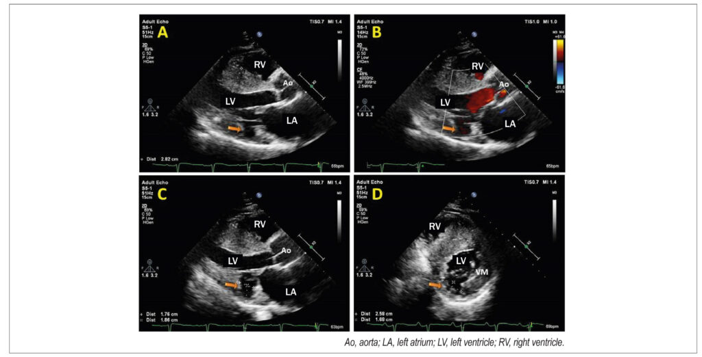
(A, B, and C) Transthoracic echocardiogram; longitudinal parasternal window. The arrow identifies a protrusion located in the basal segment of the inferolateral wall compatible with a diverticulum. (D) Short-axis view of the LV.
Comments are moderated by the editorial team before publication. Discussions should be evidence-based and use scientific language. Conflicts of interest should be declared.
Comments are moderated by the editorial team before publication. Discussions should be evidence-based and use scientific language. Conflicts of interest should be declared.