ABC Family Arquivos Brasileiros de Cardiologia International Journal of Cardiovascular Sciences ABC Heart Failure & Cardiomyopathy ABC Imagem Cardiovascular
Português English
ABC Family
Português English
  • About
    • About ABC Family
    • Internal Regulation
    • Editorial Board
    • Brazilian Society of Cardiology (SBC)
  • Issues
  • SBC Guidelines
  • Content
    • Videos
    • Podcasts
    • News
  • About
    • About ABC Family
    • Internal Regulation
    • Editorial Board
    • Brazilian Society of Cardiology (SBC)
  • Issues
  • SBC Guidelines
  • Content
    • Videos
    • Podcasts
    • News

ABC Family

  • ABC Family
  • Arquivos Brasileiros de Cardiologia
  • International Journal of Cardiovascular Sciences
  • ABC Heart Failure & Cardiomyopathy
  • ABC Imagem Cardiovascular
Search in

No content found.

Journals
Ordering
Article types
Others

  1. Home
  2. Keyword: Cardiac magnetic resonance
Figure 1
ABC
Original Article

Cardiac Magnetic Resonance as an Etiological Diagnosis Tool in Recovered Sudden Cardiac Death or Unstable Ventricular Arrhythmia Patients

Paula C. Marçal et al. Maria Fernanda Braggion-Santos Danilo Tadao Wada Marcel Koenigkam Santos … Henrique Turin Moreira Gustavo Jardim Volpe André Schmidt

Abstract

Background

Cardiac magnetic resonance (CMR) has an increasing diagnostic relevance in survivors of sudden cardiac death (SCD) or unstable ventricular arrhythmia (UVA) in developed countries.

Objective

To evaluate retrospectively the additional role of CMR in a developing country where few resources are available, and should be used more effectively.

Methods

The study included SCD or UVA survivors admitted between 2009 and 2019 at a tertiary academic institution referred to CMR. Demographic, clinical, and laboratory data were collected from the medical records. CMR images and reports were reviewed and their impact on the final etiological diagnosis was determined. A descriptive analysis was performed and p<0.05 established as significant.

Results

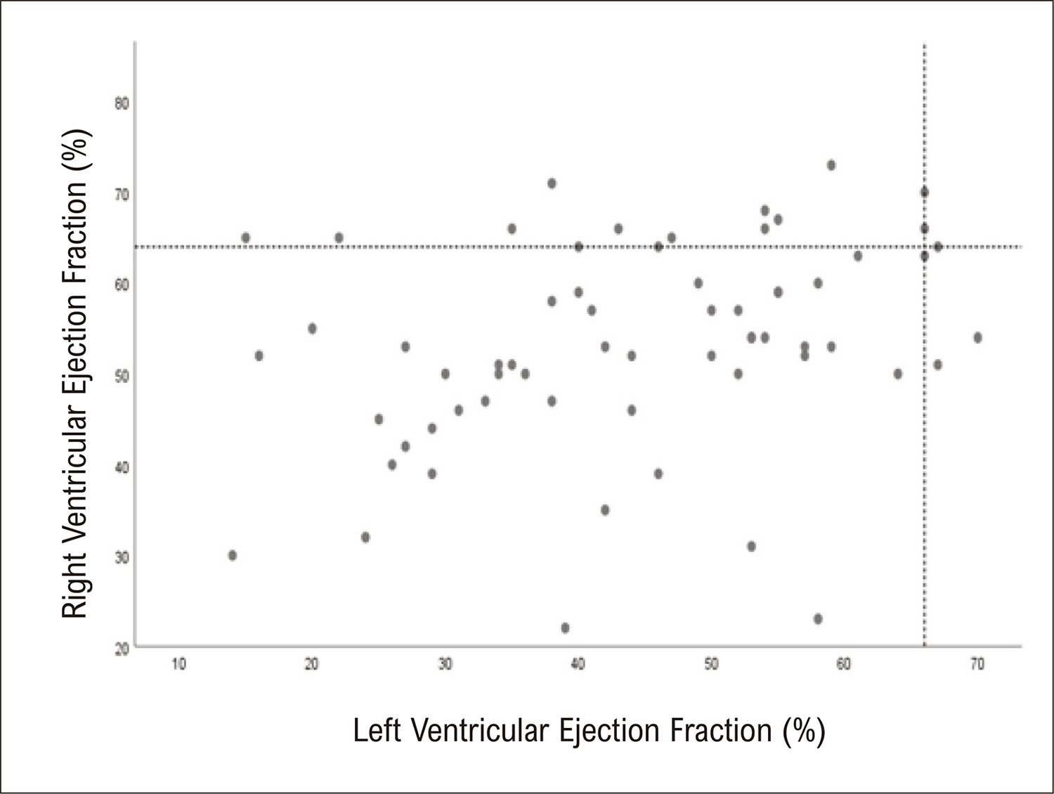
Sixty-four patients, 54.9±15.4 years old, and 42 (71.9%) males. Most events (81.3%) were out of the hospital and ventricular tachycardia was the most common rhythm. Cardiovascular medications were previously used by 55 patients, and beta-blockers were the most used medications (37.5%). Electrocardiogram had electrical inactive areas in 21.9% and all of them had fibrosis at CMR. Mean left ventricular ejection fraction (LVEF) was 44±14%, with 60.9% ≤50% and only 29.7% ≤35%. Late gadolinium enhancement was identified in 71.9%, with a transmural pattern in 43.8%. Chagas cardiomyopathy was the most common etiology (28.1%), followed by ischemic cardiomyopathy (17.2%). Among 26 without a previously identified etiology, CMR could define it (15 patients – 57%).

Conclusion

In accordance with previous studies in developed countries, CMR was capable of increasing etiological diagnosis and identifying the arrhythmogenic substrate, allowing better care in half of the underdiagnosed patients.
  • Cardiac magnetic resonance
  • Sudden Cardiac Death
  • Unstable Ventricular Tachycardia
  • ventricular arrhythmia
Read Full Article
Creative Commons Attribution (CC BY) Creative Commons license
21/04/2023 DOI: 10.36660/abc.20220411
Figure 1
ABCIC
Original Article

Myocardial Deformation (Strain) and Myocardial Fibrosis in Mild Cardiac Chagas Disease

Polyana Evangelista Lima et al. Rafael de Castro da Silva André Maurício Souza Fernandes Marta Silva Menezes Edmundo José Nassri Camara

Abstract

Background:

The early identification of myocardial damage seems important in the management of patients with Chagas disease. However, it is unknown whether speckle tracking echocardiography strain and cardiac magnetic resonance imaging are promising methods for assessing myocardial involvement and fibrosis, respectively.

Objective:

To evaluate myocardial involvement in the mild chronic cardiac form of Chagas disease using speckle tracking strain and myocardial fibrosis by cardiac magnetic resonance and assess their correlations.

Method:

This cross-sectional study analyzed patients with the mild chronic cardiac form of Chagas disease (preserved ejection fraction) using speckle tracking echocardiography and cardiac magnetic resonance.

Results:

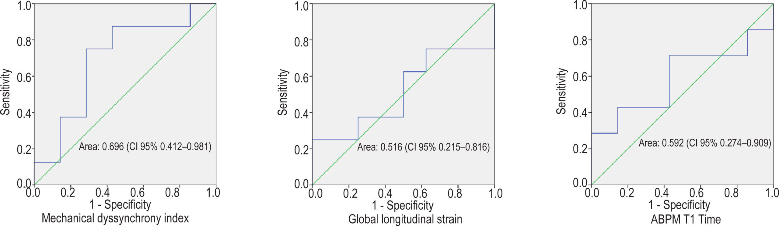
The study included 21 participants (women: 62%; age: 54 ± 5 years). The prevalence of myocardial fibrosis was 50% on delayed myocardial enhancement. The median global longitudinal strain was decreased (14.1%; interquartile range, 12.1–16.3%) in 17 patients (81%). The mean T1 mapping value was high in patients with Chagas disease (993 ± 163 ms). The T1 map was significantly correlated with the global longitudinal strain (r = 0.634; p = 0.015). In addition, the mechanical dispersion index obtained by strain was increased (>55 ms) by 84%, with the largest area under the receiver operating characteristic curve (area under the curve, 0.696; 95% confidence interval, 0.412–0.981) for fibrosis discrimination by delayed myocardial enhancement.

Conclusion:

Myocardial strain and T1 mapping are early markers of myocardial damage in mild chronic Chagas heart disease. The mechanical dispersion index was high and the most closely correlated with delayed myocardial enhancement.

  • Cardiac magnetic resonance
  • Chagas disease
  • Myocardial fibrosis
  • Myocardial strain
  • Speckle tracking echocardiography
Read Full Article
Creative Commons Attribution (CC BY) Creative Commons license
26/11/2021 DOI: 10.47593/2675-312X/20213403eabc173
ABC
Editorial

New Paradigms in the Evaluation of Diastolic Function by Cardiac Magnetic Resonance Imaging in Aortic Valvopathy

Vera Maria Cury Salemi et al. Marcelo Dantas Tavares de Melo José de Arimatéia Batista Araujo Filho

The noninvasive analysis of left ventricular diastolic function is a challenge in clinical practice. Estimation of the time constant of isovolumetric relaxation – or Tau constant – is the best and most established parameter for ventricular diastolic function analysis. However, this measure is obtained through an invasive assessment and it is very difficult to acquire. In clinical practice, echocardiography is considered a key tool, among all the complementary exams that can be used to evaluate diastolic function, validated in relation to the pressure-volume curves by catheterization, restricting the invasive evaluation to be used in exceptional cases. Thus, echocardiography remains the first-line noninvasive method, providing data on early diastolic dysfunction through indices that reflect relaxation, compliance, and also indirect measurements of ventricular filling pressures.

The latest recommendations for diastolic dysfunction assessment by echocardiography basically include analysis of the left ventricle without structural changes and preserved ejection fraction, as well as a flowchart for cases of left ventricular ejection fraction reduction/structural change. However, the classification of the degree of diastolic dysfunction and its application in the therapeutic management of valvular heart disease remains controversial., Moreover, aortic stenosis may be associated with mitral annulus calcification, which may lead to a reduction of the mitral orifice area, with an increase in early transmitral diastolic velocity (E), while the Doppler lateral mitral annulus velocity (e’) may be reduced due to limitation of the posterior cusp excursion, which may lead to an artificial increase in the E/e’ ratio. In aortic regurgitation, however, the aortic reflux jet may interfere with the mitral flow, which, when significant, can lead to a restrictive ventricular filling pattern; however, the accuracy of E/e’ ratio is questionable.

[…]

  • Aortic Stenosis
  • Aortic Valvopathy
  • Cardiac magnetic resonance
  • Diastolic Function
  • Echocardiography
Read Full Article
Creative Commons Attribution (CC BY) Creative Commons license
20/03/2020 DOI: 10.36660/abc.20190903
Figure 1
ABC
Image

Challenging Diagnosis of Myocardial Infarction Due to Anomalous Left Circumflex Artery

Maksymilian P. Opolski et al. Kajetan Grodecki Mateusz Spiewak Mariusz Furmanek Ilona Michalowska

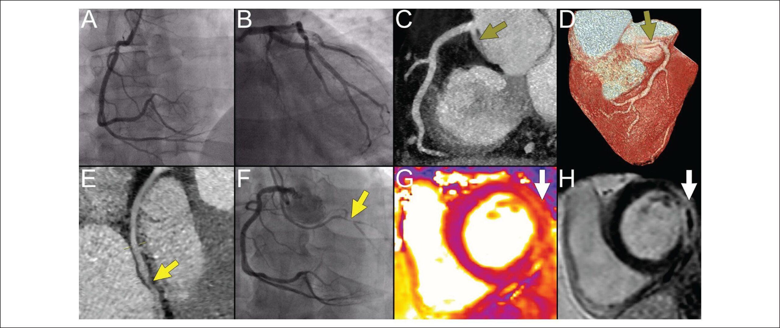
A 45-year-old male without past medical history presented with retrosternal chest pain and ST-segment elevation in inferolateral leads at ECG. Invasive coronary angiography, along with optical coherence tomography performed as part of the clinical study, showed normal coronaries, and myocardial infarction with non-obstructive coronary arteries (MINOCA) was diagnosed (-). Due to ongoing chest pain, triple-rule-out computed tomography angiography (CTA) was undertaken to exclude aortic dissection and pulmonary embolism. Incidentally, anomalous left circumflex artery (LCx) originating from the right sinus of Valsalva with a suspicion on severe stenosis was detected (-). Selective angiography of the LCx confirmed severe lesion in the distal vessel segment (), however given the resolution of patient’s symptoms, a decision on medical therapy with dual antiplatelet agents was undertaken. At discharge, cardiac magnetic resonance disclosed mildly reduced left ventricular ejection fraction (53%) with myocardial edema and transmural infarction of the basal-to-mid lateral wall (-).

[…]

  • Cardiac magnetic resonance
  • Coronary Angiography
  • Coronary Artery Anomalies
  • Myocardial Infarction / Diagnosis
Read Full Article
Creative Commons Attribution (CC BY) Creative Commons license
01/06/2018 DOI: 10.5935/abc.20180093
Latent obstructive hypertrophic cardiomyopathy: is echocardiography enough?
ABC
Case Report

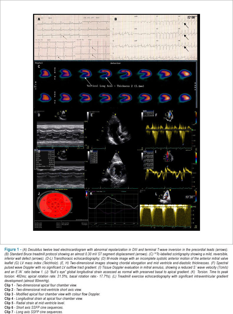
Latent obstructive hypertrophic cardiomyopathy: is echocardiography enough?

João Abecasis et al. Regina Ribeiras Antonio Ferreira Raquel Gouveia Miguel Mendes

CASE REPORT

Latent obstructive hypertrophic cardiomyopathy: is echocardiography enough?

[…]

  • Cardiac magnetic resonance
  • cardiomyopathy
  • Echocardiography
  • hypertrophic
Read Full Article
Creative Commons Attribution (CC BY) Creative Commons license
04/09/2012 DOI: 10.1590/S0066-782X2012001000016 Views 1

How to cite this article

EndNote RIS

Electronic Document Format (Vancouver)

Marçal PC, Braggion-Santos MF, Wada DT, Santos MK, Moreira HT, Volpe GJ, Schmidt A, et al. Cardiac Magnetic Resonance as an Etiological Diagnosis Tool in Recovered Sudden Cardiac Death or Unstable Ventricular Arrhythmia Patients. Arquivos Brasileiros de Cardiologia 2023;120(4):e20220411.

Electronic Document Format (ABNT)

Marçal, Paula C.; Braggion-Santos, Maria Fernanda; Wada, Danilo Tadao; Santos, Marcel Koenigkam; Moreira, Henrique Turin; Volpe, Gustavo Jardim; Schmidt, André. Cardiac Magnetic Resonance as an Etiological Diagnosis Tool in Recovered Sudden Cardiac Death or Unstable Ventricular Arrhythmia Patients. Arquivos Brasileiros de Cardiologia, v. 120, n. 4, e20220411, Apr. 2023.

Electronic Document Format (APA)

Marçal, P. C., Braggion-Santos, M. F., Wada, D. T., Santos, M. K., Moreira, H. T., Volpe, G. J., & Schmidt, A. (2023). Cardiac Magnetic Resonance as an Etiological Diagnosis Tool in Recovered Sudden Cardiac Death or Unstable Ventricular Arrhythmia Patients. Arquivos Brasileiros de Cardiologia, 120(4), e20220411.

Electronic Document Format (ISO)

Marçal, Paula C. and Braggion-Santos, Maria Fernanda and Wada, Danilo Tadao and Santos, Marcel Koenigkam and Moreira, Henrique Turin and Volpe, Gustavo Jardim and Schmidt, André. Cardiac Magnetic Resonance as an Etiological Diagnosis Tool in Recovered Sudden Cardiac Death or Unstable Ventricular Arrhythmia Patients. Arquivos Brasileiros de Cardiologia [online]. 2023, vol. 120, n. 4, [cited 2026-10-14], e20220411. Available from: <https://journals.cardiol.br/en/article/cardiac-magnetic-resonance-as-an-etiological-diagnosis-tool-in-recovered-sudden-cardiac-death-or-unstable-ventricular-arrhythmia-patients/>. ISSN 0066-782X

EndNote RIS

Electronic Document Format (Vancouver)

Lima PE, Silva RC, Fernandes AMS, Menezes MS, Camara EJN. Myocardial Deformation (Strain) and Myocardial Fibrosis in Mild Cardiac Chagas Disease. ABC Imagem Cardiovascular 2021;34(3):eabc173.

Electronic Document Format (ABNT)

Lima, Polyana Evangelista; Silva, Rafael de Castro da; Fernandes, André Maurício Souza; Menezes, Marta Silva; Camara, Edmundo José Nassri. Myocardial Deformation (Strain) and Myocardial Fibrosis in Mild Cardiac Chagas Disease. ABC Imagem Cardiovascular, v. 34, n. 3, eabc173, Nov. 2021.

Electronic Document Format (APA)

Lima, P. E., Silva, R. C. , Fernandes, A. M. S., Menezes, M. S., & Camara, E. J. N. (2021). Myocardial Deformation (Strain) and Myocardial Fibrosis in Mild Cardiac Chagas Disease. ABC Imagem Cardiovascular, 34(3), eabc173.

Electronic Document Format (ISO)

Lima, Polyana Evangelista and Silva, Rafael de Castro da and Fernandes, André Maurício Souza and Menezes, Marta Silva and Camara, Edmundo José Nassri. Myocardial Deformation (Strain) and Myocardial Fibrosis in Mild Cardiac Chagas Disease. ABC Imagem Cardiovascular [online]. 2021, vol. 34, n. 3, [cited 2026-10-14], eabc173. Available from: <https://journals.cardiol.br/en/article/myocardial-deformation-strain-and-myocardial-fibrosis-in-mild-cardiac-chagas-disease/>. ISSN 2318-8219

EndNote RIS

Electronic Document Format (Vancouver)

Salemi VMC, Melo MDT, Araujo Filho JAB. New Paradigms in the Evaluation of Diastolic Function by Cardiac Magnetic Resonance Imaging in Aortic Valvopathy. Arquivos Brasileiros de Cardiologia 2020;114(2):293-4.

Electronic Document Format (ABNT)

Salemi, Vera Maria Cury; Melo, Marcelo Dantas Tavares de; Araujo Filho, José de Arimatéia Batista. New Paradigms in the Evaluation of Diastolic Function by Cardiac Magnetic Resonance Imaging in Aortic Valvopathy. Arquivos Brasileiros de Cardiologia, v. 114, n. 2, p. 293-294, Mar. 2020.

Electronic Document Format (APA)

Salemi, V. M. C., Melo, M. D. T. , & Araujo Filho, J. A. B. (2020). New Paradigms in the Evaluation of Diastolic Function by Cardiac Magnetic Resonance Imaging in Aortic Valvopathy. Arquivos Brasileiros de Cardiologia, 114(2), 293-294.

Electronic Document Format (ISO)

Salemi, Vera Maria Cury and Melo, Marcelo Dantas Tavares de and Araujo Filho, José de Arimatéia Batista. New Paradigms in the Evaluation of Diastolic Function by Cardiac Magnetic Resonance Imaging in Aortic Valvopathy. Arquivos Brasileiros de Cardiologia [online]. 2020, vol. 114, n. 2, [cited 2026-10-14], pp. 293-294. Available from: <https://journals.cardiol.br/en/article/new-paradigms-in-the-evaluation-of-diastolic-function-by-cardiac-magnetic-resonance-imaging-in-aortic-valvopathy/>. ISSN 0066-782X

EndNote RIS

Electronic Document Format (Vancouver)

Opolski MP, Grodecki K, Spiewak M, Furmanek M, Michalowska I. Challenging Diagnosis of Myocardial Infarction Due to Anomalous Left Circumflex Artery. Arquivos Brasileiros de Cardiologia 2018;110(6):600-1.

Electronic Document Format (ABNT)

Opolski, Maksymilian P.; Grodecki, Kajetan; Spiewak, Mateusz; Furmanek, Mariusz; Michalowska, Ilona. Challenging Diagnosis of Myocardial Infarction Due to Anomalous Left Circumflex Artery. Arquivos Brasileiros de Cardiologia, v. 110, n. 6, p. 600-601, May. 2018.

Electronic Document Format (APA)

Opolski, M. P., Grodecki, K., Spiewak, M., Furmanek, M., & Michalowska, I. (2018). Challenging Diagnosis of Myocardial Infarction Due to Anomalous Left Circumflex Artery. Arquivos Brasileiros de Cardiologia, 110(6), 600-601.

Electronic Document Format (ISO)

Opolski, Maksymilian P. and Grodecki, Kajetan and Spiewak, Mateusz and Furmanek, Mariusz and Michalowska, Ilona. Challenging Diagnosis of Myocardial Infarction Due to Anomalous Left Circumflex Artery. Arquivos Brasileiros de Cardiologia [online]. 2018, vol. 110, n. 6, [cited 2026-10-14], pp. 600-601. Available from: <https://journals.cardiol.br/en/article/challenging-diagnosis-of-myocardial-infarction-due-to-anomalous-left-circumflex-artery/>. ISSN 0066-782X

EndNote RIS

Electronic Document Format (Vancouver)

Abecasis J, Ribeiras R, Ferreira A, Gouveia R, Mendes M. Latent obstructive hypertrophic cardiomyopathy: is echocardiography enough?. Arquivos Brasileiros de Cardiologia 2012;99(1):e108-11.

Electronic Document Format (ABNT)

Abecasis, João; Ribeiras, Regina; Ferreira, Antonio; Gouveia, Raquel; Mendes, Miguel. Latent obstructive hypertrophic cardiomyopathy: is echocardiography enough?. Arquivos Brasileiros de Cardiologia, v. 99, n. 1, p. e108-e111, Sep. 2012.

Electronic Document Format (APA)

Abecasis, J., Ribeiras, R., Ferreira, A., Gouveia, R., & Mendes, M. (2012). Latent obstructive hypertrophic cardiomyopathy: is echocardiography enough?. Arquivos Brasileiros de Cardiologia, 99(1), e108-e111.

Electronic Document Format (ISO)

Abecasis, João and Ribeiras, Regina and Ferreira, Antonio and Gouveia, Raquel and Mendes, Miguel. Latent obstructive hypertrophic cardiomyopathy: is echocardiography enough?. Arquivos Brasileiros de Cardiologia [online]. 2012, vol. 99, n. 1, [cited 2026-10-14], pp. e108-e111. Available from: <https://journals.cardiol.br/en/article/latent-obstructive-hypertrophic-cardiomyopathy-is-echocardiography-enough/>. ISSN 0066-782X

  • About ABC Family
  • Internal Regulation
  • Editorial Board
  • Brazilian Society of Cardiology (SBC)

Issues

SBC Guidelines

  • Videos
  • Podcasts
  • News