| ABCIC
ABC Family Arquivos Brasileiros de Cardiologia International Journal of Cardiovascular Sciences ABC Heart Failure & Cardiomyopathy ABC Imagem Cardiovascular
Português English
ABC Family | ABCIC
Português English
  • About
    • About the Journal
    • Internal Regulation
    • Editors Section
    • Editorial Board
    • SBC Award
  • Instructions to Authors
    • Guidelines for Publication
    • Editorial Policy
    • Ethics Policy
    • Policy on the Use of Artificial Intelligence
    • Mandatory Publication Form
  • Issues
  • SBC Guidelines
  • About
    • About the Journal
    • Internal Regulation
    • Editors Section
    • Editorial Board
    • SBC Award
  • Instructions to Authors
    • Guidelines for Publication
    • Editorial Policy
    • Ethics Policy
    • Policy on the Use of Artificial Intelligence
    • Mandatory Publication Form
  • Issues
  • SBC Guidelines

ABC Family

  • ABC Family
  • Arquivos Brasileiros de Cardiologia
  • International Journal of Cardiovascular Sciences
  • ABC Heart Failure & Cardiomyopathy
  • ABC Imagem Cardiovascular
Search in

No content found.

Journals
Ordering
Article types
Others

  1. Home
  2. ABCIC
  3. Issues
  4. 2023, Volume 36, Number 2
Figure 1
Review Article

My Approach to Echocardiographic Evaluation after Pediatric Heart Transplantation to Control Rejection and/or Graft Vascular Disease

Adailson Wagner da Silva Siqueira et al. Mirela Frederico de Almeida

Abstract

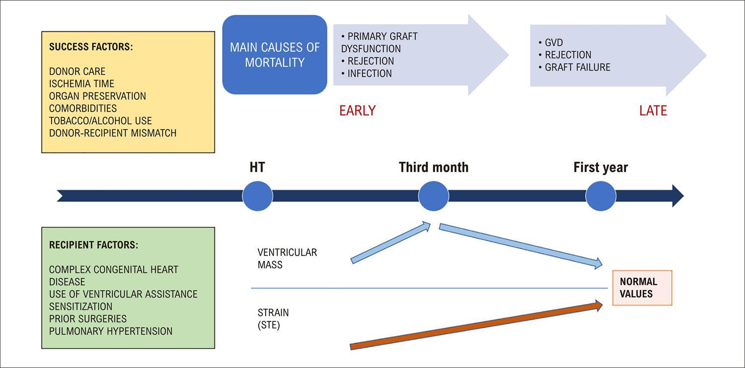
Pediatric heart transplantation is the definitive therapeutic option for patients with heart failure refractory to optimized clinical treatment, due either to cardiomyopathies or congenital heart disease. Morbidity and mortality remain concerning factors during evolution, and primary dysfunction, rejection, and graft vascular disease (GVD) are the main causes of death in the first 5 years after transplantation. As a surveillance method, transthoracic echocardiography has significant benefits in assisting diagnosis when there is clinical suspicion of rejection or GVD. The objective of this article is to present, in a clear and objective manner, which echocardiographic data assist pediatric cardiologists in follow-up of transplanted patients. The use of echocardiography, either through conventional tools or advanced methods, carefully focusing on different early and late post-cardiac transplantation phases, with information from current and comparative examination of evolution, mainly regarding tissue Doppler and longitudinal strain, can assist pediatric cardiologists in the decision to anticipate endomyocardial biopsy and coronary angiography (gold standard) for early diagnosis and immediate intervention, allowing greater graft durability.

  • Coronary Disease
  • Echocardiography
  • Heart Defects, Congenital
  • Heart Transplantation
Read Full Article
Creative Commons Attribution (CC BY) Creative Commons license
30/06/2023 DOI: 10.36660/abcimg.20230045i
Figure 1
Review Article

My Approach to Three-Dimensional Echocardiography for Pathophysiological Classification of Tricuspid Regurgitation

Alex dos Santos Felix et al. Monica Luiza de Alcantara Konstantinos Papadopoulos

Introduction

Although it was known as the “forgotten valve” in the past, there has been increasing interest in studying the tricuspid valve (TV) during the last two decades. Tricuspid regurgitation (TR) has been identified as a prognostic marker not only when associated with other cardiac diseases, like heart failure, mitral regurgitation, or aortic stenosis, – but also as an isolated entity. Understanding the pathophysiological mechanisms of this disease is of paramount importance, since new options of transcatheter devices and techniques are emerging, offering invasive treatment for high-risk patients unsuitable for surgical intervention.

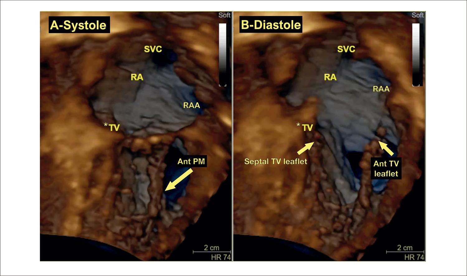
The TV is difficult to image in transesophageal echocardiography (TEE) for the following reasons: 1) the leaflets are much thinner compared to mitral leaflets, with greater anatomic variability; 2) it is an anterior structure, far away from the esophagus, with acoustic shadowing from the fibrous heart skeleton; and 3) it cannot be aligned to the esophageal probe in order to acquire en face views and requires use of lateral resolution. These restrictions may limit the ability of traditional TEE to evaluate the TV, making it necessary to complement the evaluation with special windows from the lower esophagus and transgastric views. Different from other cardiac valves, images of the TV obtained from transthoracic echocardiography (TTE) usually have better resolution than images obtained by TEE. Even though all protocols start with two-dimensional (2D) assessment of the TV, three-dimensional echocardiography (3DE) plays an important role in the evaluation of TV diseases, due not only to its ability to precisely depict the anatomy of the valve and the subvalvular apparatus, but also to its accuracy in quantitation of right ventricular (RV) and right atrial volumes and function and functional analysis of valvular dysfunction, especially for grading TR and evaluating dynamics of the tricuspid annulus (TA) through dedicated software. All of this information is of unparalleled importance for patient management and pre-procedural planning in surgical and transcatheter approaches.

[…]

  • Echocardiography, Three-Dimensional
  • Tricuspid Valve
  • Tricuspid Valve Insufficiency
Read Full Article
Creative Commons Attribution (CC BY) Creative Commons license
30/06/2023 DOI: 10.36660/abcimg.20230055i Views 3
Figure 1
Case Report

Dual Mechanism of Mitral Valve Injury: Additional Value of Three-Dimensional Transesophageal Echocardiography

Larissa Neto Espíndola et al. Angele Azevedo Alves Mattoso Geórgia dos Santos Couto Pompílio Sampaio Britto

Abstract

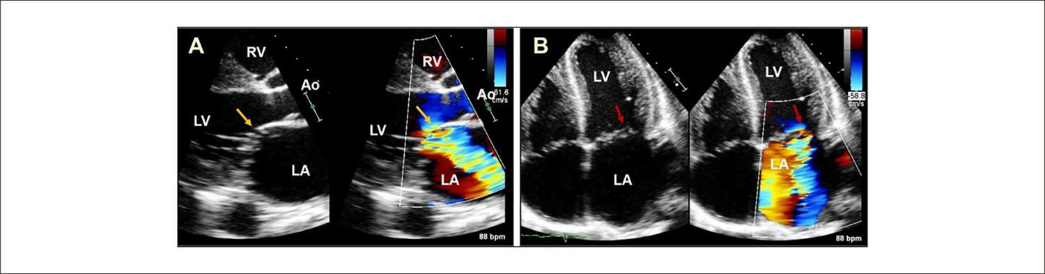
Infective endocarditis (IE) is a life-threatening condition and during its course several complications may occur, including embolic events, perivalvular extension and valvular destruction. Echocardiography is fundamental across all stages of the disease, from the diagnosis until the follow-up after completion of the definitive therapy. Three-dimensional transesophageal echocardiography (TEE) can demonstrate a better anatomical definition and it is especially useful in the assessment of perivalvular extension of the infection, prosthetic valve dehiscence and valve perforation. In this article we report a case of double damage in the same mitral valve: leaflet perforation and rupture of chords in a patient with IE.

  • Echocardiography, Three-Dimensional
  • Endocarditis
  • Mitral Valve
Read Full Article
Creative Commons Attribution (CC BY) Creative Commons license
30/06/2023 DOI: 10.36660/abcimg.2023322i Views 1
Review Article

My Approach To: Vascular Ultrasonography in Dolichoarteriopathies of the Carotid Arteries

Armando Luis Cantisano et al. Catarina Schiavo Grubert

Abstract

This article provides step-by-step guidance on how I investigate, classify and evaluate dolichoarteriopathies anatomically and hemodynamically, often found in the carotid and vertebral arteries. Dolichoarteriopathies may be present in children, disappearing during growth and reappearing with increasing age. Previously considered benign anatomic variations (AVs), discussion has arisen on the topic in view of new implications due to associations with cardiovascular events, risk factors, and various pathologies.

  • Anatomic Variation
  • Carotid Arteries
  • Ultrasonography, Carotid Arteries
Read Full Article
Creative Commons Attribution (CC BY) Creative Commons license
30/06/2023 DOI: 10.36660/abcimg.20230034i Views 1
Figure 1
Original Article

Right Ventricle Diastolic Function: Correlation with Age

Arthur Nascimento de Moura et al. Ana Cristina Camarozano Cintia Rocha Fortes de Sá Daniela de Castro Carmo … Jerônimo Antonio Fortunato Rubens Zenóbio Darwich Liz Andréa Villela Baroncini

Abstract

Introduction

Right ventricle (RV) elasticity tends to decrease with aging and may alter its diastolic function.

Objective

To evaluate the correlation between echocardiographic changes in RV diastolic function and advancing age in individuals with normal biventricular systolic function.

Methods

This retrospective observational cohort study included 110 patients: 66 (60%) women (62 [SD, 13.7] years) and 44 (40%) men (60.6 [SD, 10.8] years). The transthoracic echocardiography data included: spectral Doppler of tricuspid inflow E wave, A wave, E/A ratio, and E wave deceleration time; tissue Doppler of lateral tricuspid annulus e’ wave, a’ wave, and E/e’ ratio. Correlations were calculated using Pearson’s and Spearman’s correlation coefficients.

Results

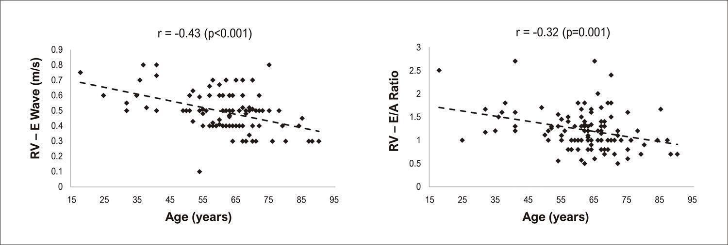
Only 35.2% of the sample had RV diastolic dysfunction. Of the evaluated echocardiographic variables, the E wave was negatively correlated with age in both women (r 0.473; p < 0.001) and men (r -0.37, p = 0.015). The E/A ratio was negatively correlated with age only in women (r -0.32; p = 0.001), and E wave deceleration time was positively correlated with age only in women (r 0.31; p = 0.014). The other variables were not significantly correlated with age in men or women.

Conclusions

We found a significant correlation between age and echocardiographic parameters for RV diastolic dysfunction, which was more evident in women.

  • Echocardiography
  • Heart Failure, Diastolic
  • Heart Ventricles
Read Full Article
Creative Commons Attribution (CC BY) Creative Commons license
27/06/2023 DOI: 10.36660/abcimg.2023333i Views 1
Figure 1
Review Article

My Approach to Echocardiographic Assessment of the Premature Newborn

Karen Saori Shiraishi Sawamura et al. Márcio Miranda Brito

Abstract

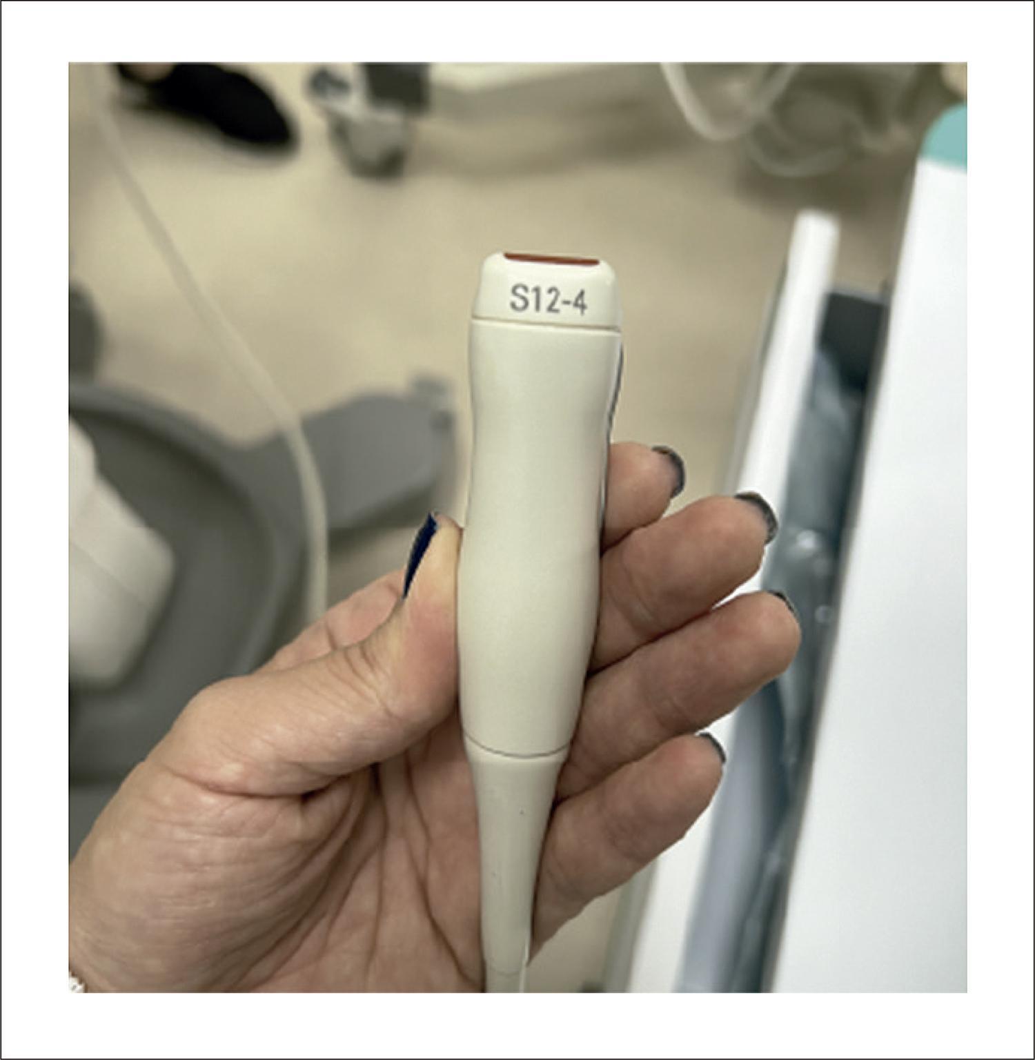
Echocardiographic assessment of preterm infants, why do it? The incidence of premature births has been increasing worldwide and already affects about 10% of live births. Extremely preterm newborns (PTNB) are at increased risk of developing pulmonary immaturity, leading to pulmonary hypertension (PH), in addition to cardiovascular immaturity and patent ductus arteriosus (PDA).Hemodynamic monitoring of this population is challenging and differs from critically ill pediatric patients due to hemodynamic lability. Thus, minimal manipulation is essential to avoid complications. In this context, non-invasive monitoring methods such as transthoracic echocardiography (TTE) gain importance for hemodynamic assessment. It is a low-cost, radiation-free test that offers good anatomical and functional visualization in pediatric patients, making it a useful tool in the clinical management of severe preterm newborns.What you will find in this article:We will take an objective, didactic, and at times humorous approach to the use of echocardiography in premature newborns in the neonatal intensive care unit (NICU), and we will discuss the echocardiographic particularities of premature infants, especially regarding equipment adjustments, PH, and persistence of the ductus arteriosus (DA).

  • Ductus Arteriosus
  • Echocardiography
  • Neonatology
  • Premature Infant
  • Pulmonary Hypertension
Read Full Article
Creative Commons Attribution (CC BY) Creative Commons license
27/06/2023 DOI: 10.36660/abcimg.20230035i Views 1
Figure 1
Case Report

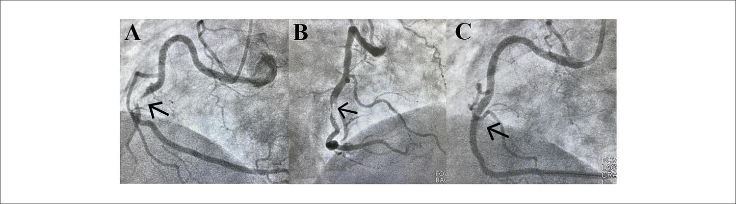
Acute Coronary Syndrome Followed by Pulmonary Thromboembolism and Identification of a Large Fixed Thrombus between Atria in a Patient with Severe COVID-19: A Case Report

Alice Mirane Malta Carrijo et al. Veronica Perius de Brito Gabriel Alvarenga Santos Samuel Gomes Tomaz da Silva João Lucas O’Connell

Introduction

Patent foramen ovale (PFO) is a relatively frequent congenital heart defect; however, the identification of large thrombi between the atria through a PFO is a rare condition. When visualized, they represent a major clinical challenge and require emergency treatment.

The severity of this condition becomes even more accentuated when associated with other thrombotic events, such as pulmonary embolism. In turn, these events may have their genesis in hyperinflammatory states with high thrombogenic and angiopathic potential, as in the case of severe acute respiratory syndrome coronavirus 2 (SARS-CoV-2) infection.

[…]

  • Case Reports
  • Patent Oval Foramen
  • Pulmonary embolism
Read Full Article
Creative Commons Attribution (CC BY) Creative Commons license
27/06/2023 DOI: 10.36660/abcimg.2023351i
Figure 1
Case Report

Patent Forame Ovale: Contribution of Transcranial Doppler in Patient Diagnosis

Eliza de Almeida Gripp et al. Ana Carolina de Freitas Portela Flávio Luis da Costa Junior Rafael Rabishoffsky … Jéssica Rizkalla Corrêa Medeiros Arnaldo Rabischoffsky

Introduction

Patent forame ovale (PFO) is the most common type of interatrial shunt, with a prevalence between 25% and 27% of the adult population. It has a flap-like functioning and is generally asymptomatic. In some cases, it can be associated with clinical complications, such as brain strokes, paradoxical embolism, platypnea-orthodeoxia syndrome, and obstructive sleep apnea. The diagnosis is mainly found in young patients who present brain strokes, with no other apparent risk factor.

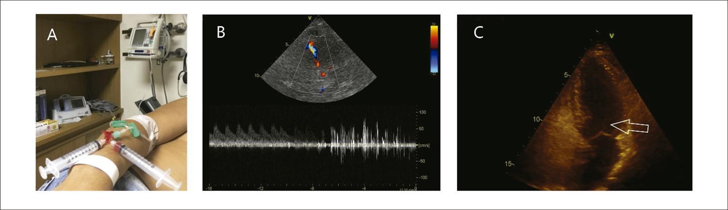
The evaluation of PFO is commonly performed by means of transthoracic echocardiogram (TTE) and transesophageal echocardiogram (TEE). However, another important method of evaluation that is rarely used in our area is the Transcranial Doppler (TCD), which evaluates the presence of an indirect shunt in an awake patient and with high sensitivity.

[…]

  • Echocardiography, Doppler
  • Foramen Ovale
  • Patente
  • Ultrasonography, Doppler, Transcranial
Read Full Article
Creative Commons Attribution (CC BY) Creative Commons license
27/06/2023 DOI: 10.36660/abcimg.20230020i Views 1
Editorial

Lung Ultrasound in Outpatients with Heart Failure

Marco Stephan Lofrano-Alves

Over the last decade, we have witnessed the widespread use of lung ultrasound (LU) in the assessment of pulmonary congestion. In addition to the well-known technique of auscultation of adventitious lung sounds, LU makes an important contribution by sharpening our vision to the diagnosis of pulmonary interstitial edema by quantification of B-lines. LU has emerged as a relevant and updated method, since it is a safe, low-cost, rapid and available at bedside, being an alternative to imaging methods associated with ionizing radiation, such as chest tomography and X-ray.

LU is a standardized tool in diagnosis and treatment monitoring today, and has been studied in different clinical scenarios of pulmonary congestion of cardiogenic origin.– In outpatients with heart failure and reduced ejection fraction (HFrEF), B-line counting by LU showed an 89% accuracy with a cutoff ≥ 15 B-lines. In another study with patients hospitalized for HFrEF, the risk of an adverse in-hospital event increased with the rising number of B-lines at admission, and the risk of HF hospitalization and all-cause death was greater in patients with a higher number of B-lines at discharge.

[…]

  • Heart Failure
  • Lung
  • Ultrasonics
Read Full Article
Creative Commons Attribution (CC BY) Creative Commons license
27/06/2023 DOI: 10.36660/abcimg.20230049i
Editorial

The Use of Echocardiography in Schistosomiasis

Daniela do Carmo Rassi

Schistosomiasis, a neglected tropical disease, affects 240 million people worldwide, with more than 90% of cases reported in Africa, according to the World Health Organization. The disease is endemic to South America, the Caribbean, Southeast Asia, and Africa, in areas with little infrastructure, especially lack of drinking water and basic sanitation. The infection is caused by helminths of the genus Schistosoma. Contamination occurs through contact with water where host snails are present. After penetrating human skin, the worms can migrate and produce eggs in the liver, lungs, and intestines.

During the acute phase, patients may be asymptomatic or develop myocarditis and pericarditis related to an allergic reaction to the schistosomes, in which eosinophils play an important role. Myocardial ischemia may rarely occur, and the exact mechanism remains unknown.

[…]

  • Cardiovascular Diseases
  • Echocardiography
  • Schistosomiasis
Read Full Article
Creative Commons Attribution (CC BY) Creative Commons license
27/06/2023 DOI: 10.36660/abcimg.20230037i
Figure 1
Case Report

Expandable Airway Stents: Success in the Extrinsic Bronchial Compression Approach After Reconstruction of the Aortic Arch

Camila Magalhães Silva et al. Adriana Furletti Machado Guimarães Ricardo Wang Carla Maria Silva e Alves Edmundo Clarindo Oliveira

Introduction

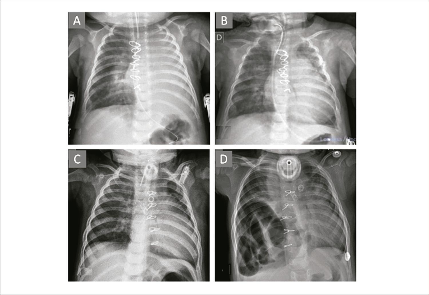
The interruption of the aortic arch is a rare congenital cardiopathy, whose surgical correction generally requires reconstruction of the aortic arch. This intervention can cause distortion of the aortopulmonary space and results in bronchial compression with a consequent pulmonary atelectasis and dependence on ventilatory assistance. The most commonly used therapeutic options for bronchial decompression are not always enough to make pulmonary expansion possible. Our study presents a case of the implant of an endoluminal stent in an infant who, in the post-operative stage of the correction of the interruption of the aortic arch, developed complete atelectasis of the left lung secondary to bronchial compression, with no response after posterior aortopexy

[…]

  • Cardiology Service, Hospital
  • Pediatrics
  • Stents
Read Full Article
Creative Commons Attribution (CC BY) Creative Commons license
27/06/2023 DOI: 10.36660/abcimg.20230023i
Figure 1
Case Report

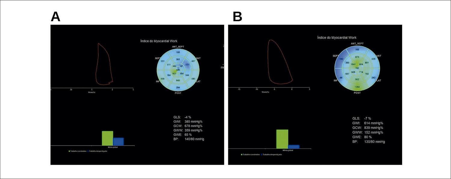
Comparative Analysis of Myocardial Work After Decongestion Therapy in a Patient With Acutely Decompensated HFrEF

Alexandre Costa Souza et al. Stephanie de Azevedo Drubi Bruna de Mattos Ivo Junqueira Ricardo André Sales Pereira Guedes … Adriano Chaves de Almeida Filho Carolina Thé Macêdo

Introduction

Acutely decompensated reduced ejection fraction heart failure (HFrEF) is one of the leading causes of hospitalization in our setting, with a 90-day readmission rate of 50%. The use of additional tools in the assessment of the left ventricular function, such as the measurement of myocardial deformation through the Global Longitudinal Strain of the left ventricle (GLS) has greater prognostic value than the evaluation of the left ventricular ejection fraction (LVEF) by the Simpson method. Despite the analysis of the myocardial deformity through the GLS being considered an independent predictor of mortality in patients with HFrEF, it is a parameter that experiences changes in pre- and after-load and, therefore, has limitations regarding the assessment of ventricular performance.– Myocardial work (MW) has stood out in recent years as a complementary tool to access parameters of myocardial function from the GLS-derived analysis, with the benefit of incorporating afterload information through the interpretation of the dynamic curve of deformity due to non-invasive left ventricular (LV) filling pressure. Hence, the analysis of MW has been encouraging in patients with HFrHF, and, when analyzed in combination with classic hemodynamic parameters, it can add information with greater accuracy and prognostic value in the cardiac decompensation scenario.–

[…]

  • Echocardiography
  • Heart Failure
  • Ventricular Dysfunction, Left
Read Full Article
Creative Commons Attribution (CC BY) Creative Commons license
27/06/2023 DOI: 10.36660/abcimg.20230008i
Editorial

How to Proceed in a Multimodality Analysis for the Diagnosis and Risk Stratification of Pulmonary Hypertension?

Pedro Gutiérrez-Fajardo

Transthoracic echocardiography

TTE is considered a highly useful, inexpensive, safe, and available imaging modality for initial approach to differential diagnosis of PH. Although a normal TTE does not rule out diagonsis, it decreases the likelihood of PH. Neither CT nor CMRI are part of the initial line of PH; however, in selected patients they can provide additional information before proceeding to right heart catheterization. ,

It is important to be familiar with normal ranges of RV size and function. , TTE showing right heart structure and function abnormalities increases suspicion of PH. It also allows detection of left heart (systolic and diastolic dysfunction, valvular heart disease) and congenital heart disease as potential causes of PH. RV function surrogates such as RV fractional area change, right atrial size, pericardial effusion, tricuspid annular plane systolic excursion (TAPSE), systolic pulmonary artery pressure (SPAP)/TAPSE ratio, and eccentricity index are recommended in follow-up and assessment of therapy response. However, it is important to note that these surrogates should be interpreted in the context of other clinical and imaging parameters, as they are not standalone diagnostic tools. Doppler can be used to estimate the possibility of PH by measuring tricuspid regurgitant jet velocity (TR) and calculating SPAP using the modified Bernoulli equation. When no TR is present or the signal is inadequate, pulmonary acceleration time can be used to calculate mPAP obtained from pulse wave Doppler signal of pulmonary flow. Pulmonary artery diastolic pressure can be estimated if pulmonary regurgitation is present. The velocity at end-diastole is converted into a pressure gradient, adding the estimated right atrial pressure to the resulting value. Finally, both left ventricle and RV output can be calculated. In clinical practice, differentiating acute, chronic, and chronic acute PH can be challenging ( ).

[…]

  • Diagnosis
  • Pulmonary Hypertension
  • Risk Factors
Read Full Article
Creative Commons Attribution (CC BY) Creative Commons license
21/06/2023 DOI: 10.36660/abcimg.20230041i Views 1
Figure 1
Case Report

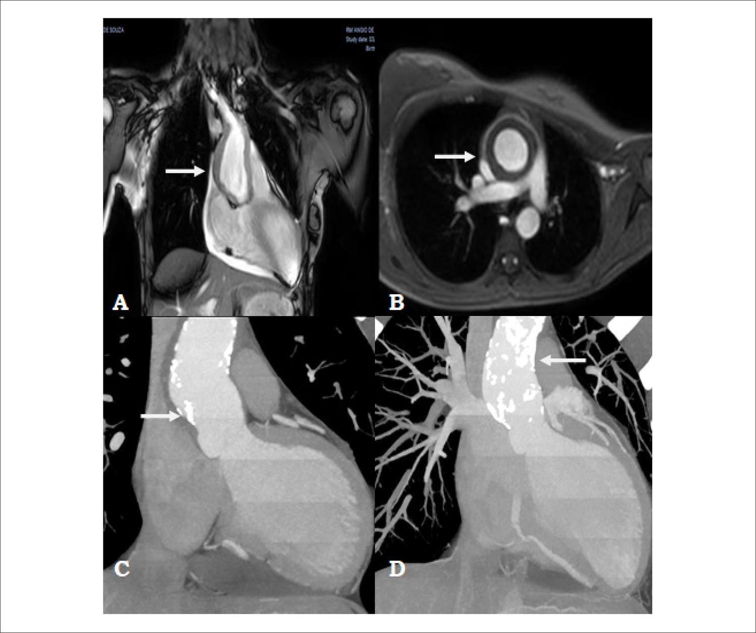
Takayasu’s Arteritis: Ascending Aortic Aneurysm and Coronary Artery Disease in a 19-year-old Young Adult

Iuri Betuel Gomes António et al. Adnaldo da Silveira Maia Janayna Rabelato Aloysio Abdo Silva Campos … Marilia Prudente Menezes Mário Issa

Abstract

Takayasu’s Arteritis (TA) is a disease of multiple presentation, which can involve many different systems and mainly affects women in the first decades of life. The present case reports on a 19-year-old patient with TA, with a diagnosis of ascending aortic aneurysm and severe coronary artery disease in the right coronary artery (RCA) and left coronary trunk (LCT), who underwent an implant of a supracoronary Dacron graft in the ascending aorta and myocardial revascularization with a double internal thoracic artery. The patient evolved satisfactorily in post-op and is today undergoing clinical follow-up in our medical services.

  • Aneurysm
  • Coronary Disease
  • Takayasu Arteritis
Read Full Article
Creative Commons Attribution (CC BY) Creative Commons license
21/06/2023 DOI: 10.36660/abcimg.20230032i Views 2
Figure 1
Editorial

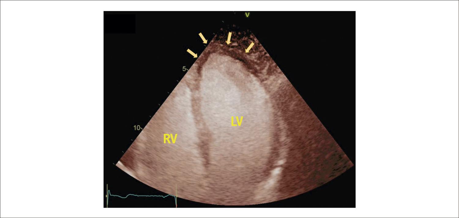
Contrast Echocardiography in Real Life: Practicality, Safety, and Cost-Effectiveness

Márcio Silva Miguel Lima

After approximately 2 decades of use in practice, based also on several clinical studies, it can be stated that ultrasound enhancing agents (UEA) are extremely safe, following recommendations for their preparation and administration.– Severe adverse events are very rare, with an approximate prevalence of around 1 in every 10,000 administrations, anaphylactoid reactions being the most frequent.,- Specifically with the contrast SonoVue® (sulfur hexafluoride) used in Brazil, Furtado, Rassi, et al., in their safety study, reported a very low incidence of only 0.6% of allergic reaction in their population of 1,099 patients who received this agent. In general, allergic reactions occur immediately or within 30 minutes after administration; they are of mild intensity and transitory. The most common presentation is the appearance of erythematous skin lesions and urticaria. In this scenario, however, special attention should be paid to respiratory signs and symptoms, such as dyspnea, laryngeal stridor, and bronchospasm, as well as signs and symptoms of progression to eventual circulatory collapse, such as dizziness, paleness, and hypotension, as these are warning signs for the onset of anaphylactic shock. Patients must be carefully evaluated and observed, and measures aimed at anaphylaxis should be promptly instituted with antihistamines, corticosteroids, and epinephrine, following the care protocol of the institution, whose personnel must be very well trained.

In October 2007, the United States Food and Drug Administration (FDA) issued a general contraindication (“black box”) for the use of Definity® and Optison® agents in acutely ill patients after analysis of 11 deaths potentially related to the use of these UEA. This restriction was later withdrawn after subsequent clinical studies provided extensive evidence of safety. Advanced pulmonary disease and pulmonary hypertension initially represented contraindications, but clinical trials confirmed their safety.– Additionally, in relation to the suspicion or presence of significant intracardiac shunt, also a previous contraindication, it was suspended by the FDA in 2016., Currently, only history of allergic reaction to any component of UEA constitutes an absolute contraindication to their use.

[…]

  • Cost-Benefit Analysis
  • Echocardiography
  • Safety
Read Full Article
Creative Commons Attribution (CC BY) Creative Commons license
21/06/2023 DOI: 10.36660/abcimg.20230033i Views 1
Fígure 1
Case Report

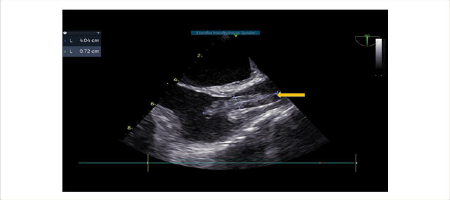
Transesophageal Echocardiogram in the Diagnosis of Superior Vena Cava Syndrome in a Patient With a Long-Term Catheter

Marcus Vinicius Silva Freire de Carvalho et al. Endy de Santana Alves Laila Caroline Gomes Carolina Thé Macêdo … Marco André Moraes Sales Alexandre Costa Souza

Introduction

The superior vena cava syndrome (SVCS) is caused by the obstruction or severe occlusion of the superior vena cava (SVC) and can result in significant morbidity or mortality., Malignancy is the most common cause of this obstruction, corresponding to approximately 70% of the cases. However, more recently, the incidence of SVCS related to devices, such as central venous catheters and electrodes of a pacemaker or defibrillator, have increased.,

These devices, together with states of hypercoagulability, can precipitate the formation of thrombi. In extreme situations, in the context of bacteremia, these can lead to the formation of a complex thrombus-vegetation association.

[…]

  • Diagnosis
  • syndrome, vein
  • Transesophageal echocardiography
Read Full Article
Creative Commons Attribution (CC BY) Creative Commons license
21/06/2023 DOI: 10.36660/abcimg.20230028i Views 2
Figure 1
Review Article

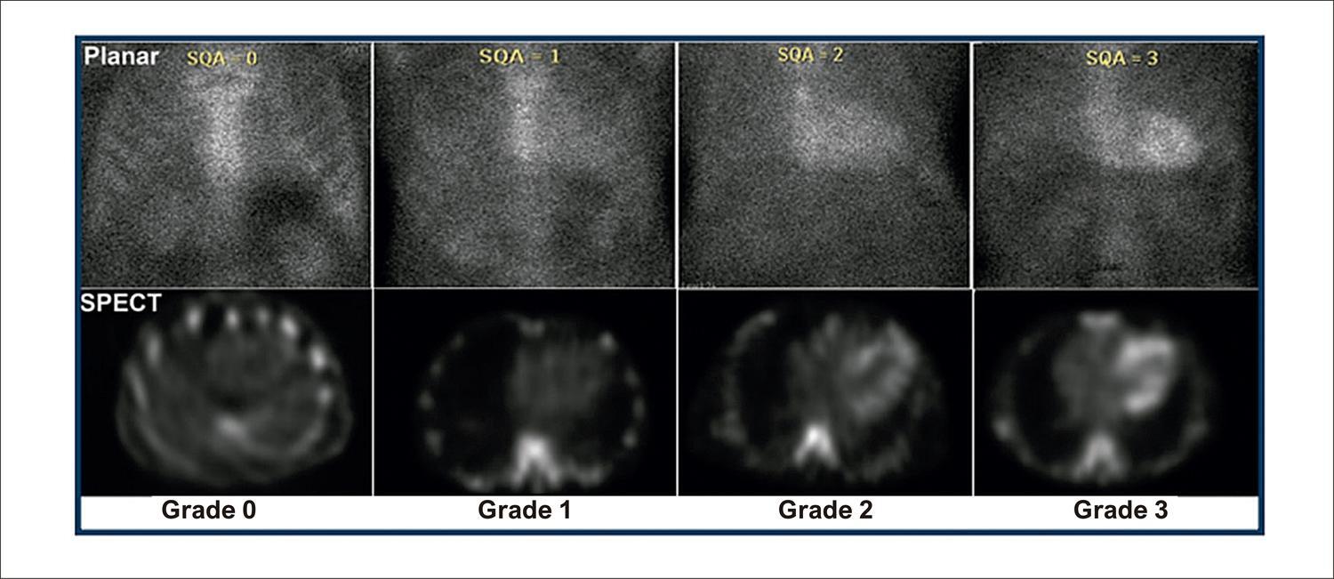
My Approach to Imaging Cardiac Amyloidosis: Role of Bone-Seeking Tracers Scintigraphy

Adriana Pereira Glavam et al. Rafael Willain Lopes Simone Cristina Soares Brandão

Abstract

Amyloidosis is a systemic infiltrative disease characterized by the extracellular deposition of amyloid fibrils. Heart involvement is common and associated with a poor prognosis. The most predominant types of cardiac amyloidosis (CA) are amyloid immunoglobulin light chain (AL) and amyloid transthyretin (ATTR). Diagnosis of CA and differentiation between the types are important for prognosis, therapy, and genetic counseling. ATTR-CA is an under-diagnosed cause of heart failure. However, great accomplishments in non-invasive imaging methods, as well as the possibility of effective clinical treatment, have shifted ATTR-CA from a rare and untreatable disease to a condition that clinicians should consider on a daily basis. The advent of scintigraphy imaging with bone-seeking tracers has allowed the early diagnosis of ATTR-CA with high accuracy once monoclonal gammopathies have been excluded. Interpretation of cardiac scintigraphy with bone-seeking tracers requires expertise, and we propose a step-by-step guide to performing this exam in clinical practice according to the most recent guidelines. Moreover, we reviewed some crucial points that we believe are of paramount importance in clinical practice and patient outcomes.

  • Amyloidosis
  • Cardiac insufficiency
  • Diphosphates
  • Pre-Albumin
  • Scintigraphy
Read Full Article
Creative Commons Attribution (CC BY) Creative Commons license
24/05/2023 DOI: 10.36660/abcimg.20230012i Views 1
Figure 1
Case Report

Behcet’s Disease with Vascular Involvement: Case Report

Isabela Rodrigues Tavares et al. Maria Cristina Miranda Ana Claudia Gomes Pereira Petisco Daniela Souto Barros … Caio Buscatti Folino Vitor Leonidas Boher Dornas Mohamed Hassan Saleh Fábio Henrique Rossi Raquel Peres Sousa Larissa Chaves Nunes Carvalho

Clinical case

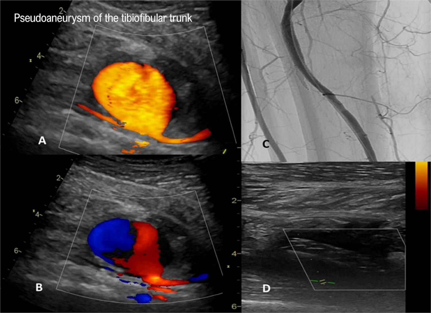
Male patient, 34 years of age, previously healthy, admitted to the Emergency Room (ER) of Instituto Dante Pazzanese de Cardiologia reporting a 3-month history of pain in the right lower limb (RLL) and limping for distances of 100 meters in the 3 weeks prior to admission. The patient evolved with worsening pain in the right calf and spontaneous pulsatile bulging a week before admission. He mentioned the use of Rivaroxaban for deep venous thrombosis (DVT) in the RLL five months before, weight loss (15kg in 6 months), and appearance of aphthous lesions in the oropharyngeal region. He had a vascular ultrasound image from another service, with signs of DVT in a partially recanalized right popliteal vein.

On the occasion of admission, physical examination indicated: posteromedial pulsatile mass on the right leg and present pulses, except posterior tibial. Heart, lungs, abdomen, and left lower limb (LLL) without alterations.

[…]

  • Behçet disease
  • Doppler ultrasonography
  • vasculitis
Read Full Article
Creative Commons Attribution (CC BY) Creative Commons license
24/05/2023 DOI: 10.36660/abcimg.20230014i
Graph 1
Original Article

Long-Term Evolution of Patients with Important Pulmonary Hypertension due to Schistosomiasis

José Maria Del Castillo et al. Katarina Barros de Oliveira Rafael Ricardo de Oliveira Travassos Ângela Maria Pontes Bandeira … Carlos Antônio da Mota Silveira Djair Brindeiro Filho

Abstract

Introduction

Hepatosplenic schistosomiasis mansoni, associated with pulmonary arterial hypertension (PAH), causes cardiac chamber remodeling. Little is known about its long-term evolution.

Objective

To evaluate the alterations caused by PAH in patients with schistosomiasis and analyze the clinical and echocardiographic evolution over a period of 10 years.

Methods

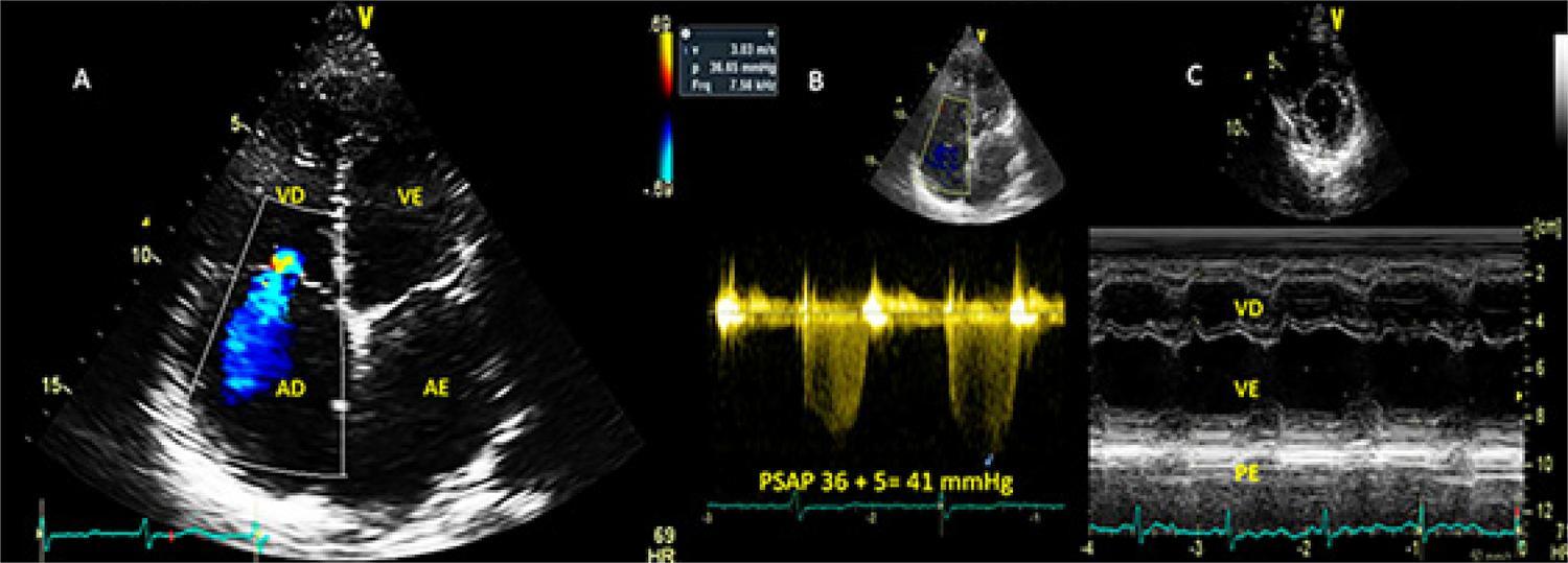
The study included 60 patients with PAH due to schistosomiasis and 50 healthy control patients. Clinical and echocardiographic data were evaluated, including dimensions, parietal thickness, function of the right and left chambers, and parameters of myocardial strain. Patients in the group with PAH due to schistosomiasis were followed for 10 years. Data were compared using Student’s t test for unpaired samples with significance level of < 5%.

Results

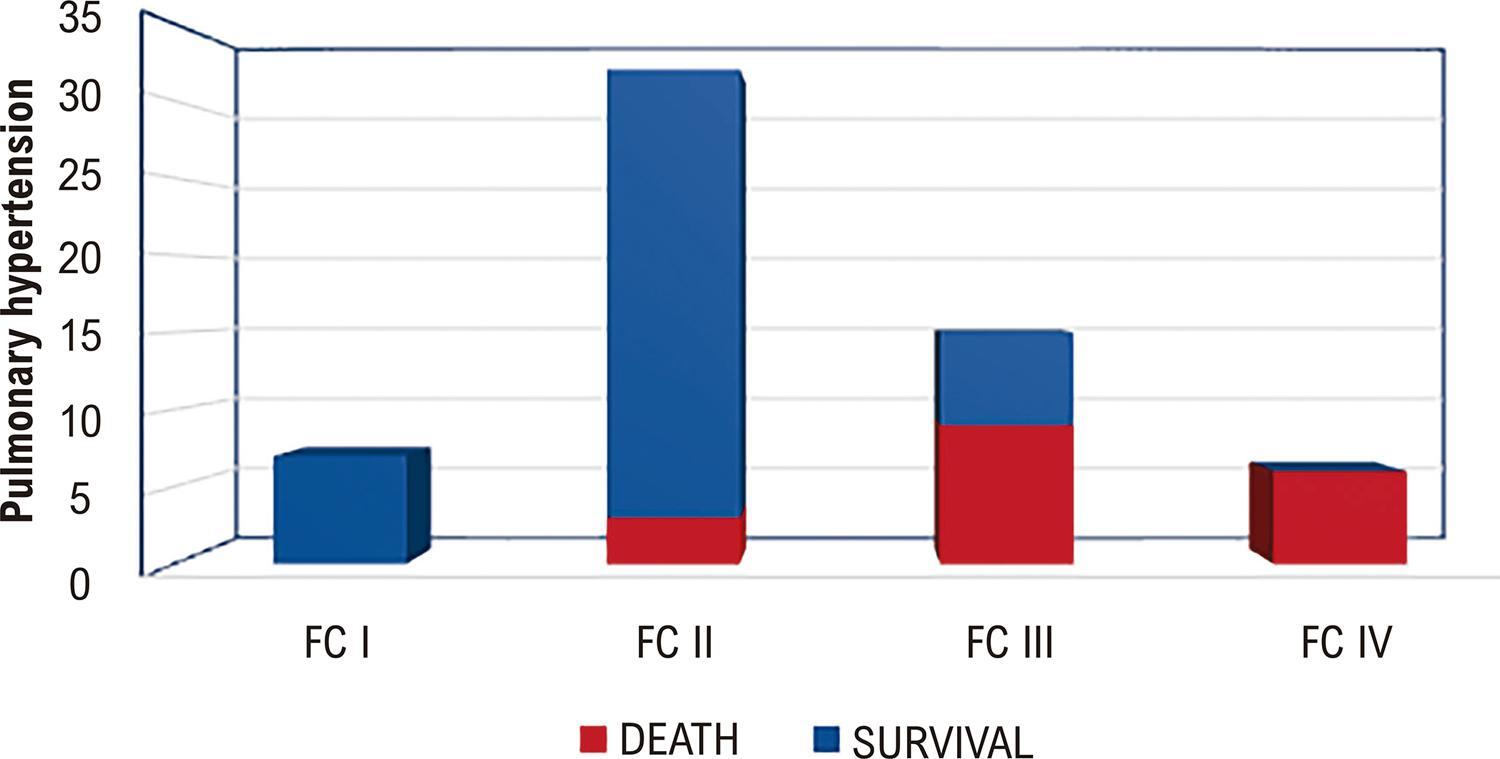
Patients with PAH had smaller left ventricular dimension without alteration in the ejection fraction, but with decreased left ventricular global longitudinal strain and left atrial reservoir strain. The right ventricular dimensions and parietal thickness were increased, and the parameters of systolic function (tricuspid annular plane systolic excursion, fractional area change, tricuspid s’ wave, and the right ventricular global longitudinal strain) were significantly reduced. During the follow-up period, 18 patients (32%) died, and death was associated with higher functional class, decreased right ventricular longitudinal strain, larger right ventricular size, decreased fractional area change, and decreased tricuspid annular s’ wave velocity.

Conclusion

PAH related to schistosomiasis causes remodeling of the right chambers, with decreased parameters of systolic function and myocardial strain. The long-term evolution, with elevated mortality, presents with larger right ventricular dimensions, lower fractional area change, lower tricuspid annular systolic velocity, lower right ventricular global longitudinal strain, and more advanced functional class.

  • Echocardiography
  • Pulmonary Hypertension
  • Schistosomiasis
Read Full Article
Creative Commons Attribution (CC BY) Creative Commons license
24/05/2023 DOI: 10.36660/abcimg.2023373i
Figure 1
Case Report

Important Constrictive Pericarditis in a Patient with Schistosomiasis: A Case Report

Djair Brindeiro Filho et al. José Maria Del Castillo Fábio Antonio Amando Granja

Introduction

Schistosomiasis mansoni (SM) is one of the most common causes of pulmonary hypertension and is often associated with the hepatosplenic form of the disease. However, constrictive pericarditis (CP) is not cited in the medical literature as a consequence of SM., One single case report, published in 1979, associating CP with schistosomiasis, presented the etiologic agent of Schistosoma haematobium. The present study described a rare case of CP in a patient with chronic hepatosplenic SM, with a significant improvement in the parameters of ventricular filling and myocardial deformation after surgery, illustrating the importance of the echocardiogram in the detection of complications caused by this disease.

[…]

  • Constrictive pericarditis
  • Echocardiography
  • Pericardiectomy
  • schistosomiais
  • Strain
Read Full Article
Creative Commons Attribution (CC BY) Creative Commons license
24/05/2023 DOI: 10.36660/abcimg.2023378i
Figure 1
Original Article

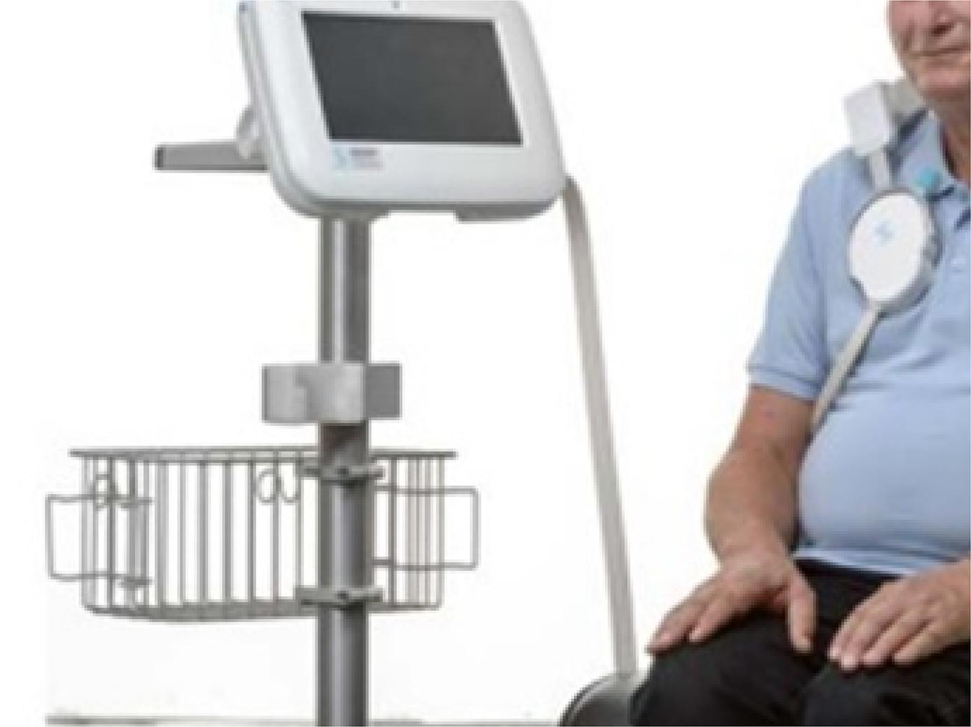
Pulmonary Congestion in Heart Failure With Reduced Ejection Fraction: Comparison Between Lung Ultrasound and Remote Dielectric Sensing

Maria Estefânia Bosco Otto et al. Vanessa Andreoli Esmanhoto Edileide de Barros Correia Ana Cristina de Souza Murta … Larissa Ventura Ribeiro Bruscky Andrea de Andrade Vilela Antonio Tito Paladino Filho Jorge Eduardo Assef

Abstract

Backgound

Outpatient assessment of pulmonary congestion in patients with heart failure with reduced ejection fraction (HFREF) can minimize hospitalizations due to decompensation and optimize the use of diuretics.

Objective

To compare the remote dielectric sensor (REDS), a validated device for detecting pulmonary extravascular fluid, with clinical parameters, transthoracic echocardiogram (TTE) and lung ultrasound (ULSP).

Methods

We included 38 patients from heart failure clinic (63±12 years; 21 men). All were submitted within 24 hours to clinical evaluation with description of paroxysmal nocturnal dyspnea, leg edema (LE), presence of dizziness; laboratory evaluation of NT-ProBNP; evaluation by REDS and TTE with analysis of parameters of systemic congestion by the inferior vena cava evaluation, function of the right ventricle and of pulmonary congestion, such as evaluation of filling pressures by average of E/e’ and indexed left atrial volume. The ULSP was performed using the 8 points anterior quadrants protocol, counting B lines in one respiratory cycle.

Results

22 patients had REDS ≥ 35% (indicative of pulmonary congestion) and 16 patients REDS<35%. Clinical and echocardiographic parameters were compared with REDS ≥ 35%. In the multivariate analysis, the variables body surface area, B lines and LE were associated with REDS ≥ 35%. NT-ProBNP was similar and elevated in both groups.

Conclusions

Outpatient monitoring of HFREF for volume control can be sensitized by the presence of B lines on the ULSP with good correlation to REDS ≥ 35%. NT pro BNP was not able to differentiate patients with congestion detected by REDS.

  • Heart Failure
  • Lung
  • Ultrasonography
Read Full Article
Creative Commons Attribution (CC BY) Creative Commons license
24/05/2023 DOI: 10.36660/abcimg.20230027i
Figure 1
Image

Aortocaval Fistula: A Rare Complication of Ruptured Abdominal Aortic Aneurysm

Karoline Evelyn Barbosa Gomes et al. Eduardo Koltun Sanvesso Edwaldo Edner Joviliano Maurício Serra Ribeiro Elisa Helena Subtil Zampieri

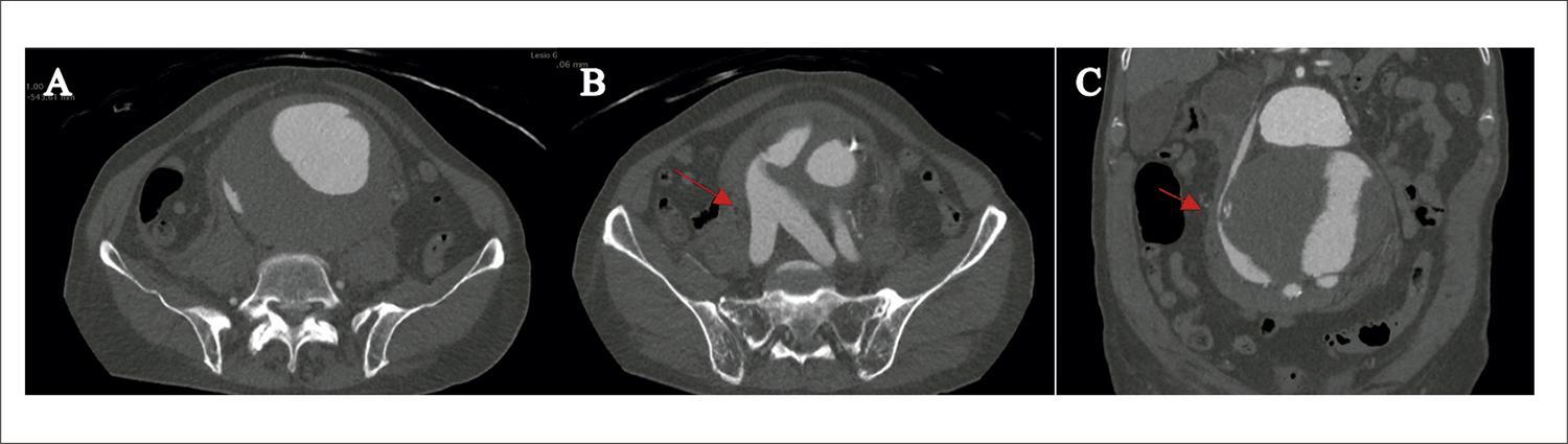
Aortocaval fistula is a rare complication of aortic aneurysm, occurring in approximately 0.2% to 6.04% of all abdominal aortic aneurysms. It may occur in the context of aortic aneurysm rupture, aortitis, Ehlers-Danlos syndrome, Marfan syndrome, or penetrating abdominal trauma., The classic clinical signs are abdominal pain, abdominal thrill, and symptoms of decompensated heart failure. It is usually diagnosed via computed tomography angiography, and typical findings include early contrast enhancement of the inferior vena cava (prior to contrast of the renal and hepatic parenchyma) and retrograde enhancement of the renal or iliac veins. Open surgical treatment has high associated morbidity and mortality, with reported rates of approximately 30%. With the advent of endovascular therapy, these rates have been reduced, with success rates as high as 96%.

An 85-year-old male patient, with hypertension and history of tobacco use, was admitted to the emergency unit with a history of sudden onset of abdominal pain four days prior, associated with a pulsating abdominal mass in the hypogastric region. Tomography angiography of the total aorta was performed, showing a pararenal abdominal aortic aneurysm with a diameter of 11.5 cm, extending from the right renal artery to the aortic bifurcation, with signs of tamponade rupture into the retroperitoneum (), associated with an aortocaval fistula with a point of communication at the level of the iliac veins. In the tomography image, retrograde and early enhancement of the iliac veins was observed in the arterial phase ( and ). During clinical evaluation, the patient presented hemodynamic instability, and an emergency surgical procedure was indicated. Intraoperatively (), an aneurysm with signs of rupture was found, showing thrill on palpation. The patient evolved with refractory hypotension and died.

[…]

  • Aortic Rupture
  • Arteriovenous Fistula
  • Vascular Surgical Procedures
Read Full Article
Creative Commons Attribution (CC BY) Creative Commons license
10/05/2023 DOI: 10.36660/abcimg.2023369i Views 1
Editorial

How to Manage a Cardiovascular Imaging Journal? Reflections and Ideas

Gerald Maurer

Cardiovascular Imaging is a rapidly growing field with an increasing impact on clinical practice. Each imaging modality has undergone remarkable development in recent years, requiring a very specialized understanding not only of physiology and disease processes, but also of technical aspects and physics. This has been reflected not only in the number but also in the complexity of published papers (as in most fields of medicine). While just two or three decades ago, cardiovascular imaging topics could be adequately covered in the major general cardiology journals, this is no longer the case and has led to the establishment of journals specializing in the field.

When managing a scientific journal, several issues need to be considered. These include its overall goals, its target audience, affiliation to an academic society, and relationship to other journals. The European Heart Journal Cardiovascular Imaging is the official journal of the European Association of Cardiovascular Imaging, which is a branch of the European Society of Cardiology. The journal’s goals are, to a great extent, influenced by these affiliations and by their policies. As stated in its title, it is a European journal, but it nevertheless aims to serve the international community, including readers, editorial board, and authors. Indeed, at this point, a large percentage of our readers are from outside Europe and we receive many submissions from other continents. Similarly, the American College of Cardiology and the American Heart Association publish dedicated imaging journals in conjunction with their more general journals. It is therefore important that a large and vital country like Brazil, with its excellent and active cardiological community, publish a journal such as ABC Imagem Cardiovascular on behalf of the Brazilian Society of Cardiology.

[…]

  • Cardiovascular therapy
  • journal ranking
  • medical publishing
  • open access
Read Full Article
Creative Commons Attribution (CC BY) Creative Commons license
10/05/2023 DOI: 10.36660/abcimg.20230030i
Figure 1
Case Report

Transient Perivascular Inflammation of the Carotid Artery (TIPIC): Vascular Ultrasonography Role

Fanilda Souto Barros et al. Simone Nascimento dos Santos Daniela Souto Barros Sérgio Salles Cunha … Ana Lopes Albricker Ana Cláudia Gomes Petisco

Case Report

Male patient, 53 years old, complaining of progressive unilateral neck pain, which appeared spontaneously, in association with edema, local redness, and great sensitivity to palpation. The patient’s past history included radiotherapy for about 5 years in the neck and face region, on the opposite side of the current complaint. The patient was a non-smoker and had no other significant risk factors for atherosclerotic disease. One year before the current clinical picture, the patient had undergone VUS of the carotid arteries, in a routine evaluation for follow-up after radiotherapy, the result was within normal limits.

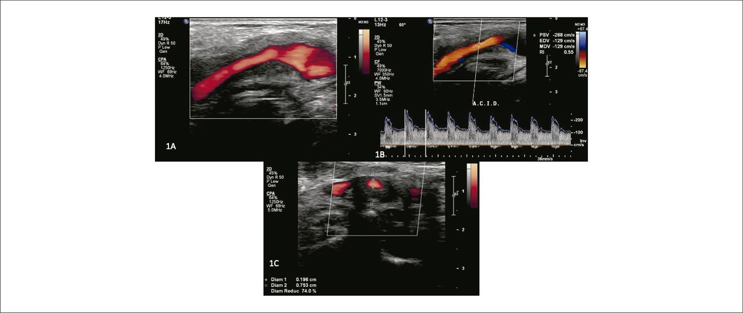
Given the clinical picture, a new VUS examination was requested, which included the standard protocol for evaluating the extracranial common, external, and internal carotid arteries (ACC, ICA and ACE). Special attention was given to extracarotid (perivascular) tissues due to the history of radiotherapy. The B-mode US showed extensive hypoechoic and homogeneous involvement of the tissues, including the arterial wall at the level of the carotid bifurcation, expanding to the proximal ICA (). The peak systolic velocity and end-diastolic velocity (PSV/EDV) were high: PSV/EDV = 288/129 cm/s (). Local stenosis measured in the transverse plane of the image was estimated at 74% (), consistent with the Doppler velocity data. The extent of the affected area was 5.57 mm compared to a lumen of 1.96 mm. Magnetic resonance imaging (MRI) and angioresonance (MRA) showed irregular thickening, with contrast enhancement, in the proximal ICA and bifurcation, corroborating the VUS findings ().

[…]

  • arteritis
  • Carotid artery
  • TIPIC
Read Full Article
Creative Commons Attribution (CC BY) Creative Commons license
10/05/2023 DOI: 10.36660/abcimg.20230019i Views 1

Could not load more articles. Please try again.

How to cite this article

EndNote RIS

Electronic Document Format (Vancouver)

Siqueira AWS, Almeida MF. My Approach to Echocardiographic Evaluation after Pediatric Heart Transplantation to Control Rejection and/or Graft Vascular Disease. ABC Imagem Cardiovascular 2023;36(2):e20230045.

Electronic Document Format (ABNT)

Siqueira, Adailson Wagner da Silva; Almeida, Mirela Frederico de. My Approach to Echocardiographic Evaluation after Pediatric Heart Transplantation to Control Rejection and/or Graft Vascular Disease. ABC Imagem Cardiovascular, v. 36, n. 2, e20230045, Jun. 2023.

Electronic Document Format (APA)

Siqueira, A. W. S., & Almeida, M. F. (2023). My Approach to Echocardiographic Evaluation after Pediatric Heart Transplantation to Control Rejection and/or Graft Vascular Disease. ABC Imagem Cardiovascular, 36(2), e20230045.

Electronic Document Format (ISO)

Siqueira, Adailson Wagner da Silva and Almeida, Mirela Frederico de. My Approach to Echocardiographic Evaluation after Pediatric Heart Transplantation to Control Rejection and/or Graft Vascular Disease. ABC Imagem Cardiovascular [online]. 2023, vol. 36, n. 2, [cited 2026-10-14], e20230045. Available from: <https://journals.cardiol.br/en/article/my-approach-to-echocardiographic-evaluation-after-pediatric-heart-transplantation-to-control-rejection-and-or-graft-vascular-disease/>. ISSN 2318-8219

EndNote RIS

Electronic Document Format (Vancouver)

Felix AS, Alcantara ML, Papadopoulos K. My Approach to Three-Dimensional Echocardiography for Pathophysiological Classification of Tricuspid Regurgitation. ABC Imagem Cardiovascular 2023;36(2):e20230055.

Electronic Document Format (ABNT)

Felix, Alex dos Santos; Alcantara, Monica Luiza de; Papadopoulos, Konstantinos. My Approach to Three-Dimensional Echocardiography for Pathophysiological Classification of Tricuspid Regurgitation. ABC Imagem Cardiovascular, v. 36, n. 2, e20230055, Jun. 2023.

Electronic Document Format (APA)

Felix, A. S., Alcantara, M. L. , & Papadopoulos, K. (2023). My Approach to Three-Dimensional Echocardiography for Pathophysiological Classification of Tricuspid Regurgitation. ABC Imagem Cardiovascular, 36(2), e20230055.

Electronic Document Format (ISO)

Felix, Alex dos Santos and Alcantara, Monica Luiza de and Papadopoulos, Konstantinos. My Approach to Three-Dimensional Echocardiography for Pathophysiological Classification of Tricuspid Regurgitation. ABC Imagem Cardiovascular [online]. 2023, vol. 36, n. 2, [cited 2026-10-14], e20230055. Available from: <https://journals.cardiol.br/en/article/my-approach-to-three-dimensional-echocardiography-for-pathophysiological-classification-of-tricuspid-regurgitation/>. ISSN 2318-8219

EndNote RIS

Electronic Document Format (Vancouver)

Espíndola LN, Mattoso AAA, Couto GS, Britto PS. Dual Mechanism of Mitral Valve Injury: Additional Value of Three-Dimensional Transesophageal Echocardiography. ABC Imagem Cardiovascular 2023;36(2):e322.

Electronic Document Format (ABNT)

Espíndola, Larissa Neto; Mattoso, Angele Azevedo Alves; Couto, Geórgia dos Santos; Britto, Pompílio Sampaio. Dual Mechanism of Mitral Valve Injury: Additional Value of Three-Dimensional Transesophageal Echocardiography. ABC Imagem Cardiovascular, v. 36, n. 2, e322, Jun. 2023.

Electronic Document Format (APA)

Espíndola, L. N., Mattoso, A. A. A., Couto, G. S., & Britto, P. S. (2023). Dual Mechanism of Mitral Valve Injury: Additional Value of Three-Dimensional Transesophageal Echocardiography. ABC Imagem Cardiovascular, 36(2), e322.

Electronic Document Format (ISO)

Espíndola, Larissa Neto and Mattoso, Angele Azevedo Alves and Couto, Geórgia dos Santos and Britto, Pompílio Sampaio. Dual Mechanism of Mitral Valve Injury: Additional Value of Three-Dimensional Transesophageal Echocardiography. ABC Imagem Cardiovascular [online]. 2023, vol. 36, n. 2, [cited 2026-10-14], e322. Available from: <https://journals.cardiol.br/en/article/dual-mechanism-of-mitral-valve-injury-additional-value-of-three-dimensional-transesophageal-echocardiography/>. ISSN 2318-8219

EndNote RIS

Electronic Document Format (Vancouver)

Cantisano AL, Grubert CS. My Approach To: Vascular Ultrasonography in Dolichoarteriopathies of the Carotid Arteries. ABC Imagem Cardiovascular 2023;36(2):e20230034.

Electronic Document Format (ABNT)

Cantisano, Armando Luis; Grubert, Catarina Schiavo. My Approach To: Vascular Ultrasonography in Dolichoarteriopathies of the Carotid Arteries. ABC Imagem Cardiovascular, v. 36, n. 2, e20230034, Jun. 2023.

Electronic Document Format (APA)

Cantisano, A. L., & Grubert, C. S. (2023). My Approach To: Vascular Ultrasonography in Dolichoarteriopathies of the Carotid Arteries. ABC Imagem Cardiovascular, 36(2), e20230034.

Electronic Document Format (ISO)

Cantisano, Armando Luis and Grubert, Catarina Schiavo. My Approach To: Vascular Ultrasonography in Dolichoarteriopathies of the Carotid Arteries. ABC Imagem Cardiovascular [online]. 2023, vol. 36, n. 2, [cited 2026-10-14], e20230034. Available from: <https://journals.cardiol.br/en/article/my-approach-to-vascular-ultrasonography-in-dolichoarteriopathies-of-the-carotid-arteries/>. ISSN 2318-8219

EndNote RIS

Electronic Document Format (Vancouver)

Moura AN, Camarozano AC, Sá CRF, Carmo DC, Fortunato JA, Darwich RZ, Baroncini LAV, et al. Right Ventricle Diastolic Function: Correlation with Age. ABC Imagem Cardiovascular 2023;36(2):e333.

Electronic Document Format (ABNT)

Moura, Arthur Nascimento de; Camarozano, Ana Cristina; Sá, Cintia Rocha Fortes de; Carmo, Daniela de Castro; Fortunato, Jerônimo Antonio; Darwich, Rubens Zenóbio; Baroncini, Liz Andréa Villela. Right Ventricle Diastolic Function: Correlation with Age. ABC Imagem Cardiovascular, v. 36, n. 2, e333, Jun. 2023.

Electronic Document Format (APA)

Moura, A. N. , Camarozano, A. C., Sá, C. R. F. , Carmo, D. C., Fortunato, J. A., Darwich, R. Z., & Baroncini, L. A. V. (2023). Right Ventricle Diastolic Function: Correlation with Age. ABC Imagem Cardiovascular, 36(2), e333.

Electronic Document Format (ISO)

Moura, Arthur Nascimento de and Camarozano, Ana Cristina and Sá, Cintia Rocha Fortes de and Carmo, Daniela de Castro and Fortunato, Jerônimo Antonio and Darwich, Rubens Zenóbio and Baroncini, Liz Andréa Villela. Right Ventricle Diastolic Function: Correlation with Age. ABC Imagem Cardiovascular [online]. 2023, vol. 36, n. 2, [cited 2026-10-14], e333. Available from: <https://journals.cardiol.br/en/article/right-ventricle-diastolic-function-correlation-with-age/>. ISSN 2318-8219

EndNote RIS

Electronic Document Format (Vancouver)

Sawamura KSS, Brito MM. My Approach to Echocardiographic Assessment of the Premature Newborn. ABC Imagem Cardiovascular 2023;36(2):e20230035.

Electronic Document Format (ABNT)

Sawamura, Karen Saori Shiraishi; Brito, Márcio Miranda. My Approach to Echocardiographic Assessment of the Premature Newborn. ABC Imagem Cardiovascular, v. 36, n. 2, e20230035, Jun. 2023.

Electronic Document Format (APA)

Sawamura, K. S. S., & Brito, M. M. (2023). My Approach to Echocardiographic Assessment of the Premature Newborn. ABC Imagem Cardiovascular, 36(2), e20230035.

Electronic Document Format (ISO)

Sawamura, Karen Saori Shiraishi and Brito, Márcio Miranda. My Approach to Echocardiographic Assessment of the Premature Newborn. ABC Imagem Cardiovascular [online]. 2023, vol. 36, n. 2, [cited 2026-10-14], e20230035. Available from: <https://journals.cardiol.br/en/article/my-approach-to-echocardiographic-assessment-of-the-premature-newborn/>. ISSN 2318-8219

EndNote RIS

Electronic Document Format (Vancouver)

Carrijo AMM, Brito VP, Santos GA, Silva SGT, O’Connell JL. Acute Coronary Syndrome Followed by Pulmonary Thromboembolism and Identification of a Large Fixed Thrombus between Atria in a Patient with Severe COVID-19: A Case Report. ABC Imagem Cardiovascular 2023;36(2):e351.

Electronic Document Format (ABNT)

Carrijo, Alice Mirane Malta; Brito, Veronica Perius de; Santos, Gabriel Alvarenga; Silva, Samuel Gomes Tomaz da; O’Connell, João Lucas. Acute Coronary Syndrome Followed by Pulmonary Thromboembolism and Identification of a Large Fixed Thrombus between Atria in a Patient with Severe COVID-19: A Case Report. ABC Imagem Cardiovascular, v. 36, n. 2, e351, Jun. 2023.

Electronic Document Format (APA)

Carrijo, A. M. M., Brito, V. P. , Santos, G. A., Silva, S. G. T. , & O’Connell, J. L. (2023). Acute Coronary Syndrome Followed by Pulmonary Thromboembolism and Identification of a Large Fixed Thrombus between Atria in a Patient with Severe COVID-19: A Case Report. ABC Imagem Cardiovascular, 36(2), e351.

Electronic Document Format (ISO)

Carrijo, Alice Mirane Malta and Brito, Veronica Perius de and Santos, Gabriel Alvarenga and Silva, Samuel Gomes Tomaz da and O’Connell, João Lucas. Acute Coronary Syndrome Followed by Pulmonary Thromboembolism and Identification of a Large Fixed Thrombus between Atria in a Patient with Severe COVID-19: A Case Report. ABC Imagem Cardiovascular [online]. 2023, vol. 36, n. 2, [cited 2026-10-14], e351. Available from: <https://journals.cardiol.br/en/article/acute-coronary-syndrome-followed-by-pulmonary-thromboembolism-and-identification-of-a-large-fixed-thrombus-between-atria-in-a-patient-with-severe-covid-19-a-case-report/>. ISSN 2318-8219

EndNote RIS

Electronic Document Format (Vancouver)

Gripp EA, Portela ACF, Costa Junior FL, Rabishoffsky R, Medeiros JRC, Rabischoffsky A. Patent Forame Ovale: Contribution of Transcranial Doppler in Patient Diagnosis. ABC Imagem Cardiovascular 2023;36(2):e20230020.

Electronic Document Format (ABNT)

Gripp, Eliza de Almeida; Portela, Ana Carolina de Freitas; Costa Junior, Flávio Luis da; Rabishoffsky, Rafael; Medeiros, Jéssica Rizkalla Corrêa; Rabischoffsky, Arnaldo. Patent Forame Ovale: Contribution of Transcranial Doppler in Patient Diagnosis. ABC Imagem Cardiovascular, v. 36, n. 2, e20230020, Jun. 2023.

Electronic Document Format (APA)

Gripp, E. A., Portela, A. C. F., Costa Junior, F. L. , Rabishoffsky, R., Medeiros, J. R. C., & Rabischoffsky, A. (2023). Patent Forame Ovale: Contribution of Transcranial Doppler in Patient Diagnosis. ABC Imagem Cardiovascular, 36(2), e20230020.

Electronic Document Format (ISO)

Gripp, Eliza de Almeida and Portela, Ana Carolina de Freitas and Costa Junior, Flávio Luis da and Rabishoffsky, Rafael and Medeiros, Jéssica Rizkalla Corrêa and Rabischoffsky, Arnaldo. Patent Forame Ovale: Contribution of Transcranial Doppler in Patient Diagnosis. ABC Imagem Cardiovascular [online]. 2023, vol. 36, n. 2, [cited 2026-10-14], e20230020. Available from: <https://journals.cardiol.br/en/article/patent-forame-ovale-contribution-of-transcranial-doppler-in-patient-diagnosis/>. ISSN 2318-8219

EndNote RIS

Electronic Document Format (Vancouver)

Lofrano-Alves MS. Lung Ultrasound in Outpatients with Heart Failure. ABC Imagem Cardiovascular 2023;36(2):e20230049.

Electronic Document Format (ABNT)

Lofrano-Alves, Marco Stephan. Lung Ultrasound in Outpatients with Heart Failure. ABC Imagem Cardiovascular, v. 36, n. 2, e20230049, Jun. 2023.

Electronic Document Format (APA)

Lofrano-Alves, M. S. (2023). Lung Ultrasound in Outpatients with Heart Failure. ABC Imagem Cardiovascular, 36(2), e20230049.

Electronic Document Format (ISO)

Lofrano-Alves, Marco Stephan. Lung Ultrasound in Outpatients with Heart Failure. ABC Imagem Cardiovascular [online]. 2023, vol. 36, n. 2, [cited 2026-10-14], e20230049. Available from: <https://journals.cardiol.br/en/article/lung-ultrasound-in-outpatients-with-heart-failure/>. ISSN 2318-8219

EndNote RIS

Electronic Document Format (Vancouver)

Rassi DC. The Use of Echocardiography in Schistosomiasis. ABC Imagem Cardiovascular 2023;36(2):e20230037.

Electronic Document Format (ABNT)

Rassi, Daniela do Carmo. The Use of Echocardiography in Schistosomiasis. ABC Imagem Cardiovascular, v. 36, n. 2, e20230037, Jun. 2023.

Electronic Document Format (APA)

Rassi, D. C. (2023). The Use of Echocardiography in Schistosomiasis. ABC Imagem Cardiovascular, 36(2), e20230037.

Electronic Document Format (ISO)

Rassi, Daniela do Carmo. The Use of Echocardiography in Schistosomiasis. ABC Imagem Cardiovascular [online]. 2023, vol. 36, n. 2, [cited 2026-10-14], e20230037. Available from: <https://journals.cardiol.br/en/article/the-use-of-echocardiography-in-schistosomiasis/>. ISSN 2318-8219

EndNote RIS

Electronic Document Format (Vancouver)

Silva CM, Guimarães AFM, Wang R, Alves CMS, Oliveira EC. Expandable Airway Stents: Success in the Extrinsic Bronchial Compression Approach After Reconstruction of the Aortic Arch. ABC Imagem Cardiovascular 2023;36(2):e20230023.

Electronic Document Format (ABNT)

Silva, Camila Magalhães; Guimarães, Adriana Furletti Machado; Wang, Ricardo; Alves, Carla Maria Silva e; Oliveira, Edmundo Clarindo. Expandable Airway Stents: Success in the Extrinsic Bronchial Compression Approach After Reconstruction of the Aortic Arch. ABC Imagem Cardiovascular, v. 36, n. 2, e20230023, Jun. 2023.

Electronic Document Format (APA)

Silva, C. M., Guimarães, A. F. M., Wang, R., Alves, C. M. S. , & Oliveira, E. C. (2023). Expandable Airway Stents: Success in the Extrinsic Bronchial Compression Approach After Reconstruction of the Aortic Arch. ABC Imagem Cardiovascular, 36(2), e20230023.

Electronic Document Format (ISO)

Silva, Camila Magalhães and Guimarães, Adriana Furletti Machado and Wang, Ricardo and Alves, Carla Maria Silva e and Oliveira, Edmundo Clarindo. Expandable Airway Stents: Success in the Extrinsic Bronchial Compression Approach After Reconstruction of the Aortic Arch. ABC Imagem Cardiovascular [online]. 2023, vol. 36, n. 2, [cited 2026-10-14], e20230023. Available from: <https://journals.cardiol.br/en/article/expandable-airway-stents-success-in-the-extrinsic-bronchial-compression-approach-after-reconstruction-of-the-aortic-arch/>. ISSN 2318-8219

EndNote RIS

Electronic Document Format (Vancouver)

Souza AC, Drubi SA, Junqueira BMI, Guedes RASP, Almeida Filho AC, Macêdo CT. Comparative Analysis of Myocardial Work After Decongestion Therapy in a Patient With Acutely Decompensated HFrEF. ABC Imagem Cardiovascular 2023;36(2):e20230008.

Electronic Document Format (ABNT)

Souza, Alexandre Costa; Drubi, Stephanie de Azevedo; Junqueira, Bruna de Mattos Ivo; Guedes, Ricardo André Sales Pereira; Almeida Filho, Adriano Chaves de; Macêdo, Carolina Thé. Comparative Analysis of Myocardial Work After Decongestion Therapy in a Patient With Acutely Decompensated HFrEF. ABC Imagem Cardiovascular, v. 36, n. 2, e20230008, Jun. 2023.

Electronic Document Format (APA)

Souza, A. C., Drubi, S. A., Junqueira, B. M. I., Guedes, R. A. S. P., Almeida Filho, A. C. , & Macêdo, C. T. (2023). Comparative Analysis of Myocardial Work After Decongestion Therapy in a Patient With Acutely Decompensated HFrEF. ABC Imagem Cardiovascular, 36(2), e20230008.

Electronic Document Format (ISO)

Souza, Alexandre Costa and Drubi, Stephanie de Azevedo and Junqueira, Bruna de Mattos Ivo and Guedes, Ricardo André Sales Pereira and Almeida Filho, Adriano Chaves de and Macêdo, Carolina Thé. Comparative Analysis of Myocardial Work After Decongestion Therapy in a Patient With Acutely Decompensated HFrEF. ABC Imagem Cardiovascular [online]. 2023, vol. 36, n. 2, [cited 2026-10-14], e20230008. Available from: <https://journals.cardiol.br/en/article/comparative-analysis-of-myocardial-work-after-decongestion-therapy-in-a-patient-with-acutely-decompensated-hfref/>. ISSN 2318-8219

EndNote RIS

Electronic Document Format (Vancouver)

Gutiérrez-Fajardo P. How to Proceed in a Multimodality Analysis for the Diagnosis and Risk Stratification of Pulmonary Hypertension?. ABC Imagem Cardiovascular 2023;36(2):e20230041.

Electronic Document Format (ABNT)

Gutiérrez-Fajardo, Pedro. How to Proceed in a Multimodality Analysis for the Diagnosis and Risk Stratification of Pulmonary Hypertension?. ABC Imagem Cardiovascular, v. 36, n. 2, e20230041, Jun. 2023.

Electronic Document Format (APA)

Gutiérrez-Fajardo, P. (2023). How to Proceed in a Multimodality Analysis for the Diagnosis and Risk Stratification of Pulmonary Hypertension?. ABC Imagem Cardiovascular, 36(2), e20230041.

Electronic Document Format (ISO)

Gutiérrez-Fajardo, Pedro. How to Proceed in a Multimodality Analysis for the Diagnosis and Risk Stratification of Pulmonary Hypertension?. ABC Imagem Cardiovascular [online]. 2023, vol. 36, n. 2, [cited 2026-10-14], e20230041. Available from: <https://journals.cardiol.br/en/article/how-to-proceed-in-a-multimodality-analysis-for-the-diagnosis-and-risk-stratification-of-pulmonary-hypertension/>. ISSN 2318-8219

EndNote RIS

Electronic Document Format (Vancouver)

António IBG, Maia AS, Rabelato J, Campos AAS, Menezes MP, Issa M. Takayasu’s Arteritis: Ascending Aortic Aneurysm and Coronary Artery Disease in a 19-year-old Young Adult. ABC Imagem Cardiovascular 2023;36(2):e20230032.

Electronic Document Format (ABNT)

António, Iuri Betuel Gomes; Maia, Adnaldo da Silveira; Rabelato, Janayna; Campos, Aloysio Abdo Silva; Menezes, Marilia Prudente; Issa, Mário. Takayasu’s Arteritis: Ascending Aortic Aneurysm and Coronary Artery Disease in a 19-year-old Young Adult. ABC Imagem Cardiovascular, v. 36, n. 2, e20230032, Jun. 2023.

Electronic Document Format (APA)

António, I. B. G., Maia, A. S., Rabelato, J., Campos, A. A. S., Menezes, M. P., & Issa, M. (2023). Takayasu’s Arteritis: Ascending Aortic Aneurysm and Coronary Artery Disease in a 19-year-old Young Adult. ABC Imagem Cardiovascular, 36(2), e20230032.

Electronic Document Format (ISO)

António, Iuri Betuel Gomes and Maia, Adnaldo da Silveira and Rabelato, Janayna and Campos, Aloysio Abdo Silva and Menezes, Marilia Prudente and Issa, Mário. Takayasu’s Arteritis: Ascending Aortic Aneurysm and Coronary Artery Disease in a 19-year-old Young Adult. ABC Imagem Cardiovascular [online]. 2023, vol. 36, n. 2, [cited 2026-10-14], e20230032. Available from: <https://journals.cardiol.br/en/article/takayasus-arteritis-ascending-aortic-aneurysm-and-coronary-artery-disease-in-a-19-year-old-young-adult/>. ISSN 2318-8219

EndNote RIS

Electronic Document Format (Vancouver)

Lima MSM. Contrast Echocardiography in Real Life: Practicality, Safety, and Cost-Effectiveness. ABC Imagem Cardiovascular 2023;36(2):e20230033.

Electronic Document Format (ABNT)

Lima, Márcio Silva Miguel. Contrast Echocardiography in Real Life: Practicality, Safety, and Cost-Effectiveness. ABC Imagem Cardiovascular, v. 36, n. 2, e20230033, Jun. 2023.

Electronic Document Format (APA)

Lima, M. S. M. (2023). Contrast Echocardiography in Real Life: Practicality, Safety, and Cost-Effectiveness. ABC Imagem Cardiovascular, 36(2), e20230033.

Electronic Document Format (ISO)

Lima, Márcio Silva Miguel. Contrast Echocardiography in Real Life: Practicality, Safety, and Cost-Effectiveness. ABC Imagem Cardiovascular [online]. 2023, vol. 36, n. 2, [cited 2026-10-14], e20230033. Available from: <https://journals.cardiol.br/en/article/contrast-echocardiography-in-real-life-practicality-safety-and-cost-effectiveness/>. ISSN 2318-8219

EndNote RIS

Electronic Document Format (Vancouver)

Carvalho MVSF, Alves ES, Gomes LC, Macêdo CT, Sales MAM, Souza AC. Transesophageal Echocardiogram in the Diagnosis of Superior Vena Cava Syndrome in a Patient With a Long-Term Catheter. ABC Imagem Cardiovascular 2023;36(2):e20230028.

Electronic Document Format (ABNT)

Carvalho, Marcus Vinicius Silva Freire de; Alves, Endy de Santana; Gomes, Laila Caroline; Macêdo, Carolina Thé; Sales, Marco André Moraes; Souza, Alexandre Costa. Transesophageal Echocardiogram in the Diagnosis of Superior Vena Cava Syndrome in a Patient With a Long-Term Catheter. ABC Imagem Cardiovascular, v. 36, n. 2, e20230028, Jun. 2023.

Electronic Document Format (APA)

Carvalho, M. V. S. F. , Alves, E. S., Gomes, L. C., Macêdo, C. T., Sales, M. A. M., & Souza, A. C. (2023). Transesophageal Echocardiogram in the Diagnosis of Superior Vena Cava Syndrome in a Patient With a Long-Term Catheter. ABC Imagem Cardiovascular, 36(2), e20230028.

Electronic Document Format (ISO)

Carvalho, Marcus Vinicius Silva Freire de and Alves, Endy de Santana and Gomes, Laila Caroline and Macêdo, Carolina Thé and Sales, Marco André Moraes and Souza, Alexandre Costa. Transesophageal Echocardiogram in the Diagnosis of Superior Vena Cava Syndrome in a Patient With a Long-Term Catheter. ABC Imagem Cardiovascular [online]. 2023, vol. 36, n. 2, [cited 2026-10-14], e20230028. Available from: <https://journals.cardiol.br/en/article/transesophageal-echocardiogram-in-the-diagnosis-of-superior-vena-cava-syndrome-in-a-patient-with-a-long-term-catheter/>. ISSN 2318-8219

EndNote RIS

Electronic Document Format (Vancouver)

Glavam AP, Lopes RW, Brandão SCS. My Approach to Imaging Cardiac Amyloidosis: Role of Bone-Seeking Tracers Scintigraphy. ABC Imagem Cardiovascular 2023;36(2):e20230012.

Electronic Document Format (ABNT)

Glavam, Adriana Pereira; Lopes, Rafael Willain; Brandão, Simone Cristina Soares. My Approach to Imaging Cardiac Amyloidosis: Role of Bone-Seeking Tracers Scintigraphy. ABC Imagem Cardiovascular, v. 36, n. 2, e20230012, May. 2023.

Electronic Document Format (APA)

Glavam, A. P., Lopes, R. W., & Brandão, S. C. S. (2023). My Approach to Imaging Cardiac Amyloidosis: Role of Bone-Seeking Tracers Scintigraphy. ABC Imagem Cardiovascular, 36(2), e20230012.

Electronic Document Format (ISO)

Glavam, Adriana Pereira and Lopes, Rafael Willain and Brandão, Simone Cristina Soares. My Approach to Imaging Cardiac Amyloidosis: Role of Bone-Seeking Tracers Scintigraphy. ABC Imagem Cardiovascular [online]. 2023, vol. 36, n. 2, [cited 2026-10-14], e20230012. Available from: <https://journals.cardiol.br/en/article/my-approach-to-imaging-cardiac-amyloidosis-role-of-bone-seeking-tracers-scintigraphy/>. ISSN 2318-8219

EndNote RIS

Electronic Document Format (Vancouver)

Tavares IR, Miranda MC, Petisco ACGP, Barros DS, Folino CB, Dornas VLB, Saleh MH, et al. Behcet’s Disease with Vascular Involvement: Case Report. ABC Imagem Cardiovascular 2023;36(2):e20230014.

Electronic Document Format (ABNT)

Tavares, Isabela Rodrigues; Miranda, Maria Cristina; Petisco, Ana Claudia Gomes Pereira; Barros, Daniela Souto; Folino, Caio Buscatti; Dornas, Vitor Leonidas Boher; Saleh, Mohamed Hassan; Rossi, Fábio Henrique; Sousa, Raquel Peres; Carvalho, Larissa Chaves Nunes. Behcet’s Disease with Vascular Involvement: Case Report. ABC Imagem Cardiovascular, v. 36, n. 2, e20230014, May. 2023.

Electronic Document Format (APA)

Tavares, I. R., Miranda, M. C., Petisco, A. C. G. P., Barros, D. S., Folino, C. B., Dornas, V. L. B., Saleh, M. H., Rossi, F. H., Sousa, R. P., & Carvalho, L. C. N. (2023). Behcet’s Disease with Vascular Involvement: Case Report. ABC Imagem Cardiovascular, 36(2), e20230014.

Electronic Document Format (ISO)

Tavares, Isabela Rodrigues and Miranda, Maria Cristina and Petisco, Ana Claudia Gomes Pereira and Barros, Daniela Souto and Folino, Caio Buscatti and Dornas, Vitor Leonidas Boher and Saleh, Mohamed Hassan and Rossi, Fábio Henrique and Sousa, Raquel Peres and Carvalho, Larissa Chaves Nunes. Behcet’s Disease with Vascular Involvement: Case Report. ABC Imagem Cardiovascular [online]. 2023, vol. 36, n. 2, [cited 2026-10-14], e20230014. Available from: <https://journals.cardiol.br/en/article/behcets-disease-with-vascular-involvement-case-report/>. ISSN 2318-8219

EndNote RIS

Electronic Document Format (Vancouver)

Castillo JMD, Oliveira KB, Travassos RRO, Bandeira MP, Silveira CAM, Brindeiro Filho D. Long-Term Evolution of Patients with Important Pulmonary Hypertension due to Schistosomiasis. ABC Imagem Cardiovascular 2023;36(2):e2023373.

Electronic Document Format (ABNT)

Castillo, José Maria Del; Oliveira, Katarina Barros de; Travassos, Rafael Ricardo de Oliveira; Bandeira, Ângela Maria Pontes; Silveira, Carlos Antônio da Mota; Brindeiro Filho, Djair. Long-Term Evolution of Patients with Important Pulmonary Hypertension due to Schistosomiasis. ABC Imagem Cardiovascular, v. 36, n. 2, e2023373, May. 2023.

Electronic Document Format (APA)

Castillo, J. M. D., Oliveira, K. B. , Travassos, R. R. O., Bandeira, M. P., Silveira, C. A. M., & Brindeiro Filho, D. (2023). Long-Term Evolution of Patients with Important Pulmonary Hypertension due to Schistosomiasis. ABC Imagem Cardiovascular, 36(2), e2023373.

Electronic Document Format (ISO)

Castillo, José Maria Del and Oliveira, Katarina Barros de and Travassos, Rafael Ricardo de Oliveira and Bandeira, Ângela Maria Pontes and Silveira, Carlos Antônio da Mota and Brindeiro Filho, Djair. Long-Term Evolution of Patients with Important Pulmonary Hypertension due to Schistosomiasis. ABC Imagem Cardiovascular [online]. 2023, vol. 36, n. 2, [cited 2026-10-14], e2023373. Available from: <https://journals.cardiol.br/en/article/long-term-evolution-of-patients-with-important-pulmonary-hypertension-due-to-schistosomiasis/>. ISSN 2318-8219

EndNote RIS

Electronic Document Format (Vancouver)

Brindeiro Filho D, Castillo JMD, Granja FAA. Important Constrictive Pericarditis in a Patient with Schistosomiasis: A Case Report. ABC Imagem Cardiovascular 2023;36(2):e2023378.

Electronic Document Format (ABNT)

Brindeiro Filho, Djair; Castillo, José Maria Del; Granja, Fábio Antonio Amando. Important Constrictive Pericarditis in a Patient with Schistosomiasis: A Case Report. ABC Imagem Cardiovascular, v. 36, n. 2, e2023378, May. 2023.

Electronic Document Format (APA)

Brindeiro Filho, D., Castillo, J. M. D., & Granja, F. A. A. (2023). Important Constrictive Pericarditis in a Patient with Schistosomiasis: A Case Report. ABC Imagem Cardiovascular, 36(2), e2023378.

Electronic Document Format (ISO)

Brindeiro Filho, Djair and Castillo, José Maria Del and Granja, Fábio Antonio Amando. Important Constrictive Pericarditis in a Patient with Schistosomiasis: A Case Report. ABC Imagem Cardiovascular [online]. 2023, vol. 36, n. 2, [cited 2026-10-14], e2023378. Available from: <https://journals.cardiol.br/en/article/important-constrictive-pericarditis-in-a-patient-with-schistosomiasis-a-case-report/>. ISSN 2318-8219

EndNote RIS

Electronic Document Format (Vancouver)

Otto MEB, Esmanhoto VA, Correia EB, Murta ACS, Bruscky LVR, Vilela AA, Paladino Filho AT, et al. Pulmonary Congestion in Heart Failure With Reduced Ejection Fraction: Comparison Between Lung Ultrasound and Remote Dielectric Sensing. ABC Imagem Cardiovascular 2023;36(2):e20230027.

Electronic Document Format (ABNT)

Otto, Maria Estefânia Bosco; Esmanhoto, Vanessa Andreoli; Correia, Edileide de Barros; Murta, Ana Cristina de Souza; Bruscky, Larissa Ventura Ribeiro; Vilela, Andrea de Andrade; Paladino Filho, Antonio Tito; Assef, Jorge Eduardo. Pulmonary Congestion in Heart Failure With Reduced Ejection Fraction: Comparison Between Lung Ultrasound and Remote Dielectric Sensing. ABC Imagem Cardiovascular, v. 36, n. 2, e20230027, May. 2023.

Electronic Document Format (APA)

Otto, M. E. B., Esmanhoto, V. A., Correia, E. B., Murta, A. C. S., Bruscky, L. V. R., Vilela, A. A., Paladino Filho, A. T., & Assef, J. E. (2023). Pulmonary Congestion in Heart Failure With Reduced Ejection Fraction: Comparison Between Lung Ultrasound and Remote Dielectric Sensing. ABC Imagem Cardiovascular, 36(2), e20230027.

Electronic Document Format (ISO)

Otto, Maria Estefânia Bosco and Esmanhoto, Vanessa Andreoli and Correia, Edileide de Barros and Murta, Ana Cristina de Souza and Bruscky, Larissa Ventura Ribeiro and Vilela, Andrea de Andrade and Paladino Filho, Antonio Tito and Assef, Jorge Eduardo. Pulmonary Congestion in Heart Failure With Reduced Ejection Fraction: Comparison Between Lung Ultrasound and Remote Dielectric Sensing. ABC Imagem Cardiovascular [online]. 2023, vol. 36, n. 2, [cited 2026-10-14], e20230027. Available from: <https://journals.cardiol.br/en/article/pulmonary-congestion-in-heart-failure-with-reduced-ejection-fraction-comparison-between-lung-ultrasound-and-remote-dielectric-sensing/>. ISSN 2318-8219

EndNote RIS

Electronic Document Format (Vancouver)

Gomes KEB, Sanvesso EK, Joviliano EE, Ribeiro MS, Zampieri EHS. Aortocaval Fistula: A Rare Complication of Ruptured Abdominal Aortic Aneurysm. ABC Imagem Cardiovascular 2023;36(2):e369.

Electronic Document Format (ABNT)

Gomes, Karoline Evelyn Barbosa; Sanvesso, Eduardo Koltun; Joviliano, Edwaldo Edner; Ribeiro, Maurício Serra; Zampieri, Elisa Helena Subtil. Aortocaval Fistula: A Rare Complication of Ruptured Abdominal Aortic Aneurysm. ABC Imagem Cardiovascular, v. 36, n. 2, e369, May. 2023.

Electronic Document Format (APA)

Gomes, K. E. B., Sanvesso, E. K., Joviliano, E. E., Ribeiro, M. S., & Zampieri, E. H. S. (2023). Aortocaval Fistula: A Rare Complication of Ruptured Abdominal Aortic Aneurysm. ABC Imagem Cardiovascular, 36(2), e369.

Electronic Document Format (ISO)

Gomes, Karoline Evelyn Barbosa and Sanvesso, Eduardo Koltun and Joviliano, Edwaldo Edner and Ribeiro, Maurício Serra and Zampieri, Elisa Helena Subtil. Aortocaval Fistula: A Rare Complication of Ruptured Abdominal Aortic Aneurysm. ABC Imagem Cardiovascular [online]. 2023, vol. 36, n. 2, [cited 2026-10-14], e369. Available from: <https://journals.cardiol.br/en/article/aortocaval-fistula-a-rare-complication-of-ruptured-abdominal-aortic-aneurysm/>. ISSN 2318-8219

EndNote RIS

Electronic Document Format (Vancouver)

Maurer G. How to Manage a Cardiovascular Imaging Journal? Reflections and Ideas. ABC Imagem Cardiovascular 2023;36(2):e20230030.

Electronic Document Format (ABNT)

Maurer, Gerald. How to Manage a Cardiovascular Imaging Journal? Reflections and Ideas. ABC Imagem Cardiovascular, v. 36, n. 2, e20230030, May. 2023.

Electronic Document Format (APA)

Maurer, G. (2023). How to Manage a Cardiovascular Imaging Journal? Reflections and Ideas. ABC Imagem Cardiovascular, 36(2), e20230030.

Electronic Document Format (ISO)

Maurer, Gerald. How to Manage a Cardiovascular Imaging Journal? Reflections and Ideas. ABC Imagem Cardiovascular [online]. 2023, vol. 36, n. 2, [cited 2026-10-14], e20230030. Available from: <https://journals.cardiol.br/en/article/how-to-manage-a-cardiovascular-imaging-journal-reflections-and-ideas/>. ISSN 2318-8219

EndNote RIS

Electronic Document Format (Vancouver)

Barros FS, Santos SN, Barros DS, Cunha SS, Albricker AL, Petisco ACG. Transient Perivascular Inflammation of the Carotid Artery (TIPIC): Vascular Ultrasonography Role. ABC Imagem Cardiovascular 2023;36(2):e20230019.

Electronic Document Format (ABNT)

Barros, Fanilda Souto; Santos, Simone Nascimento dos; Barros, Daniela Souto; Cunha, Sérgio Salles; Albricker, Ana Lopes; Petisco, Ana Cláudia Gomes. Transient Perivascular Inflammation of the Carotid Artery (TIPIC): Vascular Ultrasonography Role. ABC Imagem Cardiovascular, v. 36, n. 2, e20230019, May. 2023.

Electronic Document Format (APA)

Barros, F. S., Santos, S. N. , Barros, D. S., Cunha, S. S., Albricker, A. L., & Petisco, A. C. G. (2023). Transient Perivascular Inflammation of the Carotid Artery (TIPIC): Vascular Ultrasonography Role. ABC Imagem Cardiovascular, 36(2), e20230019.

Electronic Document Format (ISO)

Barros, Fanilda Souto and Santos, Simone Nascimento dos and Barros, Daniela Souto and Cunha, Sérgio Salles and Albricker, Ana Lopes and Petisco, Ana Cláudia Gomes. Transient Perivascular Inflammation of the Carotid Artery (TIPIC): Vascular Ultrasonography Role. ABC Imagem Cardiovascular [online]. 2023, vol. 36, n. 2, [cited 2026-10-14], e20230019. Available from: <https://journals.cardiol.br/en/article/transient-perivascular-inflammation-of-the-carotid-artery-tipic-vascular-ultrasonography-role/>. ISSN 2318-8219

Sede – São Paulo
Rua Barata Ribeiro, 380 cj 54 Bela Vista
São Paulo, Brasil – CEP: 01308-000
Tel.: +55 (11) 3120-3363
Telefax: +55 (11) 3259-2988
E-mail: depeco@cardiol.br

Sede – Rio de Janeiro
Av. Marechal Câmara, 160
3º andar – Sala: 330 – Centro
Rio de Janeiro – CEP: 20020-907
Tel.: +55 (21) 3478-2700
ou: +55 (21) 99386-3961
E-mail: abcimaging@cardiol.br

  • About the Journal
  • Internal Regulation
  • Editors Section
  • Editorial Board
  • SBC Award

  • Guidelines for Publication
  • Editorial Policy
  • Ethics Policy
  • Policy on the Use of Artificial Intelligence
  • Mandatory Publication Form

Issues

SBC Guidelines