
ABC Family Arquivos Brasileiros de Cardiologia International Journal of Cardiovascular Sciences ABC Heart Failure & Cardiomyopathy ABC Imagem Cardiovascular
Português English
ABC Family
Português English
  • About
    • About ABC Family
    • Internal Regulation
    • Editorial Board
    • Brazilian Society of Cardiology (SBC)
  • Issues
  • SBC Guidelines
  • Content
    • Videos
    • Podcasts
    • News
  • About
    • About ABC Family
    • Internal Regulation
    • Editorial Board
    • Brazilian Society of Cardiology (SBC)
  • Issues
  • SBC Guidelines
  • Content
    • Videos
    • Podcasts
    • News

ABC Family

  • ABC Family
  • Arquivos Brasileiros de Cardiologia
  • International Journal of Cardiovascular Sciences
  • ABC Heart Failure & Cardiomyopathy
  • ABC Imagem Cardiovascular
Search in

No content found.

Journals
Ordering
Article types
Others

  1. Home
  2. Keyword: Lung
ABC
Editorial

The New Frontier of Point-of-Care Ultrasound: Lung Ultrasound in Congenital Heart Disease

Maria Estefânia Bosco Otto

The bedside assessment of pulmonary congestion is undergoing conceptual consolidation: lung ultrasound (LUS) has evolved from being merely an “adjunct” tool to assuming a central role in the identification of extravascular fluid and aeration patterns, with important diagnostic, prognostic, and therapeutic implications. The article by Donmez et al. directly discusses the role of LUS in the assessment of pulmonary edema in children with congenital heart disease (CHD). In a retrospective cohort of 62 children, the study observed pulmonary edema in 53% by LUS and in 58% by radiography. Furthermore, the presence of edema by LUS was associated with clinically relevant variables in the clinical picture, such as recurrent respiratory infections, low weight, and elevated BNP levels. These findings reinforce that LUS adds objective and reproducible information to the care of children, in a context where clinical examination can be hampered by low cooperation and bedside radiography may have reduced accuracy due to positioning limitations and technical quality.

In adults with heart failure, the usefulness of LUS for pulmonary congestion is supported by several clinical studies demonstrating high diagnostic performance and correlation with outcomes. B-lines reflect increased extravascular fluid and, when diffusely present, reclassify the differential diagnosis of dyspnea. Comparison between LUS, biomarkers, and clinical assessment in an outpatient setting has shown that the burden of B-lines can detect subclinical congestion and anticipate the possibility of decompensation., In an acute setting, the systematic implementation of LUS improves diagnostic accuracy for decompensated heart failure, and prognostic studies associate congestion detected by LUS with worse short- and long-term outcomes. Reviews and perspective papers in cardiology have highlighted LUS as a rapid, reproducible, and low-cost diagnostic modality.

[…]

  • Congenital Heart Defects
  • Lung
  • Pediatrics
  • Point-of-Care Systems
  • Pulmonary Edema
  • Ultrasonography
Read Full Article
Creative Commons Attribution (CC BY) Creative Commons license
22/05/2026 DOI: 10.36660/abc.20260163i Views 2
ABC
Letter to the Editor

How to Incorporate Remote Dielectric Sensing System and Lung Ultrasound in Patients with Acute Congestive Heart Failure

Toshihide Izumida et al. Teruhiko Imamura

To Editor

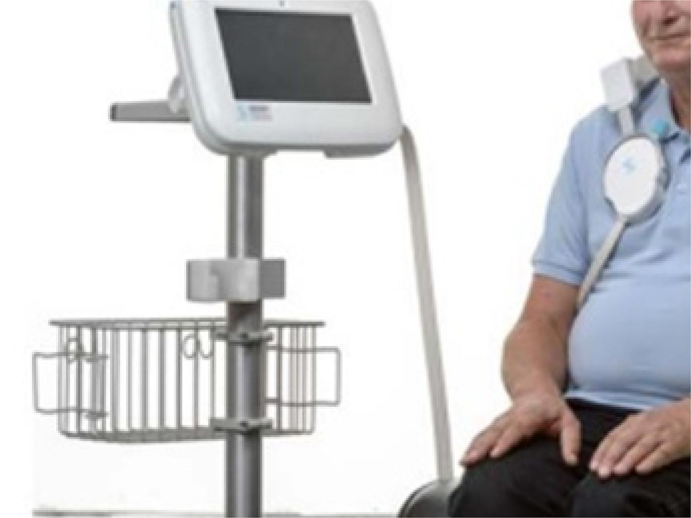
Accurate assessment of pulmonary congestion is pivotal for the optimal management of congestive heart failure. The remote dielectric sensing (ReDS) system represents a recently developed non-invasive technology designed to quantify lung fluid volume without requiring specialized expertise. Kobalava et al. demonstrated a moderate correlation between the ReDS system and lung ultrasound. However, several concerns warrant further discussion.

[…]

  • Heart Failure
  • Hemodynamics
  • Lung
Read Full Article
Creative Commons Attribution (CC BY) Creative Commons license
09/05/2025 DOI: 10.36660/abc.20250053i Views 1
ABCIC
Editorial

Lung Ultrasound in Outpatients with Heart Failure

Marco Stephan Lofrano-Alves

Over the last decade, we have witnessed the widespread use of lung ultrasound (LU) in the assessment of pulmonary congestion. In addition to the well-known technique of auscultation of adventitious lung sounds, LU makes an important contribution by sharpening our vision to the diagnosis of pulmonary interstitial edema by quantification of B-lines. LU has emerged as a relevant and updated method, since it is a safe, low-cost, rapid and available at bedside, being an alternative to imaging methods associated with ionizing radiation, such as chest tomography and X-ray.

LU is a standardized tool in diagnosis and treatment monitoring today, and has been studied in different clinical scenarios of pulmonary congestion of cardiogenic origin.– In outpatients with heart failure and reduced ejection fraction (HFrEF), B-line counting by LU showed an 89% accuracy with a cutoff ≥ 15 B-lines. In another study with patients hospitalized for HFrEF, the risk of an adverse in-hospital event increased with the rising number of B-lines at admission, and the risk of HF hospitalization and all-cause death was greater in patients with a higher number of B-lines at discharge.

[…]

  • Heart Failure
  • Lung
  • Ultrasonics
Read Full Article
Creative Commons Attribution (CC BY) Creative Commons license
27/06/2023 DOI: 10.36660/abcimg.20230049i
Figure 1
ABCIC
Original Article

Pulmonary Congestion in Heart Failure With Reduced Ejection Fraction: Comparison Between Lung Ultrasound and Remote Dielectric Sensing

Maria Estefânia Bosco Otto et al. Vanessa Andreoli Esmanhoto Edileide de Barros Correia Ana Cristina de Souza Murta … Larissa Ventura Ribeiro Bruscky Andrea de Andrade Vilela Antonio Tito Paladino Filho Jorge Eduardo Assef

Abstract

Backgound

Outpatient assessment of pulmonary congestion in patients with heart failure with reduced ejection fraction (HFREF) can minimize hospitalizations due to decompensation and optimize the use of diuretics.

Objective

To compare the remote dielectric sensor (REDS), a validated device for detecting pulmonary extravascular fluid, with clinical parameters, transthoracic echocardiogram (TTE) and lung ultrasound (ULSP).

Methods

We included 38 patients from heart failure clinic (63±12 years; 21 men). All were submitted within 24 hours to clinical evaluation with description of paroxysmal nocturnal dyspnea, leg edema (LE), presence of dizziness; laboratory evaluation of NT-ProBNP; evaluation by REDS and TTE with analysis of parameters of systemic congestion by the inferior vena cava evaluation, function of the right ventricle and of pulmonary congestion, such as evaluation of filling pressures by average of E/e’ and indexed left atrial volume. The ULSP was performed using the 8 points anterior quadrants protocol, counting B lines in one respiratory cycle.

Results

22 patients had REDS ≥ 35% (indicative of pulmonary congestion) and 16 patients REDS<35%. Clinical and echocardiographic parameters were compared with REDS ≥ 35%. In the multivariate analysis, the variables body surface area, B lines and LE were associated with REDS ≥ 35%. NT-ProBNP was similar and elevated in both groups.

Conclusions

Outpatient monitoring of HFREF for volume control can be sensitized by the presence of B lines on the ULSP with good correlation to REDS ≥ 35%. NT pro BNP was not able to differentiate patients with congestion detected by REDS.

  • Heart Failure
  • Lung
  • Ultrasonography
Read Full Article
Creative Commons Attribution (CC BY) Creative Commons license
24/05/2023 DOI: 10.36660/abcimg.20230027i
My Approach to Lung Ultrasound to Evaluate Extravascular Lung Water
ABCIC
My Approach To

My Approach to Lung Ultrasound to Evaluate Extravascular Lung Water

Maria Estefânia Bosco Otto et al. Vanessa Andreoli Esmanhoto

Introduction

A clinical history, physical examination, and focused ultrasound can accelerate the appropriate diagnosis and treatment of patients with progressive or acute dyspnea. Extravascular lung water (EVLW) is easily detected with lung ultrasound (LUS). Pivetta et al. reported excellent accuracy detecting EVLW by clinical evaluation and by the presence of “B” lines on LUS heigher than B natriuretic peptide dosage.

The usefulness of LUS for intensive care unit patients was described in the 1980s, later than that of other ultrasound modalities. LUS is useful not only for diagnosing EVLW but also for risk stratification in myocardial infarction and acute congestive heart failure (CHF).

[…]

  • Dyspneia
  • Lung
  • Ultrasonography
Read Full Article
Creative Commons Attribution (CC BY) Creative Commons license
17/08/2022 DOI: 10.47593/2675-312X/20223503ecom22
Figure 1
IJCS
Original Article

Lung Ultrasound as a Triage Tool in an Emergency Setting during the Covid-19 Outbreak: comparison with CT Findings

Monica Luiza de Alcantara et al. Marcos Paulo Lacerda Bernardo Tatiana Bagrichevsky Autran Rodolfo de Paula Lustosa … Marcelo Tayah Lucia Antunes Chagas Dequitier Carvalho Machado

Abstract

Background

Lung ultrasound (LUS) can detect interstitial alveolar changes confined to the subpleural region, like those described in Covid-19.

Objetive

To evaluate how LUS findings correlate with chest computed tomography (CT) in patients admitted to the emergency department (ED) with suspicion of Covid-19.

Methods

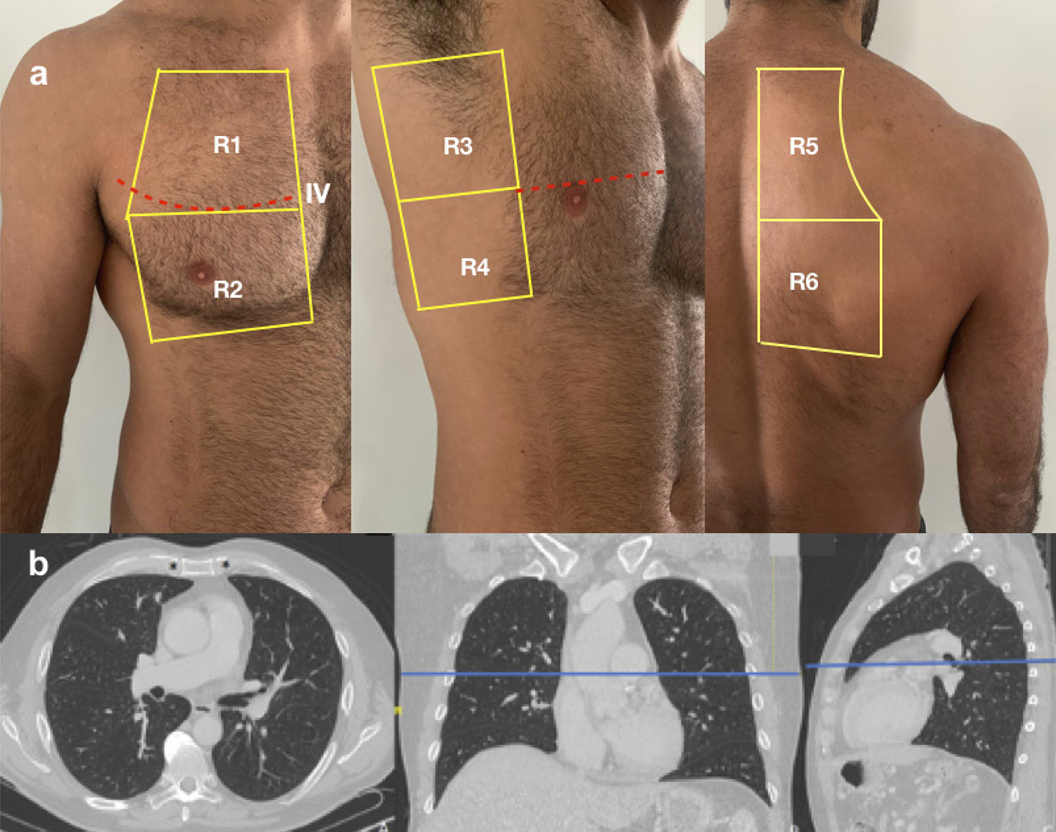
Cross-sectional study of 20 patients (median age 43 years; interquartile range, 37–63 years; 50% male). All patients underwent LUS and chest CT on the day of ED admission. Each hemithorax was divided into 6 segments with similar landmarks, and equivalent scores (sc) of lesion severity were defined for both methods. The number of affected segments on LUS (LUSseg) was divided into tertiles (0-1, 2-5, and ≥6), and compared with number of affected segments on CT (CTseg), LUSsc, CTsc, and percentage of affected lung parenchyma through visual analysis (CTvis). ANOVA or Kruskal–Wallis test for continuous variables, chi-square test for categorical variables, and receiver operating characteristic (ROC) curve analysis to define optimal cutoff points were performed. P<0.05 was considered statistically significant.

Results

Median LUSsc, CTsc, CTseg, and CTvis were significantly different between groups. A clear separation between groups was demonstrated; patients with 7 LUSsc and >3 LUSseg, respectively.

Conclusion

LUS findings correlate with chest CT, and can help identify patients with normal lung or minor pulmonary involvement secondary to Covid-19. Int J Cardiovasc Sci. 2020; [online].ahead print, PP.0-0

  • COVID-19
  • Lung
  • Pandemics
  • Pleura
  • Pulmonary Alveoli
  • Tomography, X Ray-Computed/methods
  • Triage
  • Ultrasonography/methods
Read Full Article
Creative Commons Attribution (CC BY) Creative Commons license
28/08/2020 DOI: 10.36660/ijcs.20200133 Views 1

How to cite this article

EndNote RIS

Electronic Document Format (Vancouver)

Otto MEB. The New Frontier of Point-of-Care Ultrasound: Lung Ultrasound in Congenital Heart Disease. Arquivos Brasileiros de Cardiologia 2026;123(3):e20260163.

Electronic Document Format (ABNT)

Otto, Maria Estefânia Bosco. The New Frontier of Point-of-Care Ultrasound: Lung Ultrasound in Congenital Heart Disease. Arquivos Brasileiros de Cardiologia, v. 123, n. 3, e20260163, May. 2026.

Electronic Document Format (APA)

Otto, M. E. B. (2026). The New Frontier of Point-of-Care Ultrasound: Lung Ultrasound in Congenital Heart Disease. Arquivos Brasileiros de Cardiologia, 123(3), e20260163.

Electronic Document Format (ISO)

Otto, Maria Estefânia Bosco. The New Frontier of Point-of-Care Ultrasound: Lung Ultrasound in Congenital Heart Disease. Arquivos Brasileiros de Cardiologia [online]. 2026, vol. 123, n. 3, [cited 2026-10-14], e20260163. Available from: <https://journals.cardiol.br/en/article/the-new-frontier-of-point-of-care-ultrasound-lung-ultrasound-in-congenital-heart-disease/>. ISSN 0066-782X

EndNote RIS

Electronic Document Format (Vancouver)

Izumida T, Imamura T. How to Incorporate Remote Dielectric Sensing System and Lung Ultrasound in Patients with Acute Congestive Heart Failure. Arquivos Brasileiros de Cardiologia 2025;122(4):e20250053.

Electronic Document Format (ABNT)

Izumida, Toshihide; Imamura, Teruhiko. How to Incorporate Remote Dielectric Sensing System and Lung Ultrasound in Patients with Acute Congestive Heart Failure. Arquivos Brasileiros de Cardiologia, v. 122, n. 4, e20250053, May. 2025.

Electronic Document Format (APA)

Izumida, T., & Imamura, T. (2025). How to Incorporate Remote Dielectric Sensing System and Lung Ultrasound in Patients with Acute Congestive Heart Failure. Arquivos Brasileiros de Cardiologia, 122(4), e20250053.

Electronic Document Format (ISO)

Izumida, Toshihide and Imamura, Teruhiko. How to Incorporate Remote Dielectric Sensing System and Lung Ultrasound in Patients with Acute Congestive Heart Failure. Arquivos Brasileiros de Cardiologia [online]. 2025, vol. 122, n. 4, [cited 2026-10-14], e20250053. Available from: <https://journals.cardiol.br/en/article/how-to-incorporate-remote-dielectric-sensing-system-and-lung-ultrasound-in-patients-with-acute-congestive-heart-failure/>. ISSN 0066-782X

EndNote RIS

Electronic Document Format (Vancouver)

Lofrano-Alves MS. Lung Ultrasound in Outpatients with Heart Failure. ABC Imagem Cardiovascular 2023;36(2):e20230049.

Electronic Document Format (ABNT)

Lofrano-Alves, Marco Stephan. Lung Ultrasound in Outpatients with Heart Failure. ABC Imagem Cardiovascular, v. 36, n. 2, e20230049, Jun. 2023.

Electronic Document Format (APA)

Lofrano-Alves, M. S. (2023). Lung Ultrasound in Outpatients with Heart Failure. ABC Imagem Cardiovascular, 36(2), e20230049.

Electronic Document Format (ISO)

Lofrano-Alves, Marco Stephan. Lung Ultrasound in Outpatients with Heart Failure. ABC Imagem Cardiovascular [online]. 2023, vol. 36, n. 2, [cited 2026-10-14], e20230049. Available from: <https://journals.cardiol.br/en/article/lung-ultrasound-in-outpatients-with-heart-failure/>. ISSN 2318-8219

EndNote RIS

Electronic Document Format (Vancouver)

Otto MEB, Esmanhoto VA, Correia EB, Murta ACS, Bruscky LVR, Vilela AA, Paladino Filho AT, et al. Pulmonary Congestion in Heart Failure With Reduced Ejection Fraction: Comparison Between Lung Ultrasound and Remote Dielectric Sensing. ABC Imagem Cardiovascular 2023;36(2):e20230027.

Electronic Document Format (ABNT)

Otto, Maria Estefânia Bosco; Esmanhoto, Vanessa Andreoli; Correia, Edileide de Barros; Murta, Ana Cristina de Souza; Bruscky, Larissa Ventura Ribeiro; Vilela, Andrea de Andrade; Paladino Filho, Antonio Tito; Assef, Jorge Eduardo. Pulmonary Congestion in Heart Failure With Reduced Ejection Fraction: Comparison Between Lung Ultrasound and Remote Dielectric Sensing. ABC Imagem Cardiovascular, v. 36, n. 2, e20230027, May. 2023.

Electronic Document Format (APA)

Otto, M. E. B., Esmanhoto, V. A., Correia, E. B., Murta, A. C. S., Bruscky, L. V. R., Vilela, A. A., Paladino Filho, A. T., & Assef, J. E. (2023). Pulmonary Congestion in Heart Failure With Reduced Ejection Fraction: Comparison Between Lung Ultrasound and Remote Dielectric Sensing. ABC Imagem Cardiovascular, 36(2), e20230027.

Electronic Document Format (ISO)

Otto, Maria Estefânia Bosco and Esmanhoto, Vanessa Andreoli and Correia, Edileide de Barros and Murta, Ana Cristina de Souza and Bruscky, Larissa Ventura Ribeiro and Vilela, Andrea de Andrade and Paladino Filho, Antonio Tito and Assef, Jorge Eduardo. Pulmonary Congestion in Heart Failure With Reduced Ejection Fraction: Comparison Between Lung Ultrasound and Remote Dielectric Sensing. ABC Imagem Cardiovascular [online]. 2023, vol. 36, n. 2, [cited 2026-10-14], e20230027. Available from: <https://journals.cardiol.br/en/article/pulmonary-congestion-in-heart-failure-with-reduced-ejection-fraction-comparison-between-lung-ultrasound-and-remote-dielectric-sensing/>. ISSN 2318-8219

EndNote RIS

Electronic Document Format (Vancouver)

Otto MEB, Esmanhoto VA. My Approach to Lung Ultrasound to Evaluate Extravascular Lung Water. ABC Imagem Cardiovascular 2022;35(3):ecom22.

Electronic Document Format (ABNT)

Otto, Maria Estefânia Bosco; Esmanhoto, Vanessa Andreoli. My Approach to Lung Ultrasound to Evaluate Extravascular Lung Water. ABC Imagem Cardiovascular, v. 35, n. 3, ecom22, Aug. 2022.

Electronic Document Format (APA)

Otto, M. E. B., & Esmanhoto, V. A. (2022). My Approach to Lung Ultrasound to Evaluate Extravascular Lung Water. ABC Imagem Cardiovascular, 35(3), ecom22.

Electronic Document Format (ISO)

Otto, Maria Estefânia Bosco and Esmanhoto, Vanessa Andreoli. My Approach to Lung Ultrasound to Evaluate Extravascular Lung Water. ABC Imagem Cardiovascular [online]. 2022, vol. 35, n. 3, [cited 2026-10-14], ecom22. Available from: <https://journals.cardiol.br/en/article/my-approach-to-lung-ultrasound-to-evaluate-extravascular-lung-water/>. ISSN 2318-8219

EndNote RIS

Electronic Document Format (Vancouver)

Alcantara ML, Bernardo MPL, Autran TB, Lustosa RP, Tayah M, Chagas LA, Machado DC, et al. Lung Ultrasound as a Triage Tool in an Emergency Setting during the Covid-19 Outbreak: comparison with CT Findings. International Journal of Cardiovascular Sciences 2020;33(5):479-87.

Electronic Document Format (ABNT)

Alcantara, Monica Luiza de; Bernardo, Marcos Paulo Lacerda; Autran, Tatiana Bagrichevsky; Lustosa, Rodolfo de Paula; Tayah, Marcelo; Chagas, Lucia Antunes; Machado, Dequitier Carvalho. Lung Ultrasound as a Triage Tool in an Emergency Setting during the Covid-19 Outbreak: comparison with CT Findings. International Journal of Cardiovascular Sciences, v. 33, n. 5, p. 479-487, Aug. 2020.

Electronic Document Format (APA)

Alcantara, M. L. , Bernardo, M. P. L., Autran, T. B., Lustosa, R. P., Tayah, M., Chagas, L. A., & Machado, D. C. (2020). Lung Ultrasound as a Triage Tool in an Emergency Setting during the Covid-19 Outbreak: comparison with CT Findings. International Journal of Cardiovascular Sciences, 33(5), 479-487.

Electronic Document Format (ISO)

Alcantara, Monica Luiza de and Bernardo, Marcos Paulo Lacerda and Autran, Tatiana Bagrichevsky and Lustosa, Rodolfo de Paula and Tayah, Marcelo and Chagas, Lucia Antunes and Machado, Dequitier Carvalho. Lung Ultrasound as a Triage Tool in an Emergency Setting during the Covid-19 Outbreak: comparison with CT Findings. International Journal of Cardiovascular Sciences [online]. 2020, vol. 33, n. 5, [cited 2026-10-14], pp. 479-487. Available from: <https://journals.cardiol.br/en/article/lung-ultrasound-as-a-triage-tool-in-an-emergency-setting-during-the-covid-19-outbreak-comparison-with-ct-findings/>. ISSN 2359-4802

  • About ABC Family
  • Internal Regulation
  • Editorial Board
  • Brazilian Society of Cardiology (SBC)

Issues

SBC Guidelines

  • Videos
  • Podcasts
  • News