| ABCIC
ABC Family Arquivos Brasileiros de Cardiologia International Journal of Cardiovascular Sciences ABC Heart Failure & Cardiomyopathy ABC Imagem Cardiovascular
Português English
ABC Family | ABCIC
Português English
  • About
    • About the Journal
    • Internal Regulation
    • Editors Section
    • Editorial Board
    • SBC Award
  • Instructions to Authors
    • Guidelines for Publication
    • Editorial Policy
    • Ethics Policy
    • Policy on the Use of Artificial Intelligence
    • Mandatory Publication Form
  • Issues
  • SBC Guidelines
  • About
    • About the Journal
    • Internal Regulation
    • Editors Section
    • Editorial Board
    • SBC Award
  • Instructions to Authors
    • Guidelines for Publication
    • Editorial Policy
    • Ethics Policy
    • Policy on the Use of Artificial Intelligence
    • Mandatory Publication Form
  • Issues
  • SBC Guidelines

ABC Family

  • ABC Family
  • Arquivos Brasileiros de Cardiologia
  • International Journal of Cardiovascular Sciences
  • ABC Heart Failure & Cardiomyopathy
  • ABC Imagem Cardiovascular
Search in

No content found.

Journals
Ordering
Article types
Others

  1. Home
  2. ABCIC
  3. Issues
  4. 2023, Volume 36, Number 1
Original Article

Heart Adaptation Mechanisms in Elite Female Athletes: Comparison With Healthy Individuals and Time of Training

José Maria Del Castillo et al. Thiago Boschilia Nemi Sabeh Junior Carlos Antônio da Mota Silveira Djair Brindeiro Filho

Abstract

Background:

Intense continuous exercise provokes adaptive remodeling phenotypes in athletes, the parameters of which can be evaluated through conventional echocardiography and myocardial deformation. We compared myocardial remodeling in female athletes (athlete group) with sedentary women of the same age range (control group) and between older and younger athletes.

Methods:

A total of 57 female soccer players and 25 healthy sedentary women were selected. The athlete group was subdivided into a main group and those under 17 years of age (< 17 group). The dimensions and systolic and diastolic function of the cardiac chambers and myocardial deformation (longitudinal and circumferential, as well as radial strain and rotational mechanics) was determined through echocardiography, using the Z statistic with a significance level of p< 0.05.

Results:

The mean age of the athlete, control, main, and < 17 groups was 22.1 (SD, 6.3); 21.2 (SD, 5.0); 26.5 (SD, 5.1); 16.5 (SD, 0.6) years, respectively. Weight, body mass index and heart rate were lower in the athlete group. Wall thickness, left ventricular mass index, left atrial (LA) volume, ejection fraction, and right ventricular dimensions were higher in athlete group, but remained within normal ranges. Regarding myocardial deformation, there was decreased radial strain, basal rotation, apical rotation, and twisting in the athlete group, suggesting a contractile reserve mechanism. These parameters were lesser in the main athlete group, who also had greater wall thickness, greater volume in the left atrium (LA) and larger size in the right ventricle (RV), suggesting that increased contractile reserve is related to longer time spent in the sport.

Conclusions:

In female athletes who had undergone intense long-term training, we observed adaptive remodeling of the cardiac chambers and increased contractile reserve (at rest), and these changes were more pronounced in those with longer involvement in the sport.
  • Athletes
  • Atrial Remodeling
  • Echocardiography
Read Full Article
Creative Commons Attribution (CC BY) Creative Commons license
31/03/2023 DOI: 10.36660/abcimg.2023372i Views 2
Editorial

Cardiovascular Imaging In Assessment Of Athletes

José Roberto Matos-Souza et al. Daniela do Carmo Rassi

Physical training is linked to a series of cardiac, morphological, and functional adaptations known as “athlete’s heart”., An athlete can be considered a youth or adult amateur or professional who exercises regularly and participates in official sports competitions. The changes demonstrated depend on the duration, intensity, and type of training (isotonic versus isometric).

In recent years, a large number of studies have been published, which have improved our ability to understand the characteristics of physiological cardiac remodeling in athletes. Nevertheless, there are gaps regarding the differential diagnosis of athlete’s heart, especially when it presents with a more intense expression. It can be difficult to differentiate this condition from inherited heart diseases such as hypertrophic, dilated, or arrhythmogenic cardiomyopathy and left ventricular non-compaction.,

[…]

  • Cardiac Remodeling
  • Echocardiography
  • Myocardial deformation
Read Full Article
Creative Commons Attribution (CC BY) Creative Commons license
31/03/2023 DOI: 10.36660/abcimg.20230015i Views 1
Figure 1
Editorial

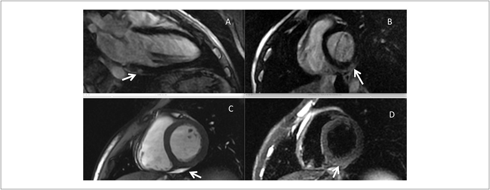
Hypothesis on the Pathogenesis of Sub-Epicardial Scar Associated with Myocarditis

João A. C. Lima et al. David A. Bluemke Joshua Hare Katherine Wu … Leonardo Sara Carlos E. Rochitte

The typical pattern of myocardial injury and subsequent fibrosis in patients with viral myocarditis is subepicardial. However, other patterns of injury and scar, as detected by contrast enhanced magnetic resonance imaging (cMRI), commonly occur, including sub-endocardial injury mimicking the ischemic heart disease pattern, and mid-wall circumferential scar seen more often in patients with advanced disease and dilated cardiomyopathies. In patients with myocarditis, who present malignant arrhythmias in the scenario of a preserved left ventricle (LV) function, it is not uncommon to find the typical subepicardial scar pattern in the presence of normal or mildly abnormal LV systolic function. However, the pathogenesis of sub-epicardial scars in patients with myocarditis accompanied or not by significant LV dysfunction remains obscure.

In addition to myocarditis, subepicardial myocardial scar is found in association with various disease processes that are not believed to be secondary to a virally induced myocardial injury. The list includes disease processes with disparate etiologies, such Duchenne and Fabry’s disease, Chagas cardiomyopathy, and rheumatic heart disease, among others.– Therefore, different theories have been proposed to explain this pattern of scar formation in patients with different pathologies. Increase in excessive regional stress and local perfusion alterations have been postulated as contributing mechanisms, but both fall short as main mechanisms. Calculated wall stress is greater at the level of the subendocardium, and reversible perfusion defects secondary to ischemia are commonly subendocardial. After scar formation, perfusion is diminished in proportion to capillary density reduction (fixed defect), but that does not implicate local ischemia as the pathogenetic mechanism underlying subepicardial scar formation.

[…]

  • Cardiac resonance
  • COVID-19
  • epicardial injury
  • Myocarditis
Read Full Article
Creative Commons Attribution (CC BY) Creative Commons license
31/03/2023 DOI: 10.36660/abcimg.20230001i Views 2
Editorial

Mitral Valve Area Quantification Using Digital Image Processing: Is That Feasible?

Edgar Daminello et al. Paulo Pinto Alves Campos Vieira Cláudio Henrique Fischer Marcelo Luiz Campos Vieira

Echocardiography, since its initial descriptions in the 1950s, has evolved every decade with the incorporation of numerous new techniques, enabling structural cardiac analysis from multiple planes of spatial observation in real time. This occurred with the advent of nanotechnology, digital technology, application of modifiable algorithms based on the concept of machine learning and deep learning through the development of artificial intelligence (AI).– The use of AI is an ever-increasing reality in medicine as a whole, mainly in operator-dependent imaging areas, such as ultrasonography imaging.

In the concept of analyzing the confluence of a very large number of pieces of information (e.g. radiomics, metabolomics, proteomics) for the formation of data clusters with the aim of integrated patient observation, the formation of precision medicine, AI is used in a rational and organized way. The use of AI allows faster, more balanced and homogeneous observation of inputs, in the sense of minimizing inter and intraobserver errors.– In the specific echocardiographic evaluation, the use of AI presents a very wide range of applications, having been used for the acquisition of echocardiographic information for the quantification of left ventricular volumes and mass, analysis of biventricular systolic function, segmentation and qualification of the cusps, annulus and mitral valve apparatus, analysis of hypertrophic phenocopy (athletes, patients with hypertrophic cardiomyopathy), to predict the outcome of percutaneous hemodynamic procedures (e.g.: mitraclip). We are currently working on a project in association with the University of Chicago for AI analysis of patients affected by transthyretin cardiac amyloidosis.

[…]

  • algorithm
  • Artificial Intelligence
  • automated diagnosis
  • Echocardiography
  • Mitral Valve
Read Full Article
Creative Commons Attribution (CC BY) Creative Commons license
31/03/2023 DOI: 10.36660/abcimg.2023376i Views 1
Review Article

Echocardiographic Evaluation of a Patient in Circulatory Shock: A Contemporary Approach

Rafael Modesto Fernandes et al. Alexandre Costa Souza Bruno de Freitas Leite Jun Ramos Kawaoka

Abstract

Circulatory shock is characterized by a state of inefficient tissue oxygen supply and multiple organ dysfunction. Patients with circulatory shock require fast and assertive diagnosis and therapies to reduce its high lethality. Echocardiography has already been established as a fundamental method in managing patients with circulatory shock. It provides crucial assistance in etiological diagnosis, prognosis, hemodynamic monitoring, and volume estimation in these patients; its potential advantages include portability, absence of contrast or radiation, low cost, and real-time serial assessment. In the intensive care unit setting, it demonstrates a high correlation with invasive (pulmonary artery catheter) and minimally invasive (transpulmonary thermodilution) forms of hemodynamic monitoring. Currently, other techniques, such as pulmonary ultrasound and VExUS score, have been added to echocardiographic assessment, making the method more comprehensive and accurate. These techniques add relevant data to blood volume estimation in critical patients, influencing the probabilistic decision of fluid responsiveness and providing additional information in the diagnostic reasoning of the causes of shock, thus optimizing these patients’ prognosis. Point of care ultrasound (POCUS) aims to make abilities to obtain information at the bedside more accessible to physicians who are not specialists in radiology, by means of ultrasound, which assists them in decision-making. This article addresses the diverse applications of echocardiography in patients with circulatory shock, including prognostic evaluation and etiological diagnosis by means of the parameters found in the main causes of shock, in addition to hemodynamic monitoring, evaluation of fluid responsiveness, and practical use of pulmonary ultrasound.

  • Circulatory shock
  • Diagnosis
  • Fluid-responsiveness
  • Lung ultrasound
  • Prognosis
Read Full Article
Creative Commons Attribution (CC BY) Creative Commons license
31/03/2023 DOI: 10.36660/abcimg.20230013i Views 2
Figure 1
Case Report

Criss-Cross Heart: A Case Report

Mateus Oliveira Potratz et al. Lorrayne Zonatele Garbo Kamila Pompermaier Pessimilio André Silveira Loss … Crislane Belisario Ambrozim Ana Luiza Teixeira Albert Lima Danielle Lopes Rocha

Abstract

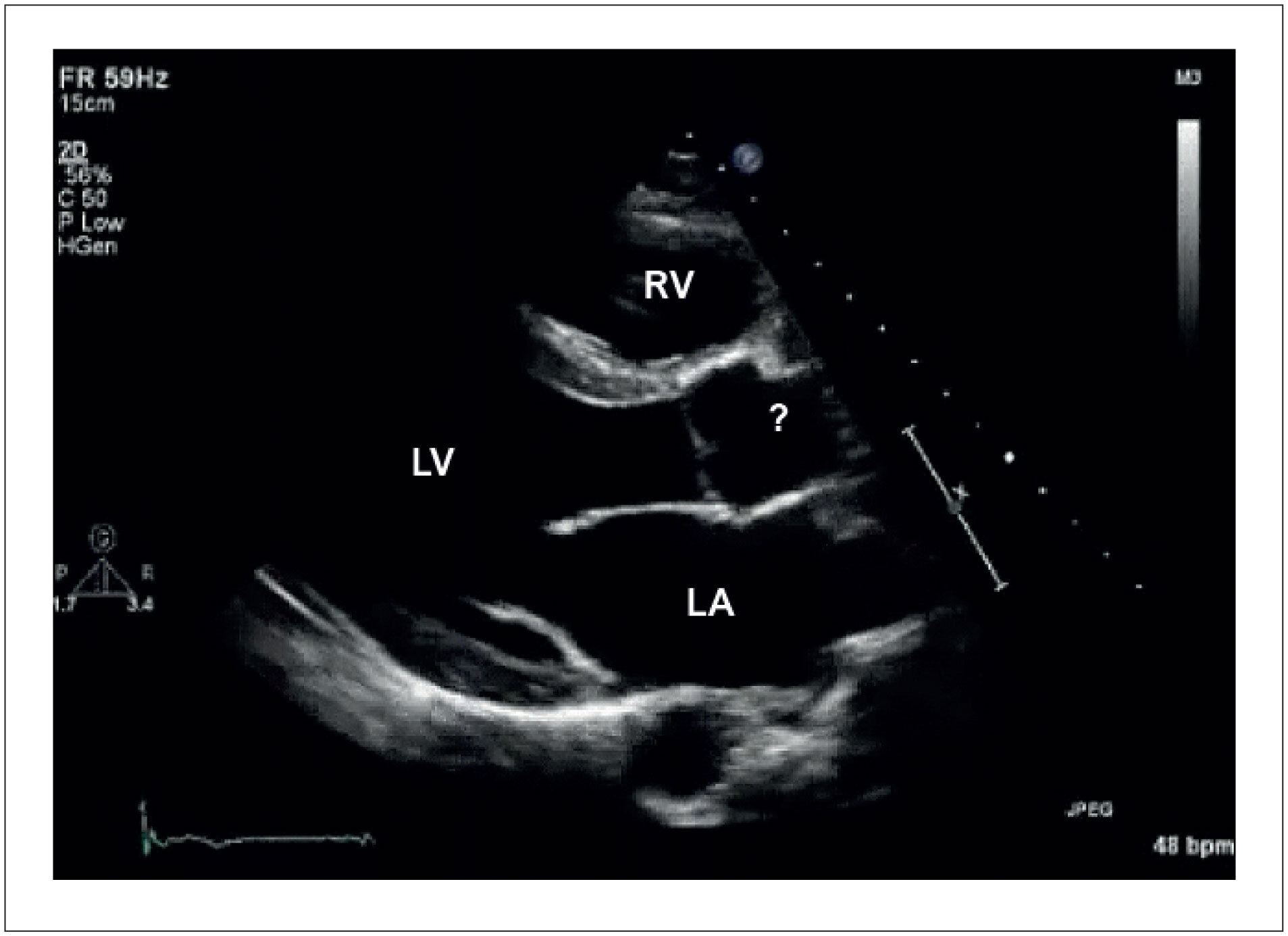
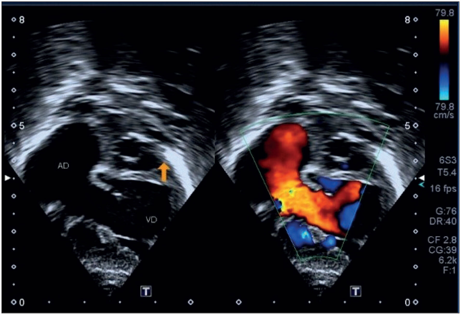
Criss-cross heart was first described in 1974. It is a rare congenital heart malformation that occurs in 8 cases per 1,000,000 children, and represents only 0.1% of congenital malformations. The diagnostic methods of choice are transthoracic echocardiography, cardiac magnetic resonance (CMR), computed tomography angiography (CT) and, sometimes, cardiac catheterization. This report describes the case of a newborn with a criss-cross heart in addition to double-outlet right ventricle (RV), with poorly positioned vessels, in addition to atrial septal defect (ASD), interventricular septal defect, tricuspid valve dysplasia and persistent left superior vena cava. The exact etiology of this malformation is not known, but it seems to occur due to rotation of the ventricles in their longitudinal axis, not accompanied by rotation of the atrial and atrioventricular (AV) valves. This movement produces abnormal ventricular inlets, determining that the RV be positioned on a superior plane and the left ventricle on an inferior plane. Although the exact cause of this anomaly is still unknown, it is believed that a genetic abnormality may be leading to these cases: mutation of the Cx43 gene. Diagnosis of the case concerned was given by transthoracic echocardiography and computed CT of the aorta and pulmonary arteries, which showed, in addition to the criss-cross heart, other abnormalities, such as double-outlet RV, large ASD and ventricular septal defect (VSD).

  • Congenital Heart Defects
  • Crisscross Heart
  • Heart Ventricles
Read Full Article
Creative Commons Attribution (CC BY) Creative Commons license
31/03/2023 DOI: 10.36660/abcimg.2023282i Views 2
Case Report

Role of Multimodality for the Diagnosis of Thrombosis at Late Follow-up of Patients Selected for TAVI: Review of a Case Series

Laila Caroline Oliveira Souza Barbosa Gomes et al. Alexandre Costa Souza Stephanie de Azevedo Drubi Bruna de Mattos Ivo Junqueira … Mariana Lins Baptista Guedes Bezerra Rodrigo Vieira de Melo

Introduction

The first transcatheter aortic valve implantation (TAVI) was performed by Alan Cribier in France in 2002 and the technique was introduced in Brazil in 2008. Since then, this modality has been consolidated and many patients have been approached using this technique. After more than 10 years of the first TAVI in Brazil, an increasing number of late complications appear in the cardiovascular imaging laboratory, with challenging anatomical and functional aspects in daily practice. Late thrombosis of aortic endoprosthesis may be one of the causes of dysfunction in the follow-up after TAVI. This clinical entity can assume a complex profile for accurate diagnosis and proper patient management, since it can have a variable presentation, ranging from subclinical leaflet thrombosis to limiting symptoms related to heart failure. Transesophageal echocardiography (TEE), the additional resource of 3D echocardiography and high-resolution computed tomography (CT) play a complementary role in the diagnosis of this etiology through anatomical valve reconstruction with evidence of hypoattenuated leaflet thickening (HALT) with or without hypoattenuation affecting motion (HAM) of one or more prosthetic valve leaflets., The risk of embolic events after diagnosis of leaflet thrombosis is still uncertain, especially for the central nervous system., This case series presents two illustrative reports of aortic endoprostheses with high gradients and complementary aspects of the multimodality that direct the etiology to the diagnosis of late prosthesis thrombosis.

[…]

  • Multimodality
  • Prosthesis thrombosis
  • Transcatheter Aortic Valve Replacement
Read Full Article
Creative Commons Attribution (CC BY) Creative Commons license
31/03/2023 DOI: 10.36660/abcimg.2023367i
Figure 1
Review Article

My Approach to “Athlete's Heart”: Evaluation of the Different Types of Adaptation to Exercise

Frederico José Neves Mancuso

Abstract

Exercise-induced adaptation may occur in amateur and professional athletes. This condition is commonly named “athlete’s heart”. The alterations observed include dilation of the heart chambers, increased myocardial thickness, improved ventricular filling, increased left ventricular trabeculation, dilation of the inferior vena cava, among others. These changes can also be observed in some heart diseases, such as dilated, hypertrophic and other cardiomyopathies (CMP). Thus, cardiac imaging tests are fundamental in identifying these alterations and in differentiating between “athlete’s heart” and possible heart disease.

  • adaptive cardiac remodeling
  • Athlete's heart
  • cardiomyopathy
  • ecocardiography
Read Full Article
Creative Commons Attribution (CC BY) Creative Commons license
31/03/2023 DOI: 10.36660/abcimg.20230002i Views 2
Figure 1
Case Report

Pulmonary Thromboembolism Associated with Paradoxical Embolism in a Patient with Patent Foramen Ovale

Arthur Barroso Vidal Vilarinho et al. Danielly Bernardes Silva Ana Rosaria Medeiros Peres Rodolfo Loureiro Borges de Souza … Carlos Eduardo de Sousa Amorelli Fábio Lemos Campedelli Luciano Moreira Alves Fabio Augusto Cypreste de Oliveira

Introduction

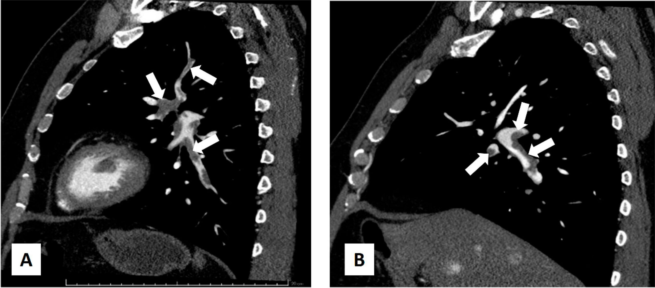
Patent foramen ovale (PFO) is a pathological condition characterized by an opening in the interatrial septum, that allows blood to flow from one atrium to the other. It is found in approximately 25% of the adult population, with no hemodynamic implications in most cases. However, due to persistence of the foramen, paradoxical embolism may occur as a cause of ischemic events; in some cases, closure of the PFO is indicated as a secondary prevention strategy for thromboembolic events.

Studies have demonstrated an association between PFO and many clinical conditions, some potentially severe, such as ischemic stroke and pulmonary thromboembolism (PTE).

[…]

  • Foramen Ovale
  • Palavras-chave Pulmonary Embolism
  • Paradoxical Embolism
  • Splenic Infarction
Read Full Article
Creative Commons Attribution (CC BY) Creative Commons license
24/03/2023 DOI: 10.36660/abcimg.2023347i Views 1
Review Article

My Approach to Imaging in Sickle Cell Anemia

João Carlos Moron Saes Braga et al. Fábio Villaça Guimarães Filho Alexandre Rodrigues Raphael Aparecido Barreto

Abstract

Sickle cell disease (SCD) is recognized as a global problem in public health, characterized by the alteration in the red blood cells to the sickle form. Moreover, chronic anemia can also be observed through the change in the rheology of the red blood cells, leading to a scenario of inflammation and oxidative stress, making SCD a multisystem disease.

Cardiac output (CO) proved to be high, leading to an overall increase in the heart chambers and an eccentric myocardial hypertrophy. These heart alterations were attributed only to adaptive reactions to chronic anemia. Recent studies have more clearly recognized an association with pulmonary hypertension (PH), left ventricular diastolic dysfunction, arrhythmias, and sudden death. Moreover, what has also arisen in this context is the hypothesis of the existence of a sickle-cell cardiomyopathy, characterized by diastolic dysfunction and restrictive physiology. The echocardiogram represents a key tool in determining cavitary volumes, diastolic dysfunction, and the estimation of pulmonary pressure, as well as constitutes a valuable resource in the diagnosis and therapeutic treatment of acute chest syndrome. The myocardial strain, rotational variables, myocardial work, and 3D echocardiography can be applied in an attempt to aid in the early detection of patients who are at a higher risk of developing complications and evolving to death related to SCD.

  • Diastolic Dysfunction
  • Pulmonary Hypertension
  • Sickle Cell Anemia
Read Full Article
Creative Commons Attribution (CC BY) Creative Commons license
24/03/2023 DOI: 10.36660/abcimg.20230009i Views 2
Original Article

Incidental Tomographic Findings of Coronary Artery Calcifications: A Prevalence Study in Southern Brazil

Karen Rafaela Okaseski Scopel et al. Tássia Machado Medeiros Bibiana Natalia Porto Maicá Maria Carbonari Velho … Mariana Motta Dias da Silva Juliane Nascimento Mattos Guilherme Galante Heuser Eliane Roseli Winkelmann

Abstract

Introduction

Coronary artery calcifications (CAC) are shown to be a predictive factor of cardiovascular diseases. Computed tomography (CT) of the chest with a low-dose acquisition protocol is accurate in identifying CAC and provides incidental findings of these calcifications, which are commonly overlooked. This study will analyze the prevalence of incidental findings of calcification in coronary arteries in non-cardiac individuals undergoing chest CT.

Methods

Consecutive cross-sectional study of an analytical and descriptive nature. Individuals of both genders who underwent chest CT by referral, over 18 years of age and without heart disease were included. Data collection was carried out using medical records and a self-applied anamnesis form. The variables referring to the CAC and the extension of the impairment were obtained from the reassessment of the chest CT images available in the institution’s system. The exams were anonymized and evaluated by two experienced radiologists. P≤0.05 was considered statistically significant.

Results

397 exams were analyzed. A prevalence of calcifications was found in 176 (44%) of the cases. The existence of these coronary calcifications is related to age (p<0.001). Calcifications are related to gender (p = 0.03) with a higher odds ratio of development in men (odds ratio [OR] = 1.55). Smoking (p<0.001), sedentary lifestyle (p<0.001), systemic arterial hypertension (p<0.001), Diabetes Mellitus (p = 0.04), and dyslipidemia (p<0.001) showed a positive association.

Conclusion

The prevalence of incidental CAC findings was 44%; vary in greater numbers between mild and severe; higher odds ratio in males and increased prevalence with age. Therefore, chest CT proves to be an effective method to assess CAC, and together with the patient’s clinical history, it can be used to measure risk factors for CVD and intervene in the outcome of the condition.

  • Coronary Vessels
  • Incidental Findings
  • Tomography, X-Ray Computed
  • Vascular Calcification
Read Full Article
Creative Commons Attribution (CC BY) Creative Commons license
24/03/2023 DOI: 10.36660/abcimg.2023368i Views 2
Editorial

A Clinician’s View of Tricuspid Regurgitation: What do I Need to Know? When to Intervene?

Tiago Bignoto

Tricuspid valve dysfunction was considered a benign clinical situation for a long time, but with the advancement of diagnostic imaging methods and better clinical stratification of patients with structural heart disease, adequate classification of tricuspid regurgitation has become fundamental to decision making in the context of a Heart Team.

The first assessment to be taken into account is the etiology of the dysfunction. With a low prevalence, the primary etiology, mainly due to rheumatic and congenital causes, has a well-established intervention flowchart, primarily based on the presence of symptoms or hemodynamic repercussions on the right side of the heart.

[…]

  • Heart Valve Diseases
  • Tricuspid Valve
  • Tricuspid Valve Insufficiency
Read Full Article
Creative Commons Attribution (CC BY) Creative Commons license
24/03/2023 DOI: 10.36660/abcimg.20230005i Views 2
Figure 1
Original Article

Automatic Measurement of the Mitral Valve Based on Echocardiography Using Digital Image Processing

Genilton de França Barros Filho et al. Israel Solha Ewerton Freitas de Medeiros Alex dos Santos Felix … André Luiz Cerqueira de Almeida José Carlos de Lima Júnior Marcelo Dantas Tavares de Melo Marcelo Cavalcanti Rodrigues

Abstract

Background

The evaluation of mitral valve area through multiplanar reconstruction in 3-dimensional echocardiography is restricted to specific software and to the experience of echocardiographers. They need to manually select the video frame that contains the maximum mitral valve opening area, as this dimension is fundamental to identification of mitral stenosis.

Objective

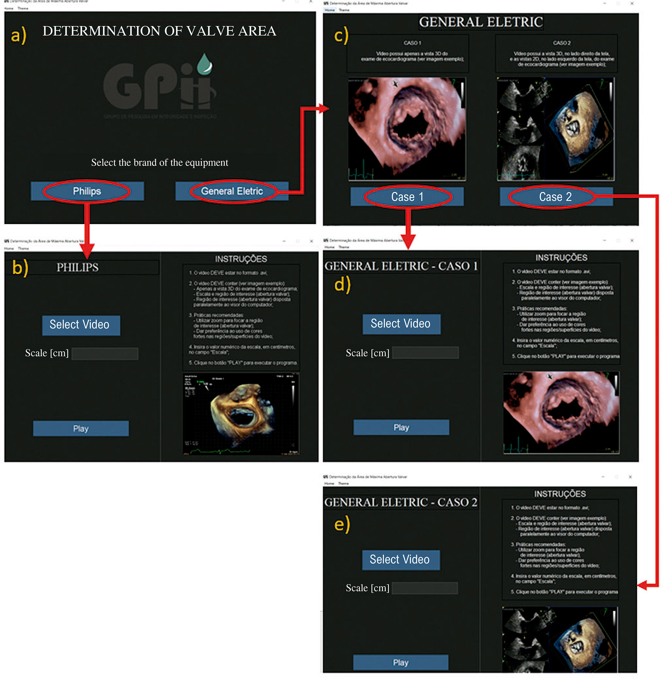
To automate the process of determining the maximum mitral valve opening area, through the application of digital image processing (DIP) in echocardiography tests, developing an open algorithm with video reading in avi format.

Method

This cross-sectional observational pilot study was conducted with 25 different echocardiography exams, 15 with normal aperture and 10 with rheumatic mitral stenosis. With the authorization of the Research Ethics Committee, all exams were performed and made available by 2 specialists who used 2 models of echocardiographic devices: Vivid E95 (GE Healthcare) and Epiq 7 (Philips), with multiplanar transesophageal probes. All videos in avi format were submitted to DIP using the image segmentation technique.

Results

The measurements obtained manually by experienced echocardiographers and the values calculated by the developed system were compared using a Bland-Altman diagram. There was greater agreement between values in the range from 0.4 to 2.7 cm2.

Conclusion

It was possible to automatically determine the maximum mitral valve opening area, for cases from both GE and Philips, using only 1 video as input data. The algorithm has been demonstrated to save time on measurements when compared to the usual method.

  • Echocardiography
  • Image Processing, Computer-Assisted
  • Mitral Valve
Read Full Article
Creative Commons Attribution (CC BY) Creative Commons license
24/03/2023 DOI: 10.36660/abcimg.2023371i Views 1
Figure 1
Editorial

Radiotherapy-Induced Heart Disease: What Can We Evaluate?

Marcelo Goulart Paiva

Radiotherapy (RT) has been part of oncological treatment since 1899 and is currently used with more than 50% of all patients. Breast, lung, esophagus, and lymphoma tumors are commonly submitted to RT treatment, showing improvements in the total survival and freedom from diseases. By contrast, when including mediastinal structures, such as the heart and blood vessels, in 10% to 30% of the patients, adverse cardiovascular events were observed after 5 and 10 years, defined as Radiation-Induced Heart Disease (RIHD).,

The RIHD spectrum includes pericardial diseases, coronary artery disease, valvar heart disease, cardiomyopathy, as well as conduction abnormality and dysautonomia, leading to an increase in the cardiovascular morbidity and mortality of the survivors.

[…]

  • Cardiotoxicity
  • Echocardiography
  • Radiotherapy
Read Full Article
Creative Commons Attribution (CC BY) Creative Commons license
24/03/2023 DOI: 10.36660/abcimg.2023358i Views 3
Figure 1
Case Report

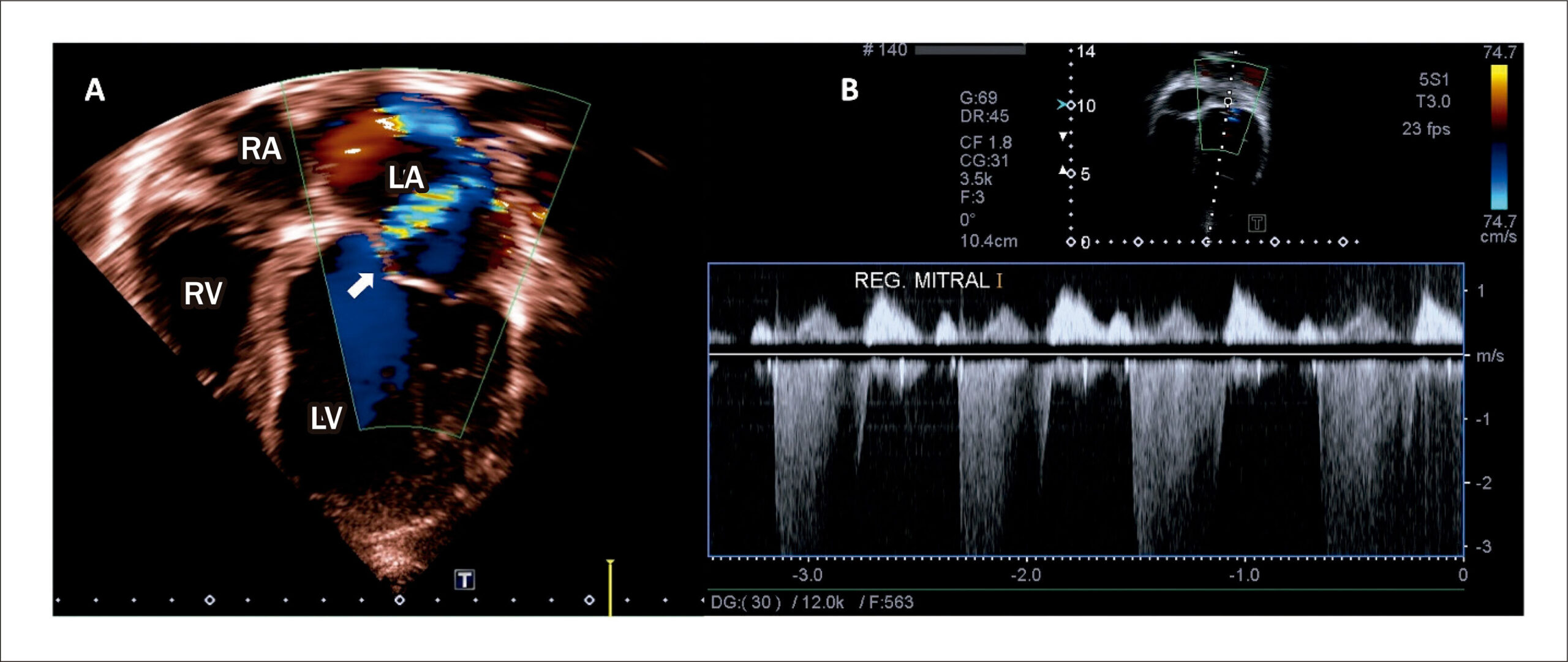
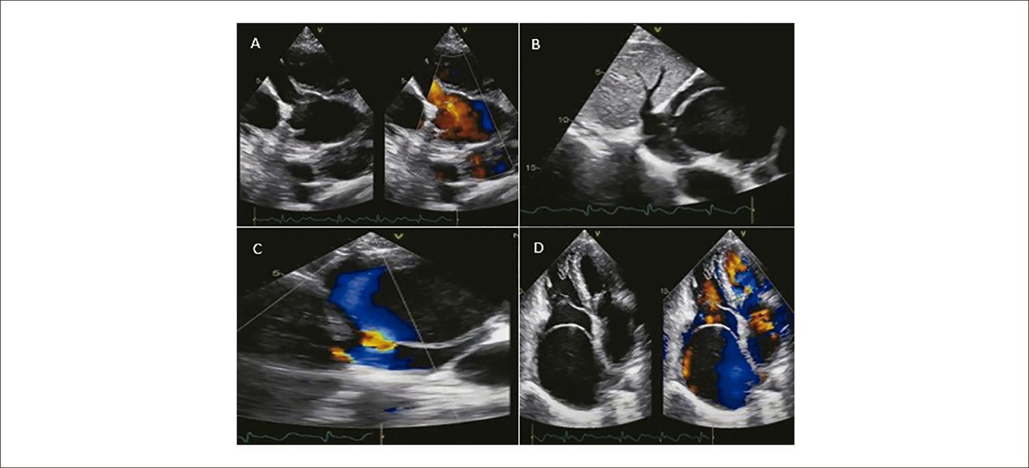
Importance of Echocardiography in the Differential Diagnosis of Rheumatic Mitral Regurgitation in Children and Adolescents

Camila Magalhães Silva et al. Maik Arantes Fátima Derlene da Rocha Araújo Adriana Furletti Machado Guimarães Zilda Maria Alves Meira

Introduction

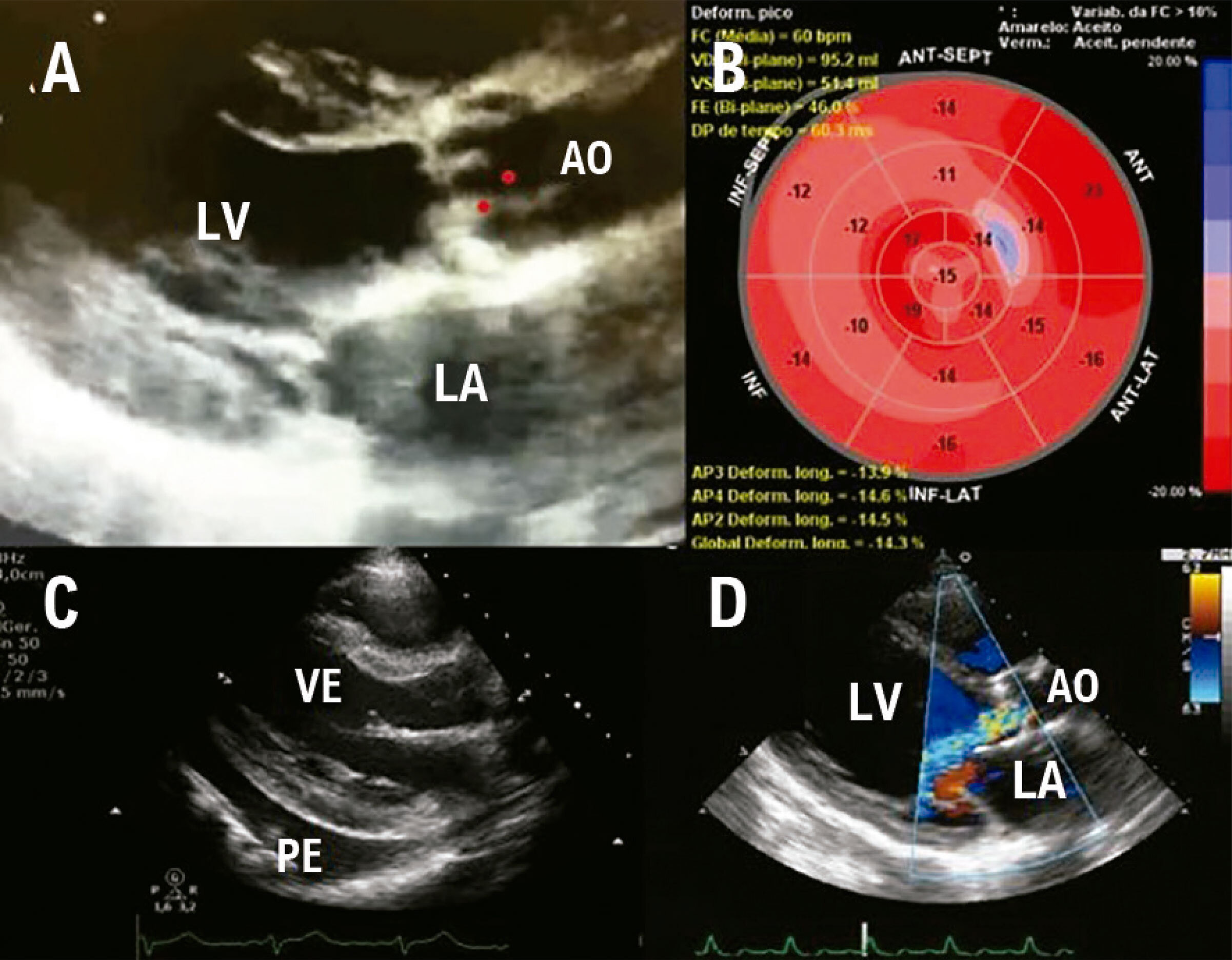
Mitral regurgitation (MR) can be a result of abnormalities in different locations of the valve apparatus (cusps, annulus, tendinous cords and papillary muscles), being of acquired or congenital etiology. MR can be classified as primary, when resulting from a deformity in the valve structure, or secondary, when related to another heart or systemic disease. In our field, the main cause of MR in students and adolescents is the acquired one. Rheumatic fever (RF) is among the causes, as well as bacterial endocarditis, which should be considered in the differential diagnosis., However, congenital anomalies, such as mitral valve (MV) prolapse, mitral cleft (MC), parachute MV and, rarely, isolated cleft MV (ICMV) should be ruled out., The etiological diagnosis of MR is very important to determine the proper treatment, once it can be clinical or surgical, with definitive correction of the lesion.

We present the report of a student whose clinical and echocardiographic scenario was first diagnosed as acute RF, showing the importance of echocardiography in the posterior differential diagnosis of MR.

[…]

  • Diagnosis, Differential
  • Echocardiography
  • Mitral Valve Insufficiency
  • Pediatrics
  • Rheumatic Fever
Read Full Article
Creative Commons Attribution (CC BY) Creative Commons license
24/03/2023 DOI: 10.36660/abcimg.2023360i Views 1
Review Article

Left Atrial Strain in the Analysis of LV Diastolic Function: Ready to Use?

Antonio Amador Calvilho Júnior et al. Jorge Eduardo Assef João Moron Saes Braga Andrea de Andrade Vilela … Antonio Tito Paladino Filho Gustavo Nishida

Abstract

Left atrial (LA) dilation is a common indicator of diastolic dysfunction, and its analysis through a volume calculation reflects the cumulative effects of the left ventricular (LV) filling pressures. However, an increase in LA volume is not exclusive to diastolic dysfunction, which has also been observed in other clinical conditions. Thus, the evaluation of the LA strain enables a functional study of this chamber, adding to the morphological analysis through the volume calculation.

The LA strain, measured using the speckle tracking technique, brings information on the reservoir, conduction, and contractile functions of the LA, and is related to the LV function. Moreover, the changes in the LA strain precede the volumetric changes by nearly a decade, and correlate inversely with the degree of LA fibrosis – this has an important relationship with the diastolic dysfunction and its grading system. Albeit insufficient to explain its totality, LA fibrosis can partially justify the functional changes of this heart chamber and can favor the use of this variable as a complement to the current protocols for the analysis of the diastolic function.

Although further study is still warranted to establish other clinical applications, the LA strain stands out in the analysis of diastolic dysfunction and can be considered ready to use, offering a great potential to improve the evaluation of the overall cardiac function.

  • Echocardiography
  • Left Ventricular Dysfunction
  • Myocardium
Read Full Article
Creative Commons Attribution (CC BY) Creative Commons license
24/03/2023 DOI: 10.36660/abcimg.2022357i Views 1
Review Article

My Approach to Assessment After Tricuspid Interventions: Tips and Tricks

Bruna Morhy Borges Leal Assunção et al. Arthur Cortez Gonçalves Lucas Velloso Dutra Renata de Sá Cassar

Abstract

Severe tricuspid regurgitation (TR) is associated with high morbidity and mortality. Given that surgical treatment of TR alone has been associated with high mortality, transcatheter interventions in the tricuspid valve (TV) have been used for its treatment, with relatively lower risk. There is a delay in intervention for TR, and this is probably related to a limited understanding of the anatomy of the TV and the right ventricle, in addition to an underestimation of the severity of TR. In this scenario, it is necessary to have comprehensive anatomical knowledge of the TV, the pathophysiology involved in the mechanism of regurgitation, and more accurate grading. The TV has anatomical, histological, and spatial peculiarities that make its assessment more complex when compared to the mitral valve, requiring knowledge and training in the various echocardiographic techniques that will often be used in combination for accurate assessment.

This review will describe the anatomy of the TV, the role of echocardiography in the diagnosis, grading, and pathophysiology involved in TR; the main transcatheter treatment options currently available for TR; and the assessment of outcomes after transcatheter intervention by means of multiple echocardiographic modalities.

  • transcatheter
  • tricuspid regurgitation
  • Tricuspid Valve
Read Full Article
Creative Commons Attribution (CC BY) Creative Commons license
07/03/2023 DOI: 10.36660/abcimg.20230006i Views 1
Figure 1
Editorial

Contemporary Non-Invasive Imaging in Chronic Coronary Syndrome: What Stress Cardiovascular Magnetic Resonance has to Offer

Isabella Leo et al. Laura Torres Araujo Chiara Bucciarelli-Ducci

Non-invasive cardiac imaging has an established diagnostic and prognostic role in patients with suspected or known ischemic heart disease (IHD) and the imaging technology has significantly evolved over the years. Cardiovascular magnetic resonance (CMR) has emerged as a robust diagnostic and prognostic imaging modality and it currently represents a valid alternative to other existing and well-validated techniques, but also as a possible first-line strategy.

Major clinical practice guidelines have been increasingly incorporating stress CMR as a clinical test indicated in patients with chest pain, receiving a Class I recommendation first in the 2014 European Society of Cardiology (ESC) guidelines for revascularisation, followed by the 2017 ESC guidelines in the diagnosis and management of ST-elevation myocardial infarction which has been recommending CMR as an alternative test for left ventricular (LV) function when echocardiography is suboptimal, but also for the evaluation of myocardial viability and ischemia. The recently published 2021 American Heart Association (AHA)/American College of Cardiology (ACC) Guidelines also indicate stress CMR with class I recommendation in patients with acute and chronic chest pain and intermediate risk of coronary artery disease (CAD) ( ).

[…]

  • Cardiovascular Imaging
  • Cardiovascular Magnetic Resonance
  • Chronic Coronary Syndrome
  • Stress Imaging
Read Full Article
Creative Commons Attribution (CC BY) Creative Commons license
06/03/2023 DOI: 10.36660/abcimg.20230004i
Review Article

My Approach to Right Ventricular Longitudinal Strain with Automated 2D and 3D Software

Lucas Arraes de França et al. Lucas Velloso Dutra

Abstract

Right ventricular strain analysis has emerged as an important diagnostic tool in the detection of early right ventricular systolic dysfunction not detected by conventional echocardiography techniques. Furthermore, it is capable of providing additional diagnostic and prognostic information to the traditional parameters for evaluating right ventricular systolic function in various pathologies. The echocardiography method of choice for its assessment is longitudinal strain derived from speckletracking. This method has been shown to be more sensitive for small changes in systolic function when compared to tricuspid annular plane systolic excursion, tissue Doppler imaging of the tricuspid annular s’ wave, and right ventricular fractional area change. Advances in artificial intelligence and software with automated analysis have been introduced to this scenario with the aim of making the method simpler and quicker to apply, with lower inter- and intra-observer variability. The objective of this review article is to demonstrate the technique step by step, from image optimization and acquisition to interpretation of results, with illustrative figures of selected cases.

  • Echocardiography
  • Left Ventricular Dysfunction
  • Myocardium
Read Full Article
Creative Commons Attribution (CC BY) Creative Commons license
06/03/2023 DOI: 10.36660/abcimg.20230010i
Figure 1
Case Report

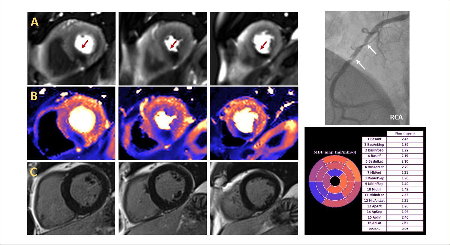
Coronary-cavitary Fistula with Aneurysm in its Path: Case Report of Percutaneous Treatment

Carlos Alberto de Jesus et al. Edileide de Barros Correia Líria Maria Lima da Silva Ibraim Masciarelli Francisco Pinto … Mercedes Del Rosario Maldonado Andrade Andrezza Lobo de Alencar Marcelo Silva Ribeiro

Abstract

The authors report the case of a 29-year-old, female patient with palpitations, chest pain, and dyspnea on exertion. The patient was diagnosed with right coronary fistula and underwent percutaneous correction of the fistulous tract with an Amplatzer prosthesis.

  • Cardiovascular Malformation
  • Coronary Aneurysm
  • Coronary Vessel Anomalies
  • vascular fistula
Read Full Article
Creative Commons Attribution (CC BY) Creative Commons license
06/03/2023 DOI: 10.36660/abcimg.2022364i Views 1
Editorial

How Can the Assessment of Myocardial Flow Reserve by Nuclear Medicine Change the Interpretation of Myocardial Perfusion Scintigraphy?

Ronaldo de Souza Leão Lima

Myocardial perfusion scintigraphy (MPS) with single-photon emission computed tomography (SPECT) is important for the diagnosis and prognostic evaluation in patients with coronary artery disease (CAD). MPS evaluates the presence, extent and degree of myocardial ischemia and/or infarction, usually through visual observation or semi-quantitative parameters. Despite its proven diagnostic and prognostic values, the relative nature of perfusion imaging may limit SPECT ability to identify patients with high-risk multivessel CAD. Limitations regarding visual or semi-quantitative assessment of regional myocardial perfusion defects may result in underestimation or misdiagnosis due to “balanced” ischemia.

This limitation can be addressed by quantifying myocardial blood flow (MBF) or myocardial flow reserve (MFR) using tracer kinetics in positron emission tomography (PET). , PET is a well-validated non-invasive method for quantification of myocardial perfusion, demonstrating an incremental diagnostic and prognostic power compared to MPS in patients with suspected or known CAD. , PET then is considered the gold standard for non-invasive quantification of MBF and MFR. – However, the production of PET tracers is costly and the technology is not yet available in many countries.

[…]

  • Coronary Artery Disease
  • Diagnosis
  • Scintigraphy
Read Full Article
Creative Commons Attribution (CC BY) Creative Commons license
06/03/2023 DOI: 10.36660/abcimg.2023363i
Case Report

Noblestitch Failure After Percutaneous Patent Foramen Ovale Closure in a Case Of Platypnea-Orthodeoxia Syndrome: Is this Device Suitable for All Patients?

Ana Rita Moura et al. Mariana Silva Alberto Rodrigues João Carlos Silva … José Ribeiro Daniel Caeiro Jorge Casanova Ricardo Fontes-Carvalho

Introduction

Patent foramen ovale (PFO) is a common congenital cardiac lesion corresponding to a normal fetal interatrial communication that typically closes after birth with the fusion of the atrial septum primum to the septum secundum . When the foramen ovale persists, it is usually asymptomatic and benign, with no need for treatment. In rare cases, it can be implicated in the pathogenesis of some medical conditions, such as platypnea-orthodeoxia syndrome (POS). In this context, PFO closure may be indicated.

Several occluder devices, traditionally based on a double-disc design, have shown that percutaneous PFO closure is possible. Despite the proven efficacy, their use is not technically feasible for some patients. In addition, they have the potential to cause some complications. NobleStitch EL (NS) is an alternative percutaneous PFO closure technique available in Europe, European Commission (EC) marked for cardiovascular suturing and PFO closure, and in the US, with Food and Drug Administration (FDA) clearance for vascular and cardiovascular suturing. It consists of a “deviceless” system with two dedicated suture delivery catheters. , To date, the experience with the NS device is limited, and we still lack information about its failure determinants, which could help select patients. –

[…]

  • Atrial Septum
  • Foramen Ovale, Patent
  • Heart Defects, Congenital
  • NobleStich/methods
  • Platypneia-Ortthodeoxia Syndrome
Read Full Article
Creative Commons Attribution (CC BY) Creative Commons license
06/03/2023 DOI: 10.36660/abcimg.2022309i
Review Article

My Approach to Echocardiographic Assessment for Constrictive Pericarditis

Débora Freire Ribeiro Rocha et al. Ana Caroline Reinaldo de Oliveira Verena Nunes e Silva João Batista Masson Silva

Abstract

  • Constrictive pericarditis
  • Heart Failure
  • Pericardium
Read Full Article
Creative Commons Attribution (CC BY) Creative Commons license
06/03/2023 DOI: 10.36660/abcimg.2022366i Views 2

How to cite this article

EndNote RIS

Electronic Document Format (Vancouver)

Castillo JMD, Boschilia T, Sabeh Junior N, Silveira CAM, Brindeiro Filho D. Heart Adaptation Mechanisms in Elite Female Athletes: Comparison With Healthy Individuals and Time of Training. ABC Imagem Cardiovascular 2023;36(1):e372.

Electronic Document Format (ABNT)

Castillo, José Maria Del; Boschilia, Thiago; Sabeh Junior, Nemi; Silveira, Carlos Antônio da Mota; Brindeiro Filho, Djair. Heart Adaptation Mechanisms in Elite Female Athletes: Comparison With Healthy Individuals and Time of Training. ABC Imagem Cardiovascular, v. 36, n. 1, e372, Mar. 2023.

Electronic Document Format (APA)

Castillo, J. M. D., Boschilia, T., Sabeh Junior, N., Silveira, C. A. M., & Brindeiro Filho, D. (2023). Heart Adaptation Mechanisms in Elite Female Athletes: Comparison With Healthy Individuals and Time of Training. ABC Imagem Cardiovascular, 36(1), e372.

Electronic Document Format (ISO)

Castillo, José Maria Del and Boschilia, Thiago and Sabeh Junior, Nemi and Silveira, Carlos Antônio da Mota and Brindeiro Filho, Djair. Heart Adaptation Mechanisms in Elite Female Athletes: Comparison With Healthy Individuals and Time of Training. ABC Imagem Cardiovascular [online]. 2023, vol. 36, n. 1, [cited 2026-10-14], e372. Available from: <https://journals.cardiol.br/en/article/heart-adaptation-mechanisms-in-elite-female-athletes-comparison-with-healthy-individuals-and-time-of-training/>. ISSN 2318-8219

EndNote RIS

Electronic Document Format (Vancouver)

Matos-Souza JR, Rassi DC. Cardiovascular Imaging In Assessment Of Athletes. ABC Imagem Cardiovascular 2023;36(1):e20230015.

Electronic Document Format (ABNT)

Matos-Souza, José Roberto; Rassi, Daniela do Carmo. Cardiovascular Imaging In Assessment Of Athletes. ABC Imagem Cardiovascular, v. 36, n. 1, e20230015, Mar. 2023.

Electronic Document Format (APA)

Matos-Souza, J. R., & Rassi, D. C. (2023). Cardiovascular Imaging In Assessment Of Athletes. ABC Imagem Cardiovascular, 36(1), e20230015.

Electronic Document Format (ISO)

Matos-Souza, José Roberto and Rassi, Daniela do Carmo. Cardiovascular Imaging In Assessment Of Athletes. ABC Imagem Cardiovascular [online]. 2023, vol. 36, n. 1, [cited 2026-10-14], e20230015. Available from: <https://journals.cardiol.br/en/article/cardiovascular-imaging-in-assessment-of-athletes/>. ISSN 2318-8219

EndNote RIS

Electronic Document Format (Vancouver)

Lima JAC, Bluemke DA, Hare J, Wu K, Sara L, Rochitte CE. Hypothesis on the Pathogenesis of Sub-Epicardial Scar Associated with Myocarditis. ABC Imagem Cardiovascular 2023;36(1):e20230001.

Electronic Document Format (ABNT)

Lima, João A. C.; Bluemke, David A.; Hare, Joshua; Wu, Katherine; Sara, Leonardo; Rochitte, Carlos E.. Hypothesis on the Pathogenesis of Sub-Epicardial Scar Associated with Myocarditis. ABC Imagem Cardiovascular, v. 36, n. 1, e20230001, Mar. 2023.

Electronic Document Format (APA)

Lima, J. A. C., Bluemke, D. A., Hare, J., Wu, K., Sara, L., & Rochitte, C. E. (2023). Hypothesis on the Pathogenesis of Sub-Epicardial Scar Associated with Myocarditis. ABC Imagem Cardiovascular, 36(1), e20230001.

Electronic Document Format (ISO)

Lima, João A. C. and Bluemke, David A. and Hare, Joshua and Wu, Katherine and Sara, Leonardo and Rochitte, Carlos E.. Hypothesis on the Pathogenesis of Sub-Epicardial Scar Associated with Myocarditis. ABC Imagem Cardiovascular [online]. 2023, vol. 36, n. 1, [cited 2026-10-14], e20230001. Available from: <https://journals.cardiol.br/en/article/hypothesis-on-the-pathogenesis-of-sub-epicardial-scar-associated-with-myocarditis/>. ISSN 2318-8219

EndNote RIS

Electronic Document Format (Vancouver)

Daminello E, Vieira PPAC, Fischer CH, Vieira MLC. Mitral Valve Area Quantification Using Digital Image Processing: Is That Feasible?. ABC Imagem Cardiovascular 2023;36(1):e376.

Electronic Document Format (ABNT)

Daminello, Edgar; Vieira, Paulo Pinto Alves Campos; Fischer, Cláudio Henrique; Vieira, Marcelo Luiz Campos. Mitral Valve Area Quantification Using Digital Image Processing: Is That Feasible?. ABC Imagem Cardiovascular, v. 36, n. 1, e376, Mar. 2023.

Electronic Document Format (APA)

Daminello, E., Vieira, P. P. A. C., Fischer, C. H., & Vieira, M. L. C. (2023). Mitral Valve Area Quantification Using Digital Image Processing: Is That Feasible?. ABC Imagem Cardiovascular, 36(1), e376.

Electronic Document Format (ISO)

Daminello, Edgar and Vieira, Paulo Pinto Alves Campos and Fischer, Cláudio Henrique and Vieira, Marcelo Luiz Campos. Mitral Valve Area Quantification Using Digital Image Processing: Is That Feasible?. ABC Imagem Cardiovascular [online]. 2023, vol. 36, n. 1, [cited 2026-10-14], e376. Available from: <https://journals.cardiol.br/en/article/mitral-valve-area-quantification-using-digital-image-processing-is-that-feasible/>. ISSN 2318-8219

EndNote RIS

Electronic Document Format (Vancouver)

Fernandes RM, Souza AC, Leite BF, Kawaoka JR. Echocardiographic Evaluation of a Patient in Circulatory Shock: A Contemporary Approach. ABC Imagem Cardiovascular 2023;36(1):e20230013.

Electronic Document Format (ABNT)

Fernandes, Rafael Modesto; Souza, Alexandre Costa; Leite, Bruno de Freitas; Kawaoka, Jun Ramos. Echocardiographic Evaluation of a Patient in Circulatory Shock: A Contemporary Approach. ABC Imagem Cardiovascular, v. 36, n. 1, e20230013, Mar. 2023.

Electronic Document Format (APA)

Fernandes, R. M., Souza, A. C., Leite, B. F., & Kawaoka, J. R. (2023). Echocardiographic Evaluation of a Patient in Circulatory Shock: A Contemporary Approach. ABC Imagem Cardiovascular, 36(1), e20230013.

Electronic Document Format (ISO)

Fernandes, Rafael Modesto and Souza, Alexandre Costa and Leite, Bruno de Freitas and Kawaoka, Jun Ramos. Echocardiographic Evaluation of a Patient in Circulatory Shock: A Contemporary Approach. ABC Imagem Cardiovascular [online]. 2023, vol. 36, n. 1, [cited 2026-10-14], e20230013. Available from: <https://journals.cardiol.br/en/article/echocardiographic-evaluation-of-a-patient-in-circulatory-shock-a-contemporary-approach/>. ISSN 2318-8219

EndNote RIS

Electronic Document Format (Vancouver)

Potratz MO, Garbo LZ, Pessimilio KP, Loss AS, Ambrozim CB, Lima ALTA, Rocha DL, et al. Criss-Cross Heart: A Case Report. ABC Imagem Cardiovascular 2023;36(1):e282.

Electronic Document Format (ABNT)

Potratz, Mateus Oliveira; Garbo, Lorrayne Zonatele; Pessimilio, Kamila Pompermaier; Loss, André Silveira; Ambrozim, Crislane Belisario; Lima, Ana Luiza Teixeira Albert; Rocha, Danielle Lopes. Criss-Cross Heart: A Case Report. ABC Imagem Cardiovascular, v. 36, n. 1, e282, Mar. 2023.

Electronic Document Format (APA)

Potratz, M. O., Garbo, L. Z., Pessimilio, K. P., Loss, A. S., Ambrozim, C. B., Lima, A. L. T. A., & Rocha, D. L. (2023). Criss-Cross Heart: A Case Report. ABC Imagem Cardiovascular, 36(1), e282.

Electronic Document Format (ISO)

Potratz, Mateus Oliveira and Garbo, Lorrayne Zonatele and Pessimilio, Kamila Pompermaier and Loss, André Silveira and Ambrozim, Crislane Belisario and Lima, Ana Luiza Teixeira Albert and Rocha, Danielle Lopes. Criss-Cross Heart: A Case Report. ABC Imagem Cardiovascular [online]. 2023, vol. 36, n. 1, [cited 2026-10-14], e282. Available from: <https://journals.cardiol.br/en/article/criss-cross-heart-a-case-report/>. ISSN 2318-8219

EndNote RIS

Electronic Document Format (Vancouver)

Gomes LCOSB, Souza AC, Drubi SA, Junqueira BMI, Bezerra MLBG, Melo RV. Role of Multimodality for the Diagnosis of Thrombosis at Late Follow-up of Patients Selected for TAVI: Review of a Case Series. ABC Imagem Cardiovascular 2023;36(1):e367.

Electronic Document Format (ABNT)

Gomes, Laila Caroline Oliveira Souza Barbosa; Souza, Alexandre Costa; Drubi, Stephanie de Azevedo; Junqueira, Bruna de Mattos Ivo; Bezerra, Mariana Lins Baptista Guedes; Melo, Rodrigo Vieira de. Role of Multimodality for the Diagnosis of Thrombosis at Late Follow-up of Patients Selected for TAVI: Review of a Case Series. ABC Imagem Cardiovascular, v. 36, n. 1, e367, Mar. 2023.

Electronic Document Format (APA)

Gomes, L. C. O. S. B., Souza, A. C., Drubi, S. A., Junqueira, B. M. I., Bezerra, M. L. B. G., & Melo, R. V. (2023). Role of Multimodality for the Diagnosis of Thrombosis at Late Follow-up of Patients Selected for TAVI: Review of a Case Series. ABC Imagem Cardiovascular, 36(1), e367.

Electronic Document Format (ISO)

Gomes, Laila Caroline Oliveira Souza Barbosa and Souza, Alexandre Costa and Drubi, Stephanie de Azevedo and Junqueira, Bruna de Mattos Ivo and Bezerra, Mariana Lins Baptista Guedes and Melo, Rodrigo Vieira de. Role of Multimodality for the Diagnosis of Thrombosis at Late Follow-up of Patients Selected for TAVI: Review of a Case Series. ABC Imagem Cardiovascular [online]. 2023, vol. 36, n. 1, [cited 2026-10-14], e367. Available from: <https://journals.cardiol.br/en/article/role-of-multimodality-for-the-diagnosis-of-thrombosis-at-late-follow-up-of-patients-selected-for-tavi-review-of-a-case-series/>. ISSN 2318-8219

EndNote RIS

Electronic Document Format (Vancouver)

Mancuso FJN. My Approach to “Athlete’s Heart”: Evaluation of the Different Types of Adaptation to Exercise. ABC Imagem Cardiovascular 2023;36(1):e20230002.

Electronic Document Format (ABNT)

Mancuso, Frederico José Neves. My Approach to “Athlete’s Heart”: Evaluation of the Different Types of Adaptation to Exercise. ABC Imagem Cardiovascular, v. 36, n. 1, e20230002, Mar. 2023.

Electronic Document Format (APA)

Mancuso, F. J. N. (2023). My Approach to “Athlete’s Heart”: Evaluation of the Different Types of Adaptation to Exercise. ABC Imagem Cardiovascular, 36(1), e20230002.

Electronic Document Format (ISO)

Mancuso, Frederico José Neves. My Approach to “Athlete’s Heart”: Evaluation of the Different Types of Adaptation to Exercise. ABC Imagem Cardiovascular [online]. 2023, vol. 36, n. 1, [cited 2026-10-14], e20230002. Available from: <https://journals.cardiol.br/en/article/my-approach-to-athletes-heart-evaluation-of-the-different-types-of-adaptation-to-exercise/>. ISSN 2318-8219

EndNote RIS

Electronic Document Format (Vancouver)

Vilarinho ABV, Silva DB, Peres ARM, Souza RLB, Amorelli CES, Campedelli FL, Alves LM, et al. Pulmonary Thromboembolism Associated with Paradoxical Embolism in a Patient with Patent Foramen Ovale. Arquivos Brasileiros de Cardiologia Imagem cardiovascular 2023;36(1):e347.

Electronic Document Format (ABNT)

Vilarinho, Arthur Barroso Vidal; Silva, Danielly Bernardes; Peres, Ana Rosaria Medeiros; Souza, Rodolfo Loureiro Borges de; Amorelli, Carlos Eduardo de Sousa; Campedelli, Fábio Lemos; Alves, Luciano Moreira; Oliveira, Fabio Augusto Cypreste de. Pulmonary Thromboembolism Associated with Paradoxical Embolism in a Patient with Patent Foramen Ovale. Arquivos Brasileiros de Cardiologia Imagem cardiovascular, v. 36, n. 1, e347, Mar. 2023.

Electronic Document Format (APA)

Vilarinho, A. B. V., Silva, D. B., Peres, A. R. M., Souza, R. L. B. , Amorelli, C. E. S., Campedelli, F. L., Alves, L. M., & Oliveira, F. A. C. (2023). Pulmonary Thromboembolism Associated with Paradoxical Embolism in a Patient with Patent Foramen Ovale. Arquivos Brasileiros de Cardiologia Imagem cardiovascular, 36(1), e347.

Electronic Document Format (ISO)

Vilarinho, Arthur Barroso Vidal and Silva, Danielly Bernardes and Peres, Ana Rosaria Medeiros and Souza, Rodolfo Loureiro Borges de and Amorelli, Carlos Eduardo de Sousa and Campedelli, Fábio Lemos and Alves, Luciano Moreira and Oliveira, Fabio Augusto Cypreste de. Pulmonary Thromboembolism Associated with Paradoxical Embolism in a Patient with Patent Foramen Ovale. Arquivos Brasileiros de Cardiologia Imagem cardiovascular [online]. 2023, vol. 36, n. 1, [cited 2026-10-14], e347. Available from: <https://journals.cardiol.br/en/article/pulmonary-thromboembolism-associated-with-paradoxical-embolism-in-a-patient-with-patent-foramen-ovale/>. ISSN 2318-8219

EndNote RIS

Electronic Document Format (Vancouver)

Braga JCMS, Guimarães Filho FV, Rodrigues A, Barreto RA. My Approach to Imaging in Sickle Cell Anemia. Arquivos Brasileiros de Cardiologia Imagem cardiovascular 2023;36(1):e20230009.

Electronic Document Format (ABNT)

Braga, João Carlos Moron Saes; Guimarães Filho, Fábio Villaça; Rodrigues, Alexandre; Barreto, Raphael Aparecido. My Approach to Imaging in Sickle Cell Anemia. Arquivos Brasileiros de Cardiologia Imagem cardiovascular, v. 36, n. 1, e20230009, Mar. 2023.

Electronic Document Format (APA)

Braga, J. C. M. S., Guimarães Filho, F. V., Rodrigues, A., & Barreto, R. A. (2023). My Approach to Imaging in Sickle Cell Anemia. Arquivos Brasileiros de Cardiologia Imagem cardiovascular, 36(1), e20230009.

Electronic Document Format (ISO)

Braga, João Carlos Moron Saes and Guimarães Filho, Fábio Villaça and Rodrigues, Alexandre and Barreto, Raphael Aparecido. My Approach to Imaging in Sickle Cell Anemia. Arquivos Brasileiros de Cardiologia Imagem cardiovascular [online]. 2023, vol. 36, n. 1, [cited 2026-10-14], e20230009. Available from: <https://journals.cardiol.br/en/article/my-approach-to-imaging-in-sickle-cell-anemia/>. ISSN 2318-8219

EndNote RIS

Electronic Document Format (Vancouver)

Scopel KRO, Medeiros TM, Maicá BNP, Velho MC, Silva MMD, Mattos JN, Heuser GG, et al. Incidental Tomographic Findings of Coronary Artery Calcifications: A Prevalence Study in Southern Brazil. Arquivos Brasileiros de Cardiologia Imagem cardiovascular 2023;36(1):e368.

Electronic Document Format (ABNT)

Scopel, Karen Rafaela Okaseski; Medeiros, Tássia Machado; Maicá, Bibiana Natalia Porto; Velho, Maria Carbonari; Silva, Mariana Motta Dias da; Mattos, Juliane Nascimento; Heuser, Guilherme Galante; Winkelmann, Eliane Roseli. Incidental Tomographic Findings of Coronary Artery Calcifications: A Prevalence Study in Southern Brazil. Arquivos Brasileiros de Cardiologia Imagem cardiovascular, v. 36, n. 1, e368, Mar. 2023.

Electronic Document Format (APA)

Scopel, K. R. O., Medeiros, T. M., Maicá, B. N. P., Velho, M. C., Silva, M. M. D. , Mattos, J. N., Heuser, G. G., & Winkelmann, E. R. (2023). Incidental Tomographic Findings of Coronary Artery Calcifications: A Prevalence Study in Southern Brazil. Arquivos Brasileiros de Cardiologia Imagem cardiovascular, 36(1), e368.

Electronic Document Format (ISO)

Scopel, Karen Rafaela Okaseski and Medeiros, Tássia Machado and Maicá, Bibiana Natalia Porto and Velho, Maria Carbonari and Silva, Mariana Motta Dias da and Mattos, Juliane Nascimento and Heuser, Guilherme Galante and Winkelmann, Eliane Roseli. Incidental Tomographic Findings of Coronary Artery Calcifications: A Prevalence Study in Southern Brazil. Arquivos Brasileiros de Cardiologia Imagem cardiovascular [online]. 2023, vol. 36, n. 1, [cited 2026-10-14], e368. Available from: <https://journals.cardiol.br/en/article/incidental-tomographic-findings-of-coronary-artery-calcifications-a-prevalence-study-in-southern-brazil/>. ISSN 2318-8219

EndNote RIS

Electronic Document Format (Vancouver)

Bignoto T. A Clinician’s View of Tricuspid Regurgitation: What do I Need to Know? When to Intervene?. Arquivos Brasileiros de Cardiologia Imagem cardiovascular 2023;36(1):e20230005.

Electronic Document Format (ABNT)

Bignoto, Tiago. A Clinician’s View of Tricuspid Regurgitation: What do I Need to Know? When to Intervene?. Arquivos Brasileiros de Cardiologia Imagem cardiovascular, v. 36, n. 1, e20230005, Mar. 2023.

Electronic Document Format (APA)

Bignoto, T. (2023). A Clinician’s View of Tricuspid Regurgitation: What do I Need to Know? When to Intervene?. Arquivos Brasileiros de Cardiologia Imagem cardiovascular, 36(1), e20230005.

Electronic Document Format (ISO)

Bignoto, Tiago. A Clinician’s View of Tricuspid Regurgitation: What do I Need to Know? When to Intervene?. Arquivos Brasileiros de Cardiologia Imagem cardiovascular [online]. 2023, vol. 36, n. 1, [cited 2026-10-14], e20230005. Available from: <https://journals.cardiol.br/en/article/a-clinicians-view-of-tricuspid-regurgitation-what-do-i-need-to-know-when-to-intervene/>. ISSN 2318-8219

EndNote RIS

Electronic Document Format (Vancouver)

Barros Filho GF, Solha I, Medeiros EF, Felix AS, Almeida ALC, Lima Júnior JC, Melo MDT, et al. Automatic Measurement of the Mitral Valve Based on Echocardiography Using Digital Image Processing. Arquivos Brasileiros de Cardiologia Imagem cardiovascular 2023;36(1):e371.

Electronic Document Format (ABNT)

Barros Filho, Genilton de França; Solha, Israel; Medeiros, Ewerton Freitas de; Felix, Alex dos Santos; Almeida, André Luiz Cerqueira de; Lima Júnior, José Carlos de; Melo, Marcelo Dantas Tavares de; Rodrigues, Marcelo Cavalcanti. Automatic Measurement of the Mitral Valve Based on Echocardiography Using Digital Image Processing. Arquivos Brasileiros de Cardiologia Imagem cardiovascular, v. 36, n. 1, e371, Mar. 2023.

Electronic Document Format (APA)

Barros Filho, G. F., Solha, I., Medeiros, E. F. , Felix, A. S., Almeida, A. L. C. , Lima Júnior, J. C. , Melo, M. D. T. , & Rodrigues, M. C. (2023). Automatic Measurement of the Mitral Valve Based on Echocardiography Using Digital Image Processing. Arquivos Brasileiros de Cardiologia Imagem cardiovascular, 36(1), e371.

Electronic Document Format (ISO)

Barros Filho, Genilton de França and Solha, Israel and Medeiros, Ewerton Freitas de and Felix, Alex dos Santos and Almeida, André Luiz Cerqueira de and Lima Júnior, José Carlos de and Melo, Marcelo Dantas Tavares de and Rodrigues, Marcelo Cavalcanti. Automatic Measurement of the Mitral Valve Based on Echocardiography Using Digital Image Processing. Arquivos Brasileiros de Cardiologia Imagem cardiovascular [online]. 2023, vol. 36, n. 1, [cited 2026-10-14], e371. Available from: <https://journals.cardiol.br/en/article/automatic-measurement-of-the-mitral-valve-based-on-echocardiography-using-digital-image-processing/>. ISSN 2318-8219

EndNote RIS

Electronic Document Format (Vancouver)

Paiva MG. Radiotherapy-Induced Heart Disease: What Can We Evaluate?. Arquivos Brasileiros de Cardiologia Imagem cardiovascular 2023;36(1):e358.

Electronic Document Format (ABNT)

Paiva, Marcelo Goulart. Radiotherapy-Induced Heart Disease: What Can We Evaluate?. Arquivos Brasileiros de Cardiologia Imagem cardiovascular, v. 36, n. 1, e358, Mar. 2023.

Electronic Document Format (APA)

Paiva, M. G. (2023). Radiotherapy-Induced Heart Disease: What Can We Evaluate?. Arquivos Brasileiros de Cardiologia Imagem cardiovascular, 36(1), e358.

Electronic Document Format (ISO)

Paiva, Marcelo Goulart. Radiotherapy-Induced Heart Disease: What Can We Evaluate?. Arquivos Brasileiros de Cardiologia Imagem cardiovascular [online]. 2023, vol. 36, n. 1, [cited 2026-10-14], e358. Available from: <https://journals.cardiol.br/en/article/radiotherapy-induced-heart-disease-what-can-we-evaluate/>. ISSN 2318-8219

EndNote RIS

Electronic Document Format (Vancouver)

Silva CM, Arantes M, Araújo FDR, Guimarães AFM, Meira ZMA. Importance of Echocardiography in the Differential Diagnosis of Rheumatic Mitral Regurgitation in Children and Adolescents. Arquivos Brasileiros de Cardiologia Imagem cardiovascular 2023;36(1):e360.

Electronic Document Format (ABNT)

Silva, Camila Magalhães; Arantes, Maik; Araújo, Fátima Derlene da Rocha; Guimarães, Adriana Furletti Machado; Meira, Zilda Maria Alves. Importance of Echocardiography in the Differential Diagnosis of Rheumatic Mitral Regurgitation in Children and Adolescents. Arquivos Brasileiros de Cardiologia Imagem cardiovascular, v. 36, n. 1, e360, Mar. 2023.

Electronic Document Format (APA)

Silva, C. M., Arantes, M., Araújo, F. D. R., Guimarães, A. F. M., & Meira, Z. M. A. (2023). Importance of Echocardiography in the Differential Diagnosis of Rheumatic Mitral Regurgitation in Children and Adolescents. Arquivos Brasileiros de Cardiologia Imagem cardiovascular, 36(1), e360.

Electronic Document Format (ISO)

Silva, Camila Magalhães and Arantes, Maik and Araújo, Fátima Derlene da Rocha and Guimarães, Adriana Furletti Machado and Meira, Zilda Maria Alves. Importance of Echocardiography in the Differential Diagnosis of Rheumatic Mitral Regurgitation in Children and Adolescents. Arquivos Brasileiros de Cardiologia Imagem cardiovascular [online]. 2023, vol. 36, n. 1, [cited 2026-10-14], e360. Available from: <https://journals.cardiol.br/en/article/importance-of-echocardiography-in-the-differential-diagnosis-of-rheumatic-mitral-regurgitation-in-children-and-adolescents/>. ISSN 2318-8219

EndNote RIS

Electronic Document Format (Vancouver)

Calvilho Júnior AA, Assef JE, Braga JMS, Vilela AA, Paladino Filho AT, Nishida G. Left Atrial Strain in the Analysis of LV Diastolic Function: Ready to Use?. Arquivos Brasileiros de Cardiologia Imagem cardiovascular 2023;36(1):e357.

Electronic Document Format (ABNT)

Calvilho Júnior, Antonio Amador; Assef, Jorge Eduardo; Braga, João Moron Saes; Vilela, Andrea de Andrade; Paladino Filho, Antonio Tito; Nishida, Gustavo. Left Atrial Strain in the Analysis of LV Diastolic Function: Ready to Use?. Arquivos Brasileiros de Cardiologia Imagem cardiovascular, v. 36, n. 1, e357, Mar. 2023.

Electronic Document Format (APA)

Calvilho Júnior, A. A., Assef, J. E., Braga, J. M. S., Vilela, A. A., Paladino Filho, A. T., & Nishida, G. (2023). Left Atrial Strain in the Analysis of LV Diastolic Function: Ready to Use?. Arquivos Brasileiros de Cardiologia Imagem cardiovascular, 36(1), e357.

Electronic Document Format (ISO)

Calvilho Júnior, Antonio Amador and Assef, Jorge Eduardo and Braga, João Moron Saes and Vilela, Andrea de Andrade and Paladino Filho, Antonio Tito and Nishida, Gustavo. Left Atrial Strain in the Analysis of LV Diastolic Function: Ready to Use?. Arquivos Brasileiros de Cardiologia Imagem cardiovascular [online]. 2023, vol. 36, n. 1, [cited 2026-10-14], e357. Available from: <https://journals.cardiol.br/en/article/left-atrial-strain-in-the-analysis-of-lv-diastolic-function-ready-to-use/>. ISSN 2318-8219

EndNote RIS

Electronic Document Format (Vancouver)

Assunção BMBL, Gonçalves AC, Dutra LV, Cassar RS. My Approach to Assessment After Tricuspid Interventions: Tips and Tricks. ABC Imagem Cardiovascular 2023;36(1):e20230006.

Electronic Document Format (ABNT)

Assunção, Bruna Morhy Borges Leal; Gonçalves, Arthur Cortez; Dutra, Lucas Velloso; Cassar, Renata de Sá. My Approach to Assessment After Tricuspid Interventions: Tips and Tricks. ABC Imagem Cardiovascular, v. 36, n. 1, e20230006, Mar. 2023.

Electronic Document Format (APA)

Assunção, B. M. B. L., Gonçalves, A. C., Dutra, L. V., & Cassar, R. S. (2023). My Approach to Assessment After Tricuspid Interventions: Tips and Tricks. ABC Imagem Cardiovascular, 36(1), e20230006.

Electronic Document Format (ISO)

Assunção, Bruna Morhy Borges Leal and Gonçalves, Arthur Cortez and Dutra, Lucas Velloso and Cassar, Renata de Sá. My Approach to Assessment After Tricuspid Interventions: Tips and Tricks. ABC Imagem Cardiovascular [online]. 2023, vol. 36, n. 1, [cited 2026-10-14], e20230006. Available from: <https://journals.cardiol.br/en/article/my-approach-to-assessment-after-tricuspid-interventions-tips-and-tricks/>. ISSN 2318-8219

EndNote RIS

Electronic Document Format (Vancouver)

Leo I, Araujo LT, Bucciarelli-Ducci C. Contemporary Non-Invasive Imaging in Chronic Coronary Syndrome: What Stress Cardiovascular Magnetic Resonance has to Offer. ABC Imagem Cardiovascular 2023;36(1):e20230004.

Electronic Document Format (ABNT)

Leo, Isabella; Araujo, Laura Torres; Bucciarelli-Ducci, Chiara. Contemporary Non-Invasive Imaging in Chronic Coronary Syndrome: What Stress Cardiovascular Magnetic Resonance has to Offer. ABC Imagem Cardiovascular, v. 36, n. 1, e20230004, Mar. 2023.

Electronic Document Format (APA)

Leo, I., Araujo, L. T., & Bucciarelli-Ducci, C. (2023). Contemporary Non-Invasive Imaging in Chronic Coronary Syndrome: What Stress Cardiovascular Magnetic Resonance has to Offer. ABC Imagem Cardiovascular, 36(1), e20230004.

Electronic Document Format (ISO)

Leo, Isabella and Araujo, Laura Torres and Bucciarelli-Ducci, Chiara. Contemporary Non-Invasive Imaging in Chronic Coronary Syndrome: What Stress Cardiovascular Magnetic Resonance has to Offer. ABC Imagem Cardiovascular [online]. 2023, vol. 36, n. 1, [cited 2026-10-14], e20230004. Available from: <https://journals.cardiol.br/en/article/contemporary-non-invasive-imaging-in-chronic-coronary-syndrome-what-stress-cardiovascular-magnetic-resonance-has-to-offer/>. ISSN 2318-8219

EndNote RIS

Electronic Document Format (Vancouver)

França LA, Dutra LV. My Approach to Right Ventricular Longitudinal Strain with Automated 2D and 3D Software. ABC Imagem Cardiovascular 2023;36(1):e20230010.

Electronic Document Format (ABNT)

França, Lucas Arraes de; Dutra, Lucas Velloso. My Approach to Right Ventricular Longitudinal Strain with Automated 2D and 3D Software. ABC Imagem Cardiovascular, v. 36, n. 1, e20230010, Mar. 2023.

Electronic Document Format (APA)

França, L. A. , & Dutra, L. V. (2023). My Approach to Right Ventricular Longitudinal Strain with Automated 2D and 3D Software. ABC Imagem Cardiovascular, 36(1), e20230010.

Electronic Document Format (ISO)

França, Lucas Arraes de and Dutra, Lucas Velloso. My Approach to Right Ventricular Longitudinal Strain with Automated 2D and 3D Software. ABC Imagem Cardiovascular [online]. 2023, vol. 36, n. 1, [cited 2026-10-14], e20230010. Available from: <https://journals.cardiol.br/en/article/my-approach-to-right-ventricular-longitudinal-strain-with-automated-2d-and-3d-software/>. ISSN 2318-8219

EndNote RIS

Electronic Document Format (Vancouver)

Jesus CA, Correia EB, Silva LML, Pinto IMF, Andrade MDRM, Alencar AL, Ribeiro MS, et al. Coronary-cavitary Fistula with Aneurysm in its Path: Case Report of Percutaneous Treatment. ABC Imagem Cardiovascular 2023;36(1):e364.

Electronic Document Format (ABNT)

Jesus, Carlos Alberto de; Correia, Edileide de Barros; Silva, Líria Maria Lima da; Pinto, Ibraim Masciarelli Francisco; Andrade, Mercedes Del Rosario Maldonado; Alencar, Andrezza Lobo de; Ribeiro, Marcelo Silva. Coronary-cavitary Fistula with Aneurysm in its Path: Case Report of Percutaneous Treatment. ABC Imagem Cardiovascular, v. 36, n. 1, e364, Mar. 2023.

Electronic Document Format (APA)

Jesus, C. A. , Correia, E. B., Silva, L. M. L. , Pinto, I. M. F., Andrade, M. D. R. M., Alencar, A. L. , & Ribeiro, M. S. (2023). Coronary-cavitary Fistula with Aneurysm in its Path: Case Report of Percutaneous Treatment. ABC Imagem Cardiovascular, 36(1), e364.

Electronic Document Format (ISO)

Jesus, Carlos Alberto de and Correia, Edileide de Barros and Silva, Líria Maria Lima da and Pinto, Ibraim Masciarelli Francisco and Andrade, Mercedes Del Rosario Maldonado and Alencar, Andrezza Lobo de and Ribeiro, Marcelo Silva. Coronary-cavitary Fistula with Aneurysm in its Path: Case Report of Percutaneous Treatment. ABC Imagem Cardiovascular [online]. 2023, vol. 36, n. 1, [cited 2026-10-14], e364. Available from: <https://journals.cardiol.br/en/article/coronary-cavitary-fistula-with-aneurysm-in-its-path-case-report-of-percutaneous-treatment/>. ISSN 2318-8219

EndNote RIS

Electronic Document Format (Vancouver)

Lima RSL. How Can the Assessment of Myocardial Flow Reserve by Nuclear Medicine Change the Interpretation of Myocardial Perfusion Scintigraphy?. ABC Imagem Cardiovascular 2023;36(1):e363.

Electronic Document Format (ABNT)

Lima, Ronaldo de Souza Leão. How Can the Assessment of Myocardial Flow Reserve by Nuclear Medicine Change the Interpretation of Myocardial Perfusion Scintigraphy?. ABC Imagem Cardiovascular, v. 36, n. 1, e363, Mar. 2023.

Electronic Document Format (APA)

Lima, R. S. L. (2023). How Can the Assessment of Myocardial Flow Reserve by Nuclear Medicine Change the Interpretation of Myocardial Perfusion Scintigraphy?. ABC Imagem Cardiovascular, 36(1), e363.

Electronic Document Format (ISO)

Lima, Ronaldo de Souza Leão. How Can the Assessment of Myocardial Flow Reserve by Nuclear Medicine Change the Interpretation of Myocardial Perfusion Scintigraphy?. ABC Imagem Cardiovascular [online]. 2023, vol. 36, n. 1, [cited 2026-10-14], e363. Available from: <https://journals.cardiol.br/en/article/how-can-the-assessment-of-myocardial-flow-reserve-by-nuclear-medicine-change-the-interpretation-of-myocardial-perfusion-scintigraphy/>. ISSN 2318-8219

EndNote RIS

Electronic Document Format (Vancouver)

Moura AR, Silva M, Rodrigues A, Silva JC, Ribeiro J, Caeiro D, Casanova J, et al. Noblestitch Failure After Percutaneous Patent Foramen Ovale Closure in a Case Of Platypnea-Orthodeoxia Syndrome: Is this Device Suitable for All Patients?. ABC Imagem Cardiovascular 2023;36(1):e309.

Electronic Document Format (ABNT)

Moura, Ana Rita; Silva, Mariana; Rodrigues, Alberto; Silva, João Carlos; Ribeiro, José; Caeiro, Daniel; Casanova, Jorge; Fontes-Carvalho, Ricardo. Noblestitch Failure After Percutaneous Patent Foramen Ovale Closure in a Case Of Platypnea-Orthodeoxia Syndrome: Is this Device Suitable for All Patients?. ABC Imagem Cardiovascular, v. 36, n. 1, e309, Mar. 2023.

Electronic Document Format (APA)

Moura, A. R., Silva, M., Rodrigues, A., Silva, J. C., Ribeiro, J., Caeiro, D., Casanova, J., & Fontes-Carvalho, R. (2023). Noblestitch Failure After Percutaneous Patent Foramen Ovale Closure in a Case Of Platypnea-Orthodeoxia Syndrome: Is this Device Suitable for All Patients?. ABC Imagem Cardiovascular, 36(1), e309.

Electronic Document Format (ISO)

Moura, Ana Rita and Silva, Mariana and Rodrigues, Alberto and Silva, João Carlos and Ribeiro, José and Caeiro, Daniel and Casanova, Jorge and Fontes-Carvalho, Ricardo. Noblestitch Failure After Percutaneous Patent Foramen Ovale Closure in a Case Of Platypnea-Orthodeoxia Syndrome: Is this Device Suitable for All Patients?. ABC Imagem Cardiovascular [online]. 2023, vol. 36, n. 1, [cited 2026-10-14], e309. Available from: <https://journals.cardiol.br/en/article/noblestitch-failure-after-percutaneous-patent-foramen-ovale-closure-in-a-case-of-platypnea-orthodeoxia-syndrome-is-this-device-suitable-for-all-patients/>. ISSN 2318-8219

EndNote RIS

Electronic Document Format (Vancouver)

Rocha DFR, Oliveira ACR, Silva VN, Silva JBM. My Approach to Echocardiographic Assessment for Constrictive Pericarditis. ABC Imagem Cardiovascular 2023;36(1):e366.

Electronic Document Format (ABNT)

Rocha, Débora Freire Ribeiro; Oliveira, Ana Caroline Reinaldo de; Silva, Verena Nunes e; Silva, João Batista Masson. My Approach to Echocardiographic Assessment for Constrictive Pericarditis. ABC Imagem Cardiovascular, v. 36, n. 1, e366, Mar. 2023.

Electronic Document Format (APA)

Rocha, D. F. R., Oliveira, A. C. R. , Silva, V. N. , & Silva, J. B. M. (2023). My Approach to Echocardiographic Assessment for Constrictive Pericarditis. ABC Imagem Cardiovascular, 36(1), e366.

Electronic Document Format (ISO)

Rocha, Débora Freire Ribeiro and Oliveira, Ana Caroline Reinaldo de and Silva, Verena Nunes e and Silva, João Batista Masson. My Approach to Echocardiographic Assessment for Constrictive Pericarditis. ABC Imagem Cardiovascular [online]. 2023, vol. 36, n. 1, [cited 2026-10-14], e366. Available from: <https://journals.cardiol.br/en/article/my-approach-to-echocardiographic-assessment-for-constrictive-pericarditis/>. ISSN 2318-8219

Sede – São Paulo
Rua Barata Ribeiro, 380 cj 54 Bela Vista
São Paulo, Brasil – CEP: 01308-000
Tel.: +55 (11) 3120-3363
Telefax: +55 (11) 3259-2988
E-mail: depeco@cardiol.br

Sede – Rio de Janeiro
Av. Marechal Câmara, 160
3º andar – Sala: 330 – Centro
Rio de Janeiro – CEP: 20020-907
Tel.: +55 (21) 3478-2700
ou: +55 (21) 99386-3961
E-mail: abcimaging@cardiol.br

  • About the Journal
  • Internal Regulation
  • Editors Section
  • Editorial Board
  • SBC Award

  • Guidelines for Publication
  • Editorial Policy
  • Ethics Policy
  • Policy on the Use of Artificial Intelligence
  • Mandatory Publication Form

Issues

SBC Guidelines