Automatic Measurement of the Mitral Valve Based on Echocardiography Using Digital Image Processing
Abstract
Background
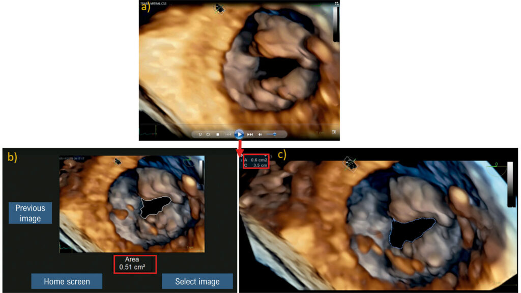
The evaluation of mitral valve area through multiplanar reconstruction in 3-dimensional echocardiography is restricted to specific software and to the experience of echocardiographers. They need to manually select the video frame that contains the maximum mitral valve opening area, as this dimension is fundamental to identification of mitral stenosis.
Objective
To automate the process of determining the maximum mitral valve opening area, through the application of digital image processing (DIP) in echocardiography tests, developing an open algorithm with video reading in avi format.
Method
This cross-sectional observational pilot study was conducted with 25 different echocardiography exams, 15 with normal aperture and 10 with rheumatic mitral stenosis. With the authorization of the Research Ethics Committee, all exams were performed and made available by 2 specialists who used 2 models of echocardiographic devices: Vivid E95 (GE Healthcare) and Epiq 7 (Philips), with multiplanar transesophageal probes. All videos in avi format were submitted to DIP using the image segmentation technique.
Results
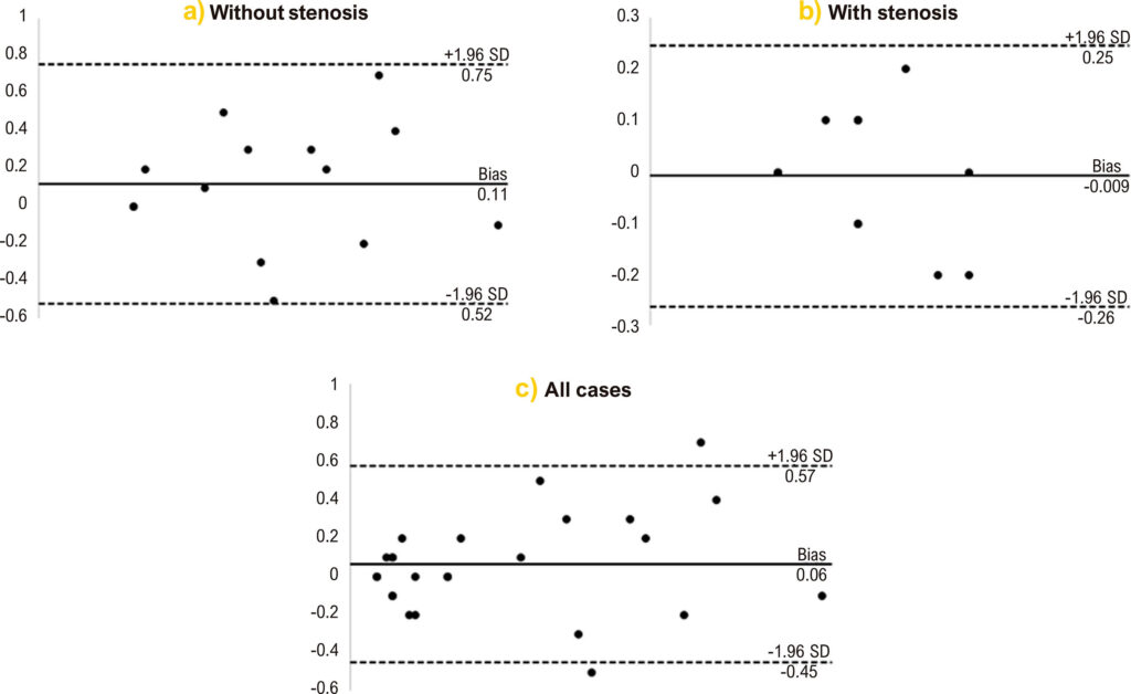
The measurements obtained manually by experienced echocardiographers and the values calculated by the developed system were compared using a Bland-Altman diagram. There was greater agreement between values in the range from 0.4 to 2.7 cm2.
Conclusion
It was possible to automatically determine the maximum mitral valve opening area, for cases from both GE and Philips, using only 1 video as input data. The algorithm has been demonstrated to save time on measurements when compared to the usual method.
Images
Image gallery fullscreen viewer
Comments are moderated by the editorial team before publication. Discussions should be evidence-based and use scientific language. Conflicts of interest should be declared.