Multimodality Images of a Mixed Atrial Septal Defect
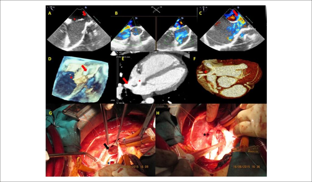
A 20-year-old male patient with dyspnea complaint was referred to our hospital. On physical examination there was grade 2/6 systolic murmur, which is best heard at the second left intercostal space and fixed split S2. ECG revealed sinus rhythm with complete right bundle brunch block. Transthoracic echocardiography demonstrated a secundum atrial septal defect (ASD) and Qp/Qs ratio was 1.6. The right ventricle was severely dilated, inconsistent with the size of the defect. Two-dimensional and color Doppler transesophageal echocardiographic examination confirmed the secundum type ASD (asterisk) and revealed an additional sinus venosus type ASD between the right atrium and the superior vena cava (SVC)(, , ; ). Three-dimensional transesophageal echocardiography verified both septal defects (). The patient underwent cardiac tomography for further anatomical outlining the. and clearly show the secundum ASD (asterisk), sinus venosus ASD (arrow) and anomalous drainage of the right superior pulmonary vein to the SVC (star). The patient underwent surgery. and show intraoperative images of the defects.
[…]
Images
Image gallery fullscreen viewer
Comments are moderated by the editorial team before publication. Discussions should be evidence-based and use scientific language. Conflicts of interest should be declared.