ABC Family Arquivos Brasileiros de Cardiologia International Journal of Cardiovascular Sciences ABC Heart Failure & Cardiomyopathy ABC Imagem Cardiovascular
Português English
ABC Family
Português English
  • About
    • About ABC Family
    • Internal Regulation
    • Editorial Board
    • Brazilian Society of Cardiology (SBC)
  • Issues
  • SBC Guidelines
  • Content
    • Videos
    • Podcasts
    • News
  • About
    • About ABC Family
    • Internal Regulation
    • Editorial Board
    • Brazilian Society of Cardiology (SBC)
  • Issues
  • SBC Guidelines
  • Content
    • Videos
    • Podcasts
    • News

ABC Family

  • ABC Family
  • Arquivos Brasileiros de Cardiologia
  • International Journal of Cardiovascular Sciences
  • ABC Heart Failure & Cardiomyopathy
  • ABC Imagem Cardiovascular
Search in

No content found.

Journals
Ordering
Article types
Others

  1. Home
  2. Keyword: Heart Septal Defects, Atrial / physiopathology
Figure 1
ABC
Image

Multimodality Images of a Mixed Atrial Septal Defect

Zafer Işılak et al. Uğur Küçük Omer Uz Murat Yalçın Veysel Temizkan

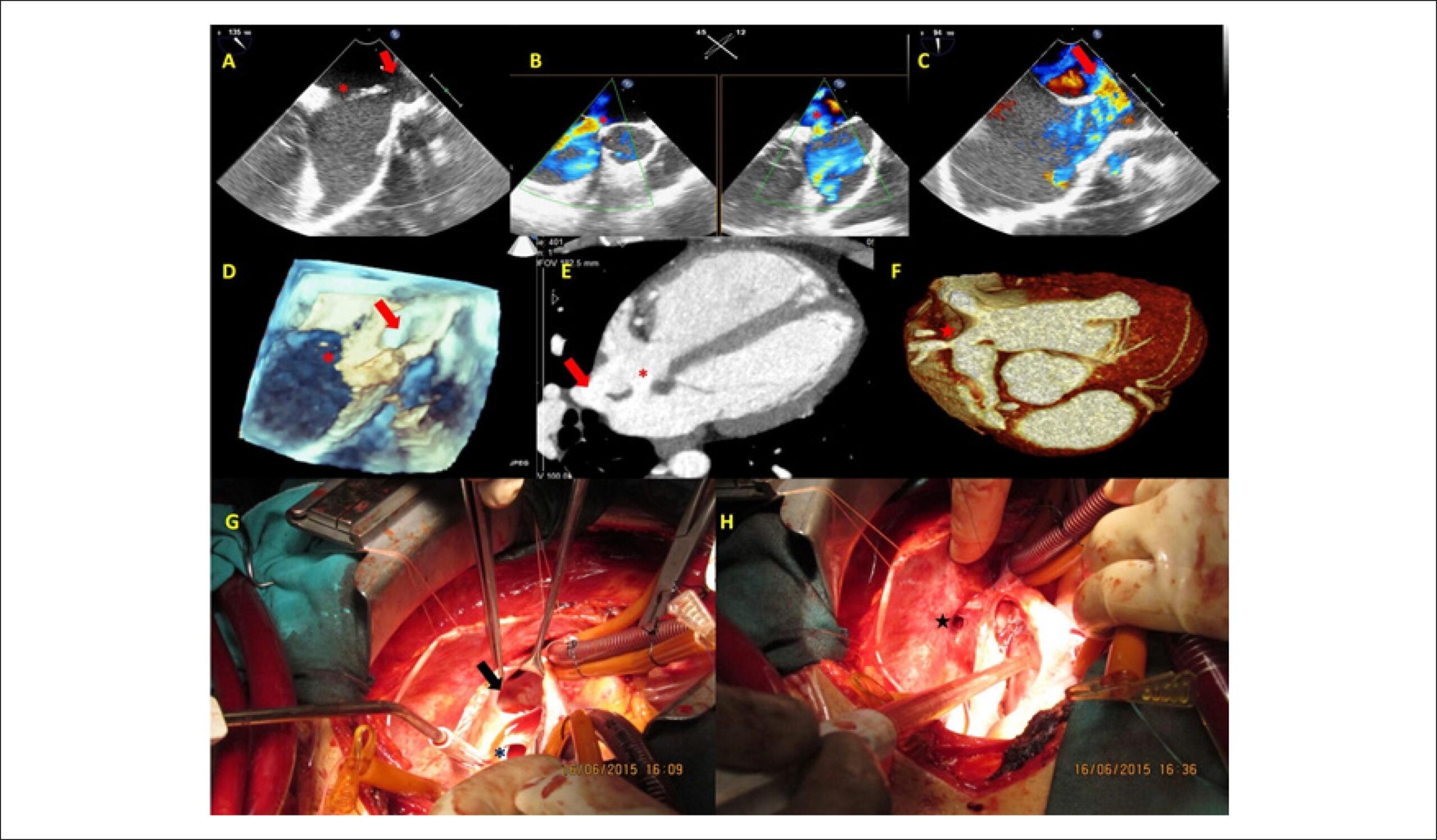
A 20-year-old male patient with dyspnea complaint was referred to our hospital. On physical examination there was grade 2/6 systolic murmur, which is best heard at the second left intercostal space and fixed split S2. ECG revealed sinus rhythm with complete right bundle brunch block. Transthoracic echocardiography demonstrated a secundum atrial septal defect (ASD) and Qp/Qs ratio was 1.6. The right ventricle was severely dilated, inconsistent with the size of the defect. Two-dimensional and color Doppler transesophageal echocardiographic examination confirmed the secundum type ASD (asterisk) and revealed an additional sinus venosus type ASD between the right atrium and the superior vena cava (SVC)(, , ; ). Three-dimensional transesophageal echocardiography verified both septal defects (). The patient underwent cardiac tomography for further anatomical outlining the. and clearly show the secundum ASD (asterisk), sinus venosus ASD (arrow) and anomalous drainage of the right superior pulmonary vein to the SVC (star). The patient underwent surgery. and show intraoperative images of the defects.

[…]

  • Atrial Septum / physiopathology
  • Echocardiography, Three-Dimensional
  • Heart Septal Defects, Atrial / physiopathology
Read Full Article
Creative Commons Attribution (CC BY) Creative Commons license
01/02/2016 DOI: 10.5935/abc.20160016

How to cite this article

EndNote RIS

Electronic Document Format (Vancouver)

Işılak Z, Küçük U, Uz O, Yalçın M, Temizkan V. Multimodality Images of a Mixed Atrial Septal Defect. Arquivos Brasileiros de Cardiologia 2016;106(2):160-1.

Electronic Document Format (ABNT)

Işılak, Zafer; Küçük, Uğur; Uz, Omer; Yalçın, Murat; Temizkan, Veysel. Multimodality Images of a Mixed Atrial Septal Defect. Arquivos Brasileiros de Cardiologia, v. 106, n. 2, p. 160-161, Jan. 2016.

Electronic Document Format (APA)

Işılak, Z., Küçük, U., Uz, O., Yalçın, M., & Temizkan, V. (2016). Multimodality Images of a Mixed Atrial Septal Defect. Arquivos Brasileiros de Cardiologia, 106(2), 160-161.

Electronic Document Format (ISO)

Işılak, Zafer and Küçük, Uğur and Uz, Omer and Yalçın, Murat and Temizkan, Veysel. Multimodality Images of a Mixed Atrial Septal Defect. Arquivos Brasileiros de Cardiologia [online]. 2016, vol. 106, n. 2, [cited 2026-10-14], pp. 160-161. Available from: <https://journals.cardiol.br/en/article/multimodality-images-of-a-mixed-atrial-septal-defect/>. ISSN 0066-782X

  • About ABC Family
  • Internal Regulation
  • Editorial Board
  • Brazilian Society of Cardiology (SBC)

Issues

SBC Guidelines

  • Videos
  • Podcasts
  • News