| ABCIC
ABC Family Arquivos Brasileiros de Cardiologia International Journal of Cardiovascular Sciences ABC Heart Failure & Cardiomyopathy ABC Imagem Cardiovascular
Português English
ABC Family | ABCIC
Português English
  • About
    • About the Journal
    • Internal Regulation
    • Editors Section
    • Editorial Board
    • SBC Award
  • Instructions to Authors
    • Guidelines for Publication
    • Editorial Policy
    • Ethics Policy
    • Policy on the Use of Artificial Intelligence
    • Mandatory Publication Form
  • Issues
  • SBC Guidelines
  • About
    • About the Journal
    • Internal Regulation
    • Editors Section
    • Editorial Board
    • SBC Award
  • Instructions to Authors
    • Guidelines for Publication
    • Editorial Policy
    • Ethics Policy
    • Policy on the Use of Artificial Intelligence
    • Mandatory Publication Form
  • Issues
  • SBC Guidelines

ABC Family

  • ABC Family
  • Arquivos Brasileiros de Cardiologia
  • International Journal of Cardiovascular Sciences
  • ABC Heart Failure & Cardiomyopathy
  • ABC Imagem Cardiovascular
Search in

No content found.

Journals
Ordering
Article types
Others

  1. Home
  2. ABCIC
  3. Issues
  4. 2021, Volume 34, Number 1
Figure 1
Case Report

Cerebrovascular Disease as the Initial Manifestation of Apical Hypertrophic Cardiomyopathy

Ana Lilia Rayas-Gómez et al. José Manuel González-Rayas Alejandro Rico-Rosas Pedro Daniel Landa-Alvarado … Ileana Maribel Ponce de León-Vargas José María Ramos-Verdugo José Manuel González-Yáñez

Introduction

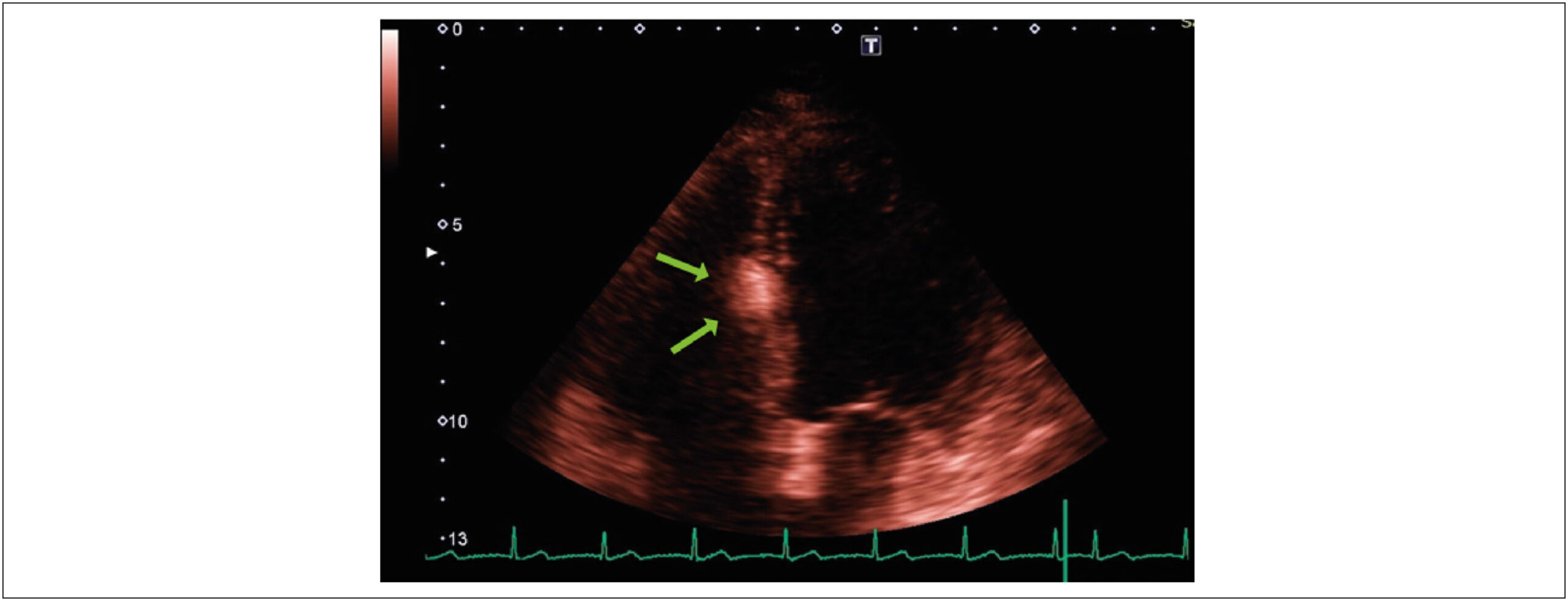
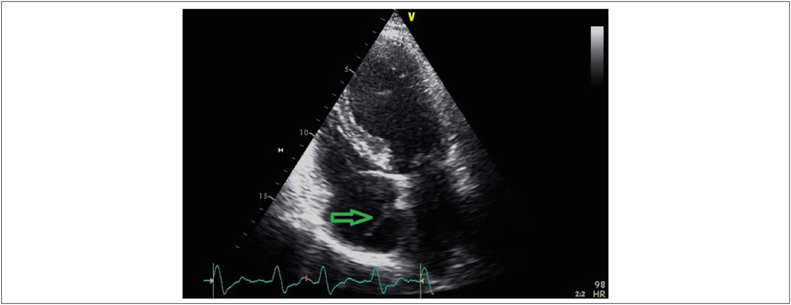
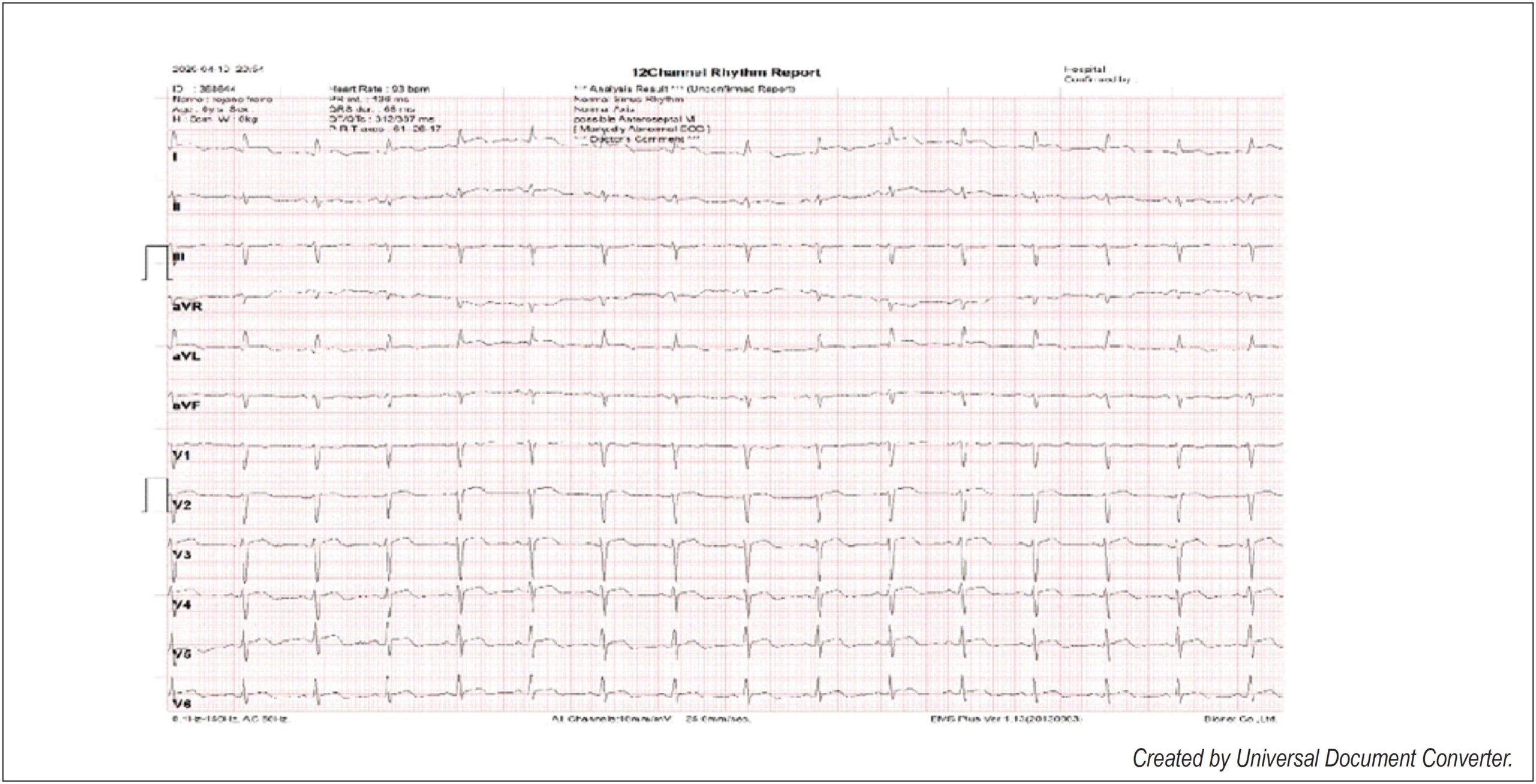
Apical hypertrophic cardiomyopathy (AHCM) is a condition that primarily involves the apex of the left ventricle. Two of its main characteristics are a non-obstructive physiology and apical hypertrophy of the left ventricle with giant negative T waves. AHCM is uncommon, and its prevalence is notably affected by ethnicity. The diagnosis is challenging to make and requires multimodal imaging techniques., Here we describe two cases of AHCM with cerebrovascular disease as the presenting condition: Patient 1 had atrial fibrillation (AF), while Patient 2 showed evidence of aortic plaques as the possible embolic source. Nonetheless, Patient 2 also presented an akinetic apex. Hence, it was impossible to rule out the previous migration of an apical thrombus as the cause of the ischemic stroke. The main clinical lesson learned from our cases is that stroke in patients with AHCM is a multifactorial complication potentially caused by AF, aortic plaques, and intracavitary thrombus. Therefore, although AHCM is uncommon, it should be considered when determining the cause of cerebrovascular embolic events.

[…]
  • Cerebrovascular Disease
  • Echocardiography
  • Embolism
  • Hypertrophic Cardiomyopathy
Read Full Article
Creative Commons Attribution (CC BY) Creative Commons license
16/06/2021 DOI: 10.47593/2675-312X/20213401eabc144 Views 1
Figure 1
Case Report

Cardiac Sarcoidosis as a Cause of Total Atrioventricular Block: Importance of Differential Diagnosis

Jorge Elias Neto et al. Márcio Augusto Silva Ricardo Ryoshim Kuniyoshi Guilherme Futuro … Erick Sessa Merçon Petherson Susano Grativvol Fátima Cristina Pedrotti

Introduction

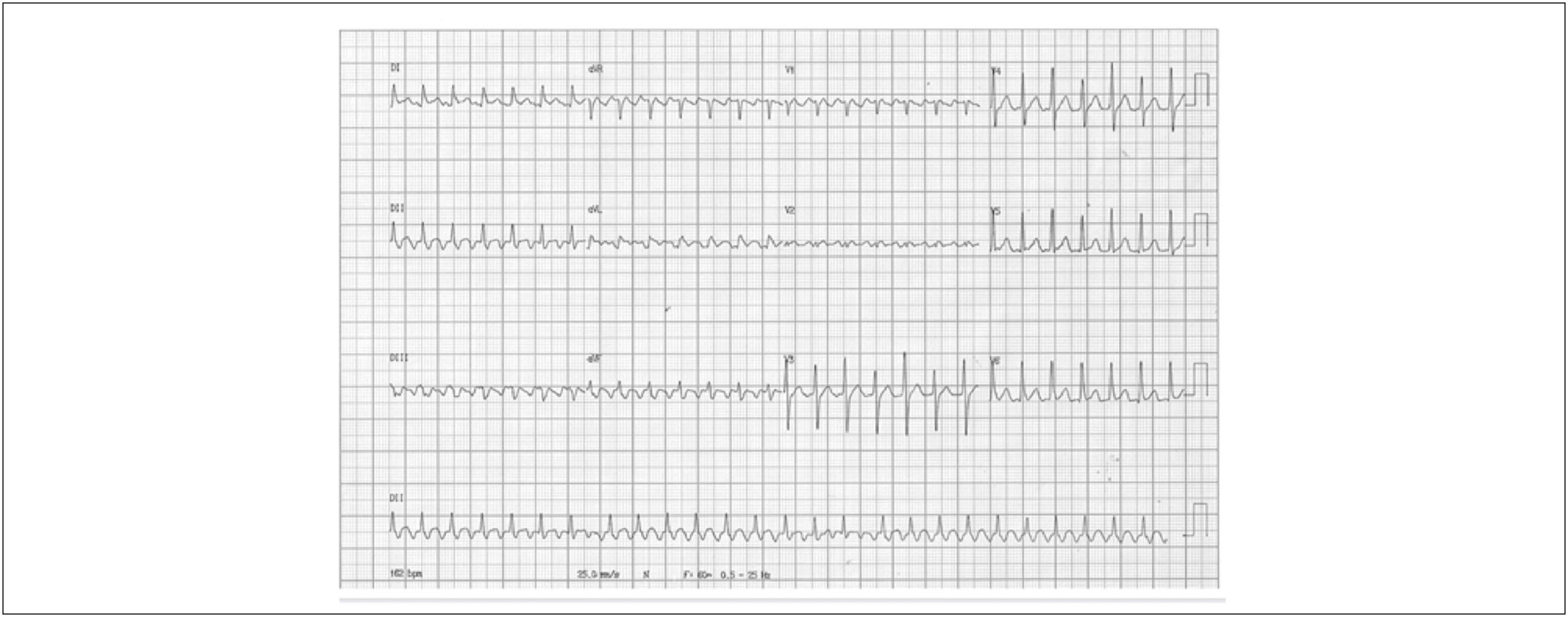
Sarcoidosis is a systemic granulomatous disorder of unknown cause that can affect virtually any organ. The extracardiac forms are usually benign and subjected to spontaneous remission. However, the prognosis may be unfavorable in case of cardiac involvement.

Symptomatic cardiac sarcoidosis (CS) is diagnosed in approximately 5% of patients with sarcoidosis. However, based on some autopsy series, the prevalence of subclinical CS can reach 25–30%.

[…]

  • Arrhythmogenic right ventricular cardiomyopathy/dysplasia
  • Atrioventricular block
  • Diagnosis
  • Sarcoidosis
  • Sudden Death
  • Ventricular tachycardia
Read Full Article
Creative Commons Attribution (CC BY) Creative Commons license
16/06/2021 DOI: 10.47593/2675-312X/20213401eabc163 Views 1
Figure 1
Image

Echocardiographic Image of Primary Mediastinal Sarcoma with Pericardial Invasion

Keyla Patrícia Barbosa Melo et al. Rafael José Coelho Maia Betty Janny Maia Siqueira Mozart Lacerda Siqueira Campos Araújo Ana Carolina Borges de Miranda Souza

Primary mediastinal sarcoma is rare, corresponding to less than 1% of all soft tissue sarcomas and less than 10% of all primary mediastinal tumors.

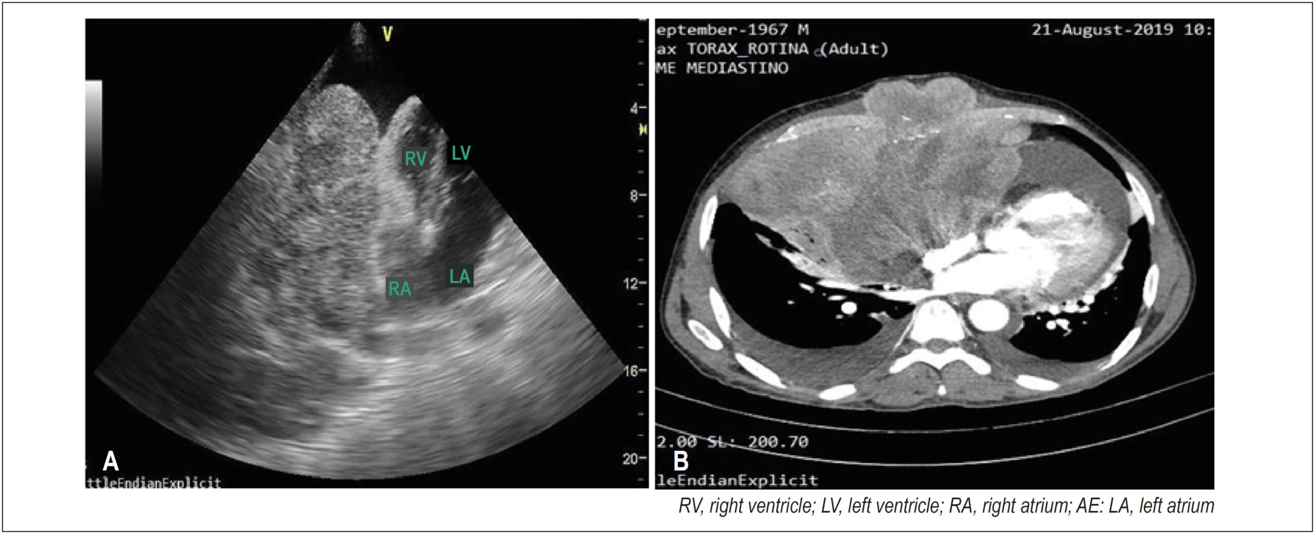
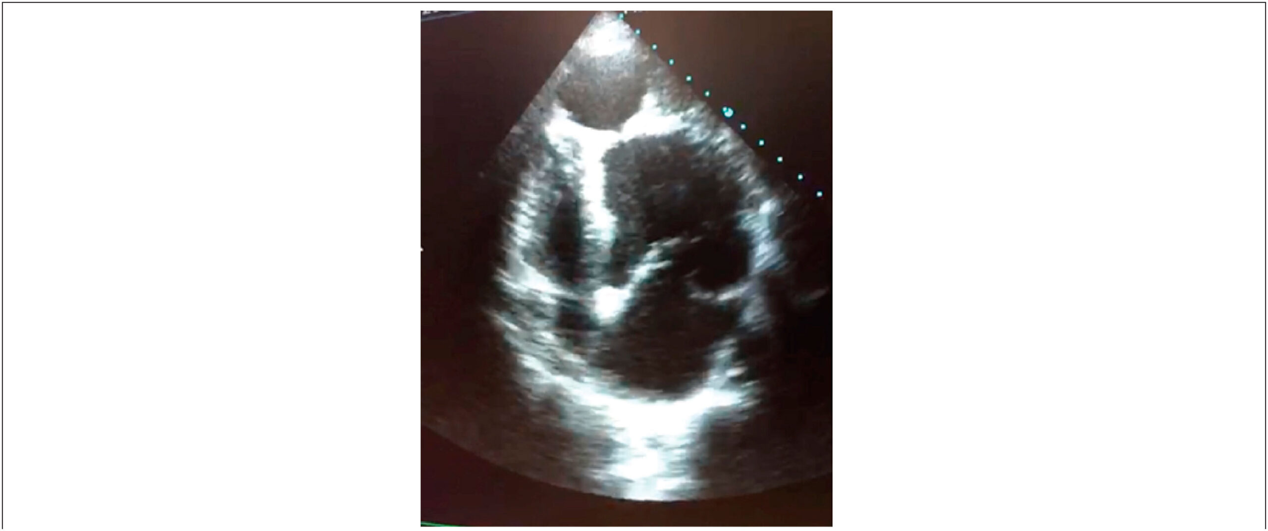
A 52-year-old man with signs and symptoms of cardiac tamponade underwent echocardiography of the right ventricle, which revealed significant pericardial effusion and an extensive heterogeneous mediastinal mass measuring approximately 15 × 8 cm in the pericardial space in close contact with the right chambers and causing diastolic collapse. The diagnostic hypothesis was a pericardial or mediastinal neoplastic process invading the pericardium. Subsequently, an immunohistochemical study confirmed a high-grade fusiform/pleomorphic cell sarcoma.

[…]

  • Echocardiography
  • Mediastinal neoplasms
  • Pericardium
  • Sarcoma
Read Full Article
Creative Commons Attribution (CC BY) Creative Commons license
16/06/2021 DOI: 10.47593/2675-312X/20213401eabc139
Figure 1
Statement

Position Statement on Indications and the Safe Reintroduction of Cardiovascular Imaging Methods in the COVID-19 Scenario – 2021

Adenalva Lima de Souza Beck et al. Silvio Henrique Barberato André Luiz Cerqueira de Almeida Claudia R. Pinheiro de Castro Grau … Marly Maria Uellendahl Lopes Ronaldo de Souza Leão Lima Rodrigo Júlio Cerci Ana Cristina Lopes Albricker Fanilda Souto Barros Alessandra Joslin Oliveira Edgar Bezerra de Lira Filho Marcelo Haertel Miglioranza Marcelo Luiz Campos Vieira José Luiz Barros Pena Tânia Mara Varejão Strabelli David Costa de Souza Le Bihan Jeane Mike Tsutsui Carlos Eduardo Rochitte

Cover Letter

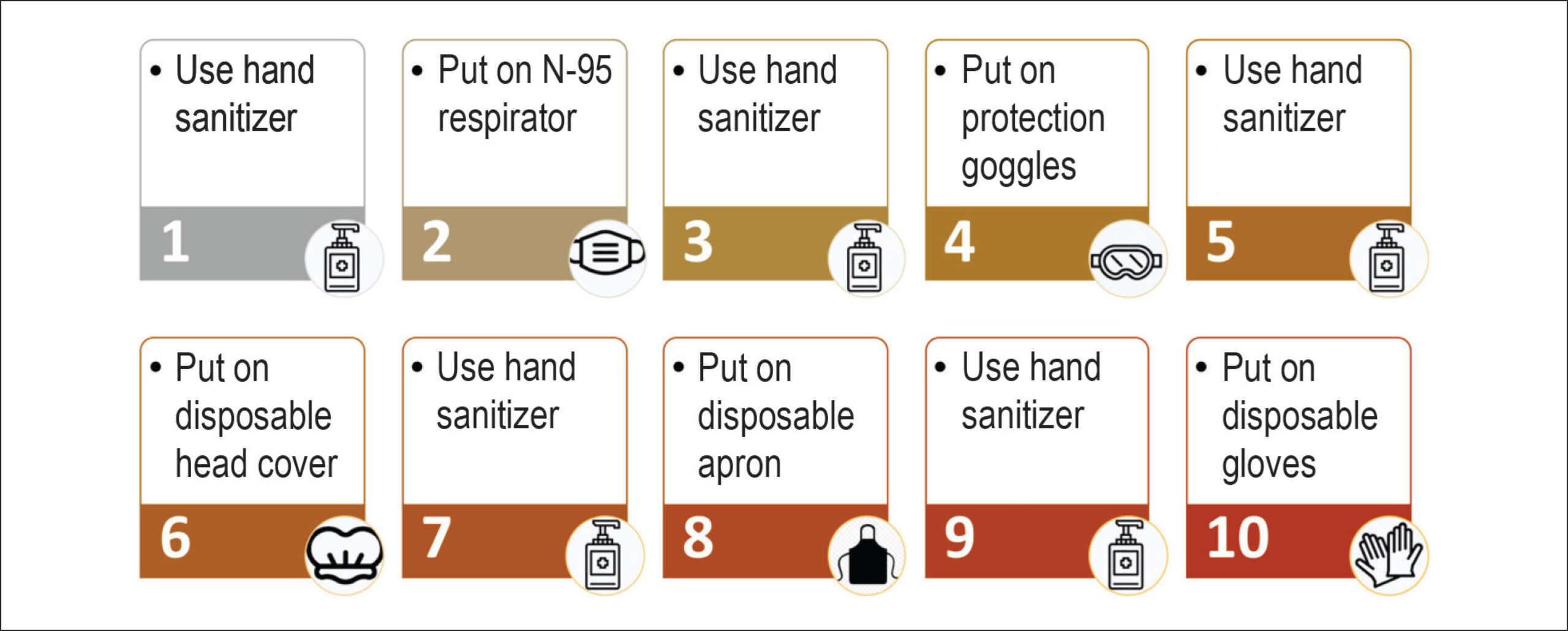
The objective of this position statement is to inform clinicians, cardiologists, and medical imaging specialists on the recommended procedures, flows, and protocols for the duration of the SARS-CoV-2 pandemic aiming at a more efficient protection of health care professionals and patients. Recommendations are based on the best available scientific evidence at the moment and on the consensus among experienced specialists. Since the first case of the new coronavirus disease (COVID-19) in Brazil, many changes in recommendations and a large scientific debate regarding some of them have occurred within various sources of scientific knowledge. This is due to our currently incomplete knowledge on COVID-19, including its pathophysiological processes and aspects of SARS-CoV-2 transmission. Another reason for a variability in recommendations is related to the epidemiological phase of the pandemic in each region of the country. Notably, in a country with continental dimensions such as Brazil, the pandemic can reach different transmission phases in a specific moment, thus requiring particular measures for each region and phase of the pandemic.

In the beginning of the pandemic, a phase of great attention and lack of knowledge on what the future held, the Cardiovascular Imaging Department (DIC) of the Brazilian Society of Cardiology (SBC) published a brief and essential version of a position statement. The document provided rapid and practical fundamental guidance regarding safety procedures during non-invasive cardiovascular imaging procedures. This document was absolutely essential at that moment for providing the best possible protection for health care professionals. After the publication of a summary document in the initial stage of the pandemic, DIC-SBC and its specialists have deemed timely to update that position statement (now published in the form of an official scientific publication); this should include not only a much wider view of the procedures in light of the data acquired since then, but also a customized orientation for each epidemiological phase of the pandemic that could be useful for professionals working in non-invasive cardiovascular imaging for the next months or possibly years of coexisting with COVID-19.

[…]

Read Full Article
Creative Commons Attribution (CC BY) Creative Commons license
16/06/2021 DOI: 10.47593/2675-312X/20213401eposi2021 Views 1
Figure 1
Case Report

Lipoma in the Interventricular Septum – Case Report

Bruna Bonamigo Thomé et al. Laura Vilela Pazzini Nathalia Regina Pavan Estevan Vieira Cabeda Tobias Sato de Almeida

Abstract

Primary cardiac tumors are a rare but mostly benign pathology found in up to 0.03% of autopsies. Among these benign tumors, with an incidence of 8.5%, lipomas are often located in the interatrial septum. When located in the interventricular septum, they are considered an even more unusual pathology with an unknown real prevalence. In most cases, the diagnosis is made incidentally on cardiovascular imaging and confirmed by histopathological examination. They are more commonly asymptomatic or feature nonspecific symptoms, but these lipomas can progress with arrhythmias, valve dysfunction, heart failure, and death, which highlights the importance of cardiovascular imaging methods in the differential diagnosis and to guide appropriate therapy. This report describes the case of a patient with an incidental echocardiographic finding of a cardiac mass located in the interventricular septum, followed by cardiac magnetic resonance imaging to better characterize the lesion and demonstrate its characteristics compatible with lipoma.

  • Diagnostic cardiovascular techniques
  • Echocardiography
  • Heart Neoplasms
  • Lipoma
Read Full Article
Creative Commons Attribution (CC BY) Creative Commons license
16/06/2021 DOI: 10.47593/2675-312X/20213401eabc152
Figure 1
Case Report

Left Ventricular-to-right Atrial Shunt (Gerbode Defect) Associated with Interventricular Communication in a 42-year-old Patient

Andressa Sardá Maiochi Takagui et al. Thales Cantelle Baggio Mariana Baretta Savariz Giulia Theilacker Thalia Elisa Baggio

Introduction

The Gerbode defect, a rare ventricular septal anomaly characterized by communication between the left ventricle (LV) and the right atrium (RA), represents approximately 0.08% of intracardiac shunts and less than 1% of congenital heart defects. Although it classically has a congenital etiology, secondary causes of infective endocarditis, myocardial infarction (MI), and iatrogenesis have been reported. The Gerbode defect was first described in 1857, and its first successful surgical treatment was reported by Kirby at the Hospital of the University of Pennsylvania.

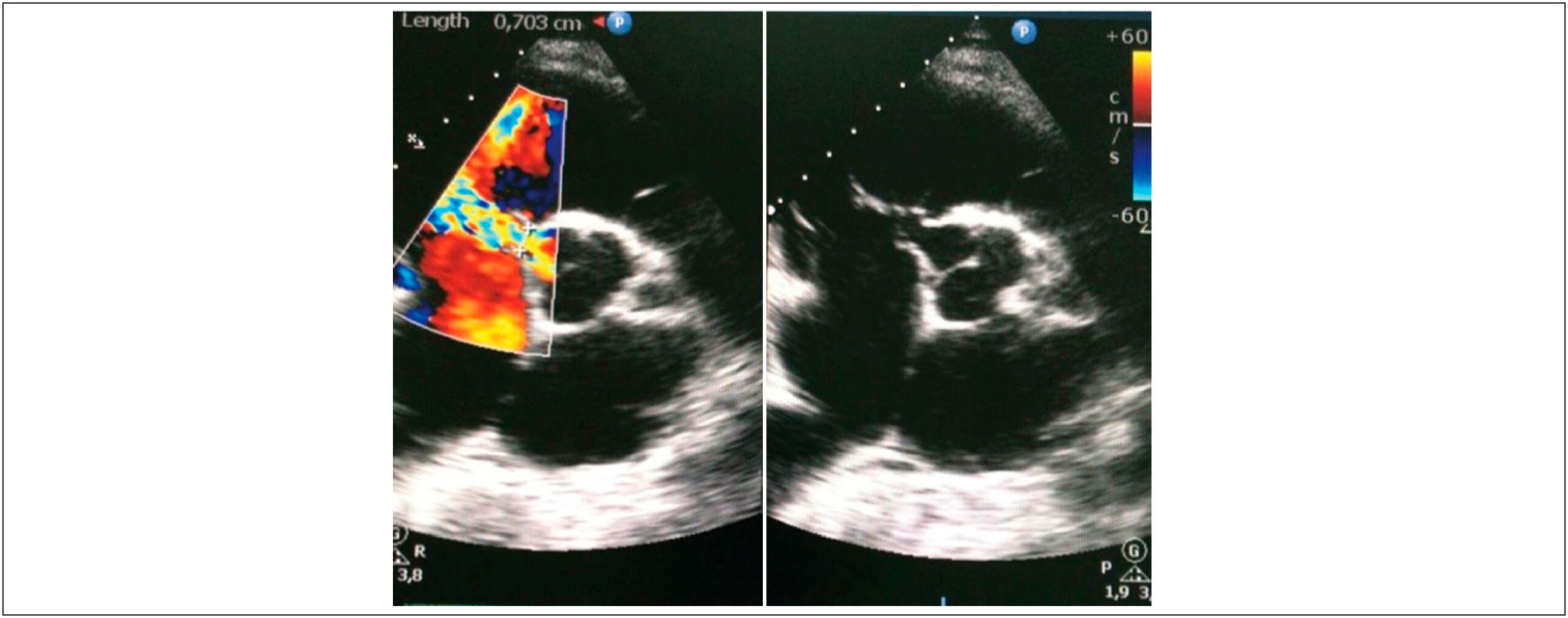
Here, we report an unusual case of congenital Gerbode defect in a 42-year-old patient whose diagnosis was challenging due to the presence of an additional shunt at the ventricular level.

[…]

  • Congenital Heart Defects
  • Heart Septal Defects, Ventricular
  • Ventricular septal defects
Read Full Article
Creative Commons Attribution (CC BY) Creative Commons license
16/06/2021 DOI: 10.47593/2675-312X/20213401eabc142 Views 1
Figure 1
Case Report

Anomalous Origin of the Right Coronary from the Left Coronary Sinus: How to approach?

Danielle Campos de Almeida et al. Alice Mirane Malta Carrijo Marcela Gomes de Souza Fernando de Melo Martinelli … Fernando Roberto de Fazzio João Lucas O’Connell

Introduction

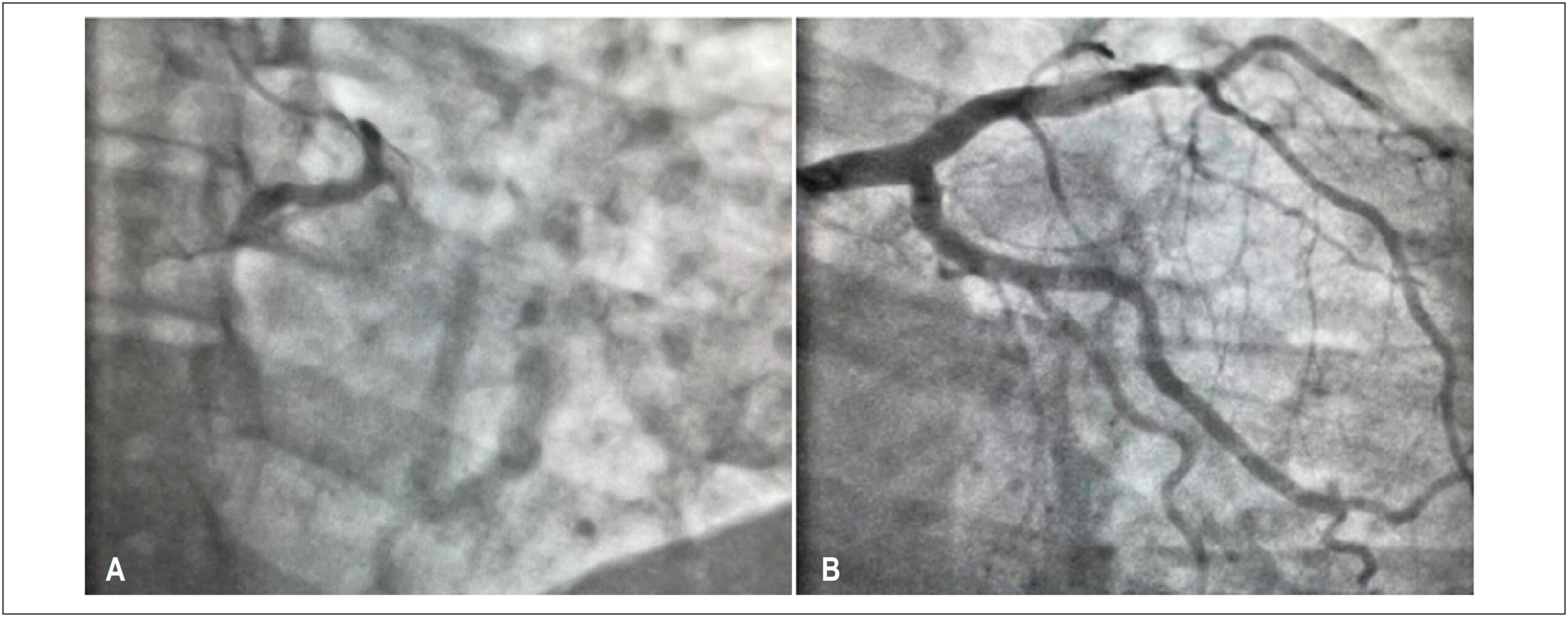
Coronary artery anomalies (CAAs) are characterized by a changed vessel origin, path, or structure. It is a rare condition, present in approximately 1% of the general population. These anomalies include the anomalous aortic origin of a coronary artery (AAOCA), which can travel retroaortic, subpulmonic, prepulmonic, and inter-arterial courses.

In general, these abnormalities remain asymptomatic until adulthood, being incidental findings on complementary tests or necropsy. However, they can also cause angina, syncope, ischemia, arrhythmias, or sudden death., Sudden death can be the first presentation of this pathology in young athletes, representing the second leading cause of death in this group.

[…]

  • Arteries
  • Coronary Vessel Anomalies
  • Coronary Vessels
  • Diagnostic Imaging
Read Full Article
Creative Commons Attribution (CC BY) Creative Commons license
16/06/2021 DOI: 10.47593/2675-312X/20213401eabc146 Views 1
Figure 1
Case Report

Isolated Fungal Pulmonary Endocarditis: Role of Transthoracic Echocardiography from the Diagnosis to Follow-up

Eduardo Gatti Pianca et al. Marcelo Nicola Branchi Bernardo Mastella Álvaro Schmidt Albrecht … Orlando Carlos Belmonte Wender Murilo Foppa Angela Barreto Santiago Santos

Introduction

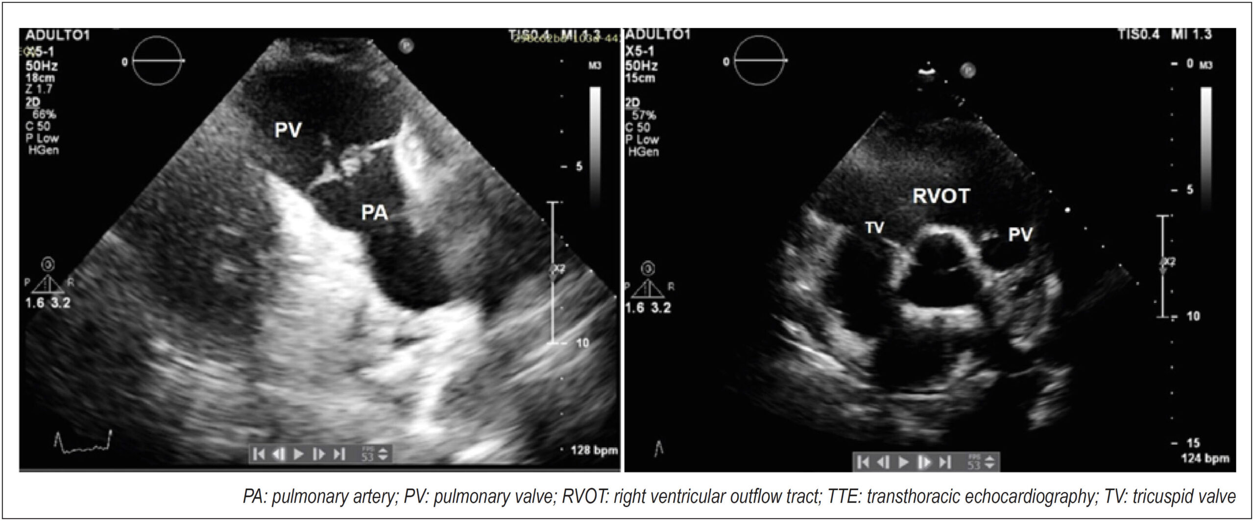
Pulmonary valve endocarditis, an uncommon entity, is associated with important complications such as pulmonary emboli, severe valvular regurgitation, and right-sided chamber dilatation and is usually concomitant with tricuspid valve endocarditis. Intravenous (IV) drug and cardiovascular implantable electronic device use are known risk factors for this condition. Staphylococcus aureus is the most prevalent causative bacteria, whereas fungi are rarely involved. Here, we present a case of Candida albicans–induced pulmonary valve endocarditis.

[…]

  • Echocardiography, Diagnosis
  • Endocarditis
Read Full Article
Creative Commons Attribution (CC BY) Creative Commons license
16/06/2021 DOI: 10.47593/2675-312X/20213401eabc143 Views 2
Figure 1
Original Article

Diagnostic yield of magnetic resonance imaging in heart failure with left ventricular dysfunction?

Ana Terra Fonseca Barreto et al. Mariana Lins Baptista Guedes Bezerra Rodrigo Morel Vieira de Melo Bruno Oliveira Isabella … Vanessa de Assis Reis Luiz Carlos Santana Passos

Abstract

Background:

Heart failure with reduced ejection fraction is responsible for half of heart failure cases worldwide and implicates in substantial morbidity and mortality. However, even with clinical history and physical examination associated with conventional complementary exams, many patients remain without etiological diagnosis. Cardiac magnetic resonance has offered the possibility to clarify a variable proportion of these cases.

Objective:

To verify how much cardiac magnetic resonance contributes to etiologic diagnosis of heart failure with left ventricular ejection fraction <50% in a specialized service.

Methods:

We included individuals referred to cardiac magnetic resonance with heart failure and left ventricular ejection fraction <50% by transthoracic echocardiogram, without defined etiology, from January, 2017 to June, 2018 in a tertiary hospital.

Results:

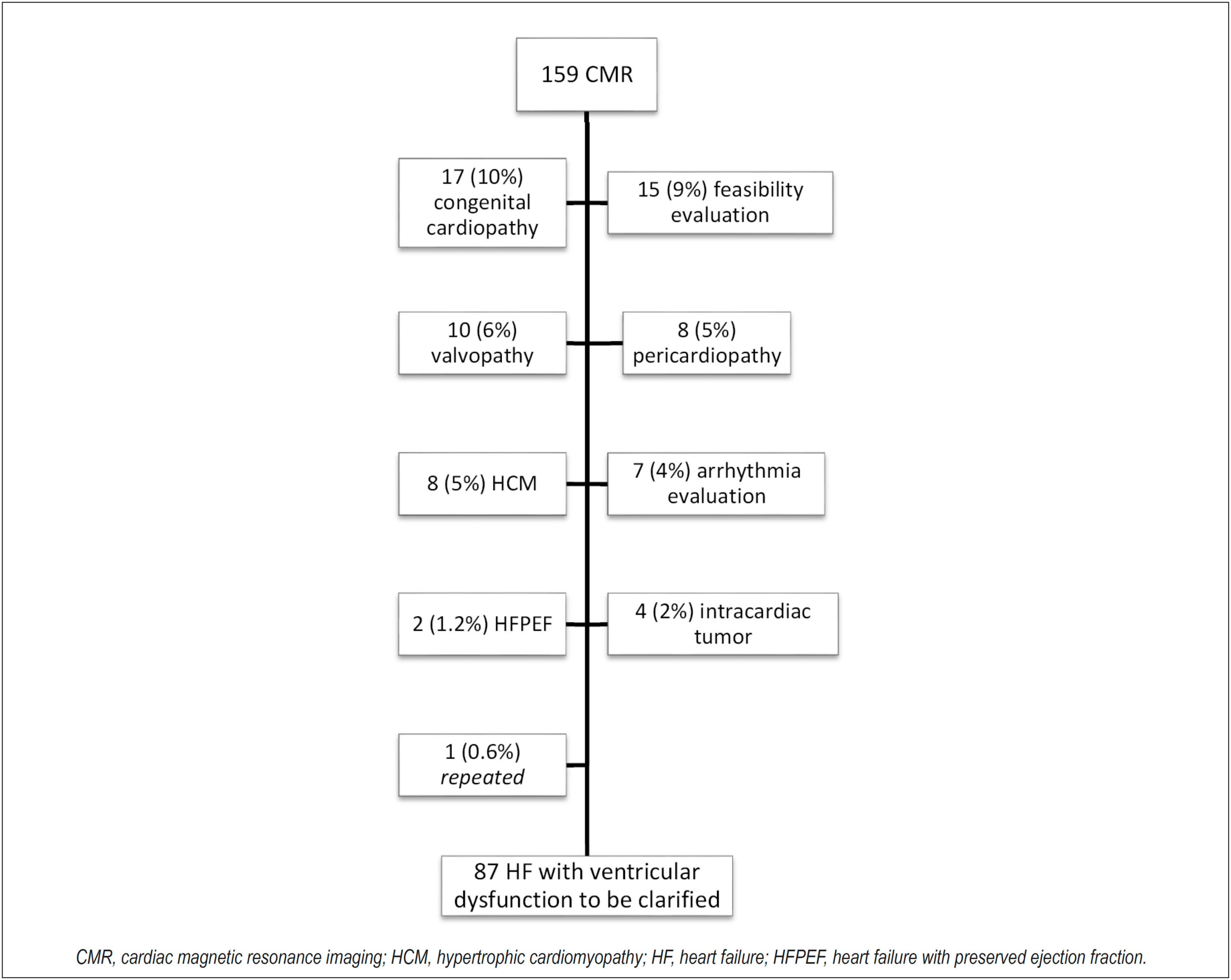
The sample consisted of 87 patients, with average age of 45±16 years, 49% male and left ventricular ejection fraction 32%±13. Of the patients, 55,3% had etiological diagnosis through cardiac magnetic resonance: 33,4% myocarditis, 11.5% non-compaction cardiomyopathy, 6.8% Chagas disease, and for hypertensive heart disease, amyloidosis and arrhythmogenic right ventricle dysplasia, 1,2% each. Late gadolinium enhancement was positive in 61% and non-ischemic pattern predominated (50,5%). Reverse remodeling occurred with normalization of ventricular function in 13% of patients.

Conclusion:

The performance of cardiac magnetic resonance in patients without etiologic diagnosis of HF with left ventricle dysfunction is clinically significant, since it contributed more than 50% of the time to the etiology and prognosis of patients. This positive impact occurred in a tertiary cardiology teaching service, so it is possible that in other circumstances the role of the cardiac magnetic resonance may be even greater than that here presented.

  • Cardiomyopathy, Dilated
  • Diagnostic Imaging
  • Heart Failure
  • Magnetic Resonance
Read Full Article
Creative Commons Attribution (CC BY) Creative Commons license
16/06/2021 DOI: 10.47593/2675-312X/20213401eabc108 Views 2
Figure 1
Case Report

Cardiac Intimal Sarcoma: A Rare Cardiac Tumor Entity

Rhavena Brasil de Andrade et al. Carlos José Mota de Lima Ingrid Alves de Freitas Larissa Chagas Corrêa … Danielli Oliveira da Costa Lino Ana Carolina Brito de Alcantâra

Introduction

Malignant cardiac neoplasia is a rare entity with an extremely low incidence that has a nonspecific clinical presentation that may mimic other conditions. It can present arrhythmias and complications such as ischemia due to coronary flow obstruction.

More than 75% of primary cardiac tumors are benign. Of the 25% of malignant tumors, 75% are sarcomas.– Primary cardiac tumors can have a primary or metastatic origin and are diagnosed by imaging methods. Primary tumors occur more frequently in the left chamber, especially in the atrial myxoma, while metastatic tumors originate from malignant melanomas, which occur more frequently in the right chambers.

[…]

  • Atrial flutter
  • Heart
  • Heart Diseases
  • Sarcoma
Read Full Article
Creative Commons Attribution (CC BY) Creative Commons license
16/06/2021 DOI: 10.47593/2675-312X/20213401eabc118 Views 2
Figure 1
Original Article

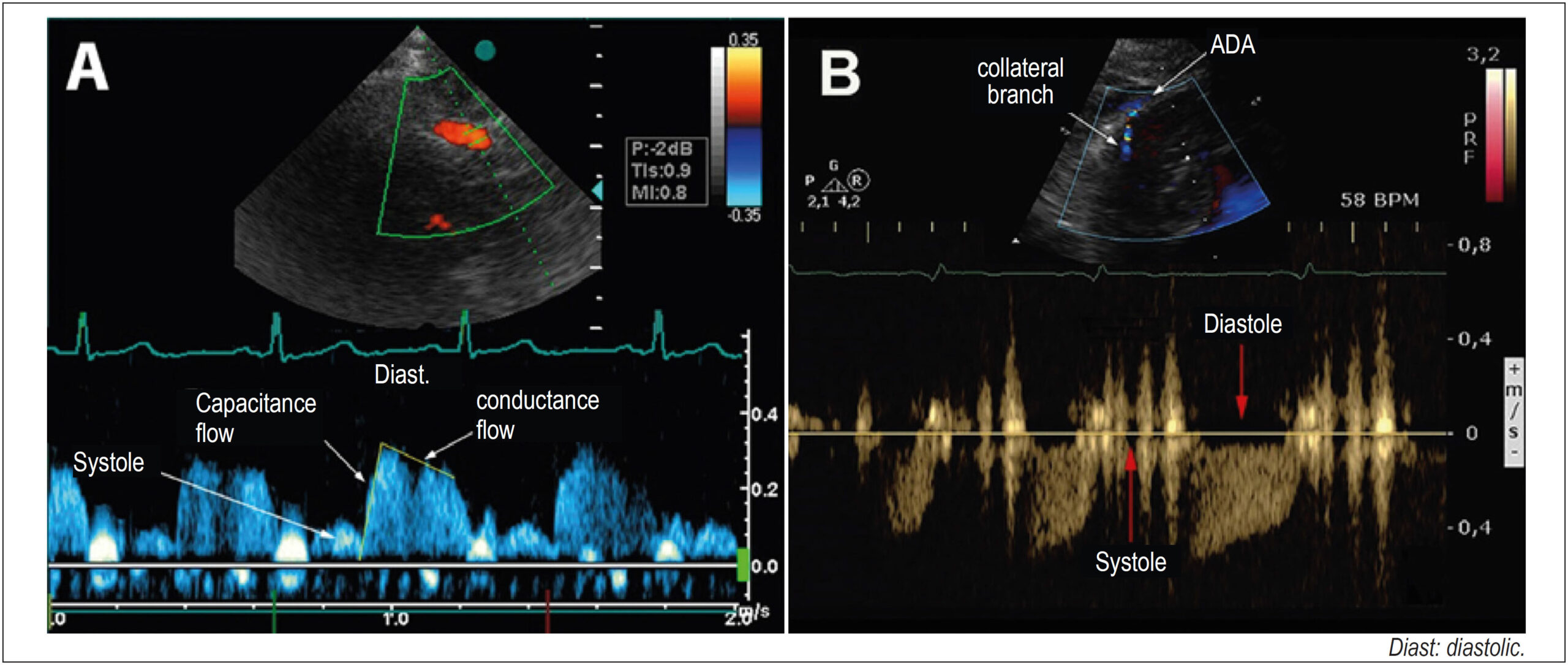
Comparative Analysis of the Coronary Arteries Flow Pattern in Secondary Myocardial Hypertrophies and by Sarcomeric Mutation

Caio Guedes de Sousa et al. José Maria Del Castillo Carlos Mazzarollo Eugenio Soares de Albuquerque … Antônia Dulcineide Medeiros Sena Djair Brindeiro Filho Carlos Antônio da Mota Silveira

Abstract

Background:

Coronary flow with a diastolic predominance increases two to five times in hyperemia, mediated by vasodilation (coronary flow reserve, CFR) and, in hypertrophy, relative ischemia may occur. In secondary hypertrophy (LVH), the flow, normal at rest, becomes ischemic due to increased demand. In hypertrophic cardiomyopathy (HCM) with perivascular fibrosis, collateral vessels appear to increase the irrigation of hypertrophied segments.

Objective:

To determine the coronary flow pattern in patients with secondary hypertrophy and hypertrophic cardiomyopathy, evaluating the coronary flow reserve.

Methods:

Coronary flow was evaluated in 34 patients with secondary hypertrophy, 24 with hypertrophic cardiomyopathy and in 16 controls. The anterior descending artery was detected with transthoracic Doppler with adequate equipment calibration. In the hypertrophic cardiomyopathy group, the flow of collaterals from the hypertrophic region was evaluated. In the control and secondary hypertrophy groups and in six patients in the hypertrophic cardiomyopathy group, the intravenous dipyridamole (0.84 mg) coronary flow reserve was calculated. The data were compared by variance with a significance of 5%.

Results:

In secondary hypertrophy there was an increase in mass index and blood pressure, and in hypertrophic cardiomyopathy an increase in relative thickness predominated. Ejection fraction and diastolic dysfunction were higher in the hypertrophic cardiomyopathy group. The coronary flow reserve was lower in the hypertrophic cardiomyopathy group, and flow of collaterals was also detected, with a reduction in the coronary flow reserve.

Conclusion:

the analysis of coronary circulation with transthoracic Doppler is possible in normal and hypertrophic individuals. Patients with secondary hypertrophy and hypertrophic cardiomyopathy have a decrease in the coronary flow reserve, and patients with hypertrophic cardiomyopathy show a hyper flow of dilated collateral vessels observed in the hypertrophic region, with a decrease in the coronary flow reserve.

  • Cardiomegaly
  • Echocardiography, Doppler
  • Fractional Flow Reserve, Myocardial
Read Full Article
Creative Commons Attribution (CC BY) Creative Commons license
16/06/2021 DOI: 10.47593/2675-312X/20213401eabc131 Views 2
Figure 1
Image

Eustachian Valve Endocarditis Associated with Complicated Cardiac Resynchronization Therapy Defibrillator Generator Pocket Infection

Maria Inês Fiuza Branco Pires et al. Inês Almeida Maria Luísa Gonçalves João Miguel Santos Miguel Correia

A

[…]

  • Cardiac resynchronization-therapy devices
  • Endocarditis
  • Heart valves
Read Full Article
Creative Commons Attribution (CC BY) Creative Commons license
16/06/2021 DOI: 10.47593/2675-312X/20213401eabc161 Views 1
Figure 1
Point of view

Echocardiographic Evaluation of Patients with Patent Foramen Ovale and Cryptogenic Stroke

Carlos Antônio da Mota Silveira et al. José Maria Del Castillo

Abstract

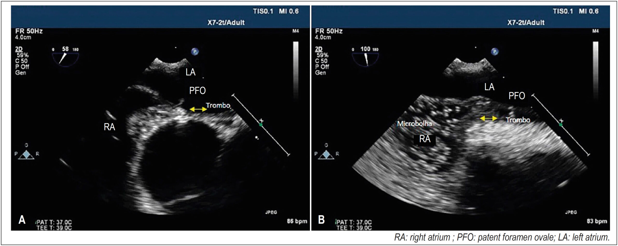
Recent studies have indicated that patent foramen ovale (PFO) may cause cryptogenic stroke in young patients presenting anatomical conditions that can favor it and that transcatheter occlusion reduces the incidence of stroke versus clinical treatment. A transesophageal echocardiographic study with agitated saline injection associated with the Valsalva maneuver can evidence right-to-left shunt with high sensitivity (89%) and specificity (92%). The Risk of Paradoxical Embolism trial evaluated the clinical characteristics of stroke patients with PFO; established a risk score for cryptogenic stroke; and used a multivariate regression model to identify six variables including age, presence of cortical ischemia, diabetes, hypertension, stroke, and previous transient ischemic attack. The highest scores were observed in young stroke patients without vascular risk factors, and the lowest scores were identified in older patients with vascular risk factors in which the PFO appeared to be incidental. Anatomical PFO conditions predispose patients to systemic embolism (PFO separation > 2 mm, PFO tunnel > 10 mm, angle between the inferior vena cava and the PFO flap < 10°, shunt intensity with Valsalva maneuver, and presence of interatrial septal aneurysm and Chiari network or prominent Eustachian valve). PFO closure can prevent paradoxical embolism by decreasing the incidence of stroke in high-risk patients.

  • Echocardiography
  • Foramen Ovale
  • Patent
  • Stroke
Read Full Article
Creative Commons Attribution (CC BY) Creative Commons license
16/06/2021 DOI: 10.47593/2675-312X/20213401eabc123
Figure 1
Case Report

Coronavirus Myocarditis: Case Report

Aída Fernanda Batista Rocha et al. José Luiz Alves de Barros Marcelo Canejo Sá Ana Claudia Maria da Silva Longo … José Gildo de Moura Monteiro Júnior José Maria Del Castillo Carlos Antônio Mota Silveira

Introduction

The first cases of coronavirus 2019 disease (COVID-19) were described in Wuhan, China, in the end of December 2019. The identified pathogen was called severe acute respiratory syndrome coronavirus 2 (SARS-CoV-2), an RNA virus of the family Coronaviridae. Due to its rapid spread, the World Health Organization declared COVID-19 a pandemic on March 11, 2020. The main clinical presentation includes respiratory symptoms, such as fever, cough, myalgia, and dyspnea, that can progress to pneumonia or severe acute respiratory syndrome. Among many other disorders, myocardial injury is widely described in the literature, especially associated with influenza and parvovirus B-19, but more clarifications are necessary with SARS-CoV-2 about the pathophysiology involved in the intense viral replication with systemic inflammatory involvement.,

SARS-CoV-2 is not only a cause of viral pneumonia, it also has important implications for the cardiovascular system, especially in men with risk factors, including older age, diabetes, hypertension, and obesity. Cardiac injury was detected in 19.7% of cases; of the patients who died, 10.6% had coronary heart disease, 4.1% had heart failure, and 5.3% had cerebrovascular disease. High cytokine concentrations are detected in the systemic inflammatory phase of COVID-19, such as interleukin 6 (IL-6), associated with an increase in troponin and other inflammatory biomarkers (D-dimer, ferritin, C-reactive protein, lactic dehydrogenase, procalcitonin, and leukocyte count), causing cardiovascular system injury.– Of these disorders, myocarditis comprises approximately 7.2% of the cardiovascular complications related to the novel coronavirus. In this phase of disease progression, transthoracic echocardiography should be the method of choice to diagnose and monitor patients, improving the therapeutic management for providing hemodynamic data since patients with ventricular dysfunction are more likely to need mechanical ventilation, which consequently results in a worse prognosis.,

[…]

  • Coronavirus
  • COVID-19
  • Echocardiography
  • Hospitalization
  • Myocarditis
Read Full Article
Creative Commons Attribution (CC BY) Creative Commons license
16/06/2021 DOI: 10.47593/2675-312X/20213401eabc120
Figure 1
Case Report

Left Ventricular Free-Wall Rupture (Pseudoaneurysm) after Acute Myocardial Infarction in an Asymptomatic Patient

Raul Serra Valério et al. Fernanda Sayuri Oshiro Alfredo Augusto Eyer Rodrigues Maria Eduarda Menezes de Siqueira Marly M. Uellendahl

Introduction

Left ventricular (LV) pseudoaneurysm after acute myocardial infarction may show nonspecific symptoms and be asymptomatic in up to 10% of cases. It demands investigation and a suspected diagnosis, with imaging methods playing a fundamental role in its diagnosis. This case report describes a pseudoaneurysm detected on a routine examination.

[…]

  • Cardiac Imaging Techniques
  • Diagnostic Imaging
  • False Aneurysm
Read Full Article
Creative Commons Attribution (CC BY) Creative Commons license
16/06/2021 DOI: 10.47593/2675-312X/20213401eabc134 Views 1

How to cite this article

EndNote RIS

Electronic Document Format (Vancouver)

Rayas-Gómez AL, González-Rayas JM, Rico-Rosas A, Landa-Alvarado PD, León-Vargas IMP, Ramos-Verdugo JM, González-Yáñez JM, et al. Cerebrovascular Disease as the Initial Manifestation of Apical Hypertrophic Cardiomyopathy. ABC Imagem Cardiovascular 2021;34(1):eabc144.

Electronic Document Format (ABNT)

Rayas-Gómez, Ana Lilia; González-Rayas, José Manuel; Rico-Rosas, Alejandro; Landa-Alvarado, Pedro Daniel; León-Vargas, Ileana Maribel Ponce de; Ramos-Verdugo, José María; González-Yáñez, José Manuel. Cerebrovascular Disease as the Initial Manifestation of Apical Hypertrophic Cardiomyopathy. ABC Imagem Cardiovascular, v. 34, n. 1, eabc144, Jun. 2021.

Electronic Document Format (APA)

Rayas-Gómez, A. L., González-Rayas, J. M., Rico-Rosas, A., Landa-Alvarado, P. D., León-Vargas, I. M. P. , Ramos-Verdugo, J. M., & González-Yáñez, J. M. (2021). Cerebrovascular Disease as the Initial Manifestation of Apical Hypertrophic Cardiomyopathy. ABC Imagem Cardiovascular, 34(1), eabc144.

Electronic Document Format (ISO)

Rayas-Gómez, Ana Lilia and González-Rayas, José Manuel and Rico-Rosas, Alejandro and Landa-Alvarado, Pedro Daniel and León-Vargas, Ileana Maribel Ponce de and Ramos-Verdugo, José María and González-Yáñez, José Manuel. Cerebrovascular Disease as the Initial Manifestation of Apical Hypertrophic Cardiomyopathy. ABC Imagem Cardiovascular [online]. 2021, vol. 34, n. 1, [cited 2026-10-14], eabc144. Available from: <https://journals.cardiol.br/en/article/cerebrovascular-disease-as-the-initial-manifestation-of-apical-hypertrophic-cardiomyopathy/>. ISSN 2318-8219

EndNote RIS

Electronic Document Format (Vancouver)

Elias Neto J, Silva MA, Kuniyoshi RR, Futuro G, Merçon ES, Grativvol PS, Pedrotti FC, et al. Cardiac Sarcoidosis as a Cause of Total Atrioventricular Block: Importance of Differential Diagnosis. ABC Imagem Cardiovascular 2021;34(1):eabc163.

Electronic Document Format (ABNT)

Elias Neto, Jorge; Silva, Márcio Augusto; Kuniyoshi, Ricardo Ryoshim; Futuro, Guilherme; Merçon, Erick Sessa; Grativvol, Petherson Susano; Pedrotti, Fátima Cristina. Cardiac Sarcoidosis as a Cause of Total Atrioventricular Block: Importance of Differential Diagnosis. ABC Imagem Cardiovascular, v. 34, n. 1, eabc163, Jun. 2021.

Electronic Document Format (APA)

Elias Neto, J., Silva, M. A., Kuniyoshi, R. R., Futuro, G., Merçon, E. S., Grativvol, P. S., & Pedrotti, F. C. (2021). Cardiac Sarcoidosis as a Cause of Total Atrioventricular Block: Importance of Differential Diagnosis. ABC Imagem Cardiovascular, 34(1), eabc163.

Electronic Document Format (ISO)

Elias Neto, Jorge and Silva, Márcio Augusto and Kuniyoshi, Ricardo Ryoshim and Futuro, Guilherme and Merçon, Erick Sessa and Grativvol, Petherson Susano and Pedrotti, Fátima Cristina. Cardiac Sarcoidosis as a Cause of Total Atrioventricular Block: Importance of Differential Diagnosis. ABC Imagem Cardiovascular [online]. 2021, vol. 34, n. 1, [cited 2026-10-14], eabc163. Available from: <https://journals.cardiol.br/en/article/cardiac-sarcoidosis-as-a-cause-of-total-atrioventricular-block-importance-of-differential-diagnosis/>. ISSN 2318-8219

EndNote RIS

Electronic Document Format (Vancouver)

Melo KPB, Maia RJC, Siqueira BJM, Araújo MLSC, Souza ACBM. Echocardiographic Image of Primary Mediastinal Sarcoma with Pericardial Invasion. ABC Imagem Cardiovascular 2021;34(1):eabc139.

Electronic Document Format (ABNT)

Melo, Keyla Patrícia Barbosa; Maia, Rafael José Coelho; Siqueira, Betty Janny Maia; Araújo, Mozart Lacerda Siqueira Campos; Souza, Ana Carolina Borges de Miranda. Echocardiographic Image of Primary Mediastinal Sarcoma with Pericardial Invasion. ABC Imagem Cardiovascular, v. 34, n. 1, eabc139, Jun. 2021.

Electronic Document Format (APA)

Melo, K. P. B., Maia, R. J. C., Siqueira, B. J. M., Araújo, M. L. S. C., & Souza, A. C. B. M. (2021). Echocardiographic Image of Primary Mediastinal Sarcoma with Pericardial Invasion. ABC Imagem Cardiovascular, 34(1), eabc139.

Electronic Document Format (ISO)

Melo, Keyla Patrícia Barbosa and Maia, Rafael José Coelho and Siqueira, Betty Janny Maia and Araújo, Mozart Lacerda Siqueira Campos and Souza, Ana Carolina Borges de Miranda. Echocardiographic Image of Primary Mediastinal Sarcoma with Pericardial Invasion. ABC Imagem Cardiovascular [online]. 2021, vol. 34, n. 1, [cited 2026-10-14], eabc139. Available from: <https://journals.cardiol.br/en/article/echocardiographic-image-of-primary-mediastinal-sarcoma-with-pericardial-invasion/>. ISSN 2318-8219

EndNote RIS

Electronic Document Format (Vancouver)

Beck ALS, Barberato SH, Almeida ALC, Grau CRPC, Lopes MMU, Lima RSL, Cerci RJ, et al. Position Statement on Indications and the Safe Reintroduction of Cardiovascular Imaging Methods in the COVID-19 Scenario – 2021. ABC Imagem Cardiovascular 2021;34(1):eposi2021.

Electronic Document Format (ABNT)

Beck, Adenalva Lima de Souza; Barberato, Silvio Henrique; Almeida, André Luiz Cerqueira de; Grau, Claudia R. Pinheiro de Castro; Lopes, Marly Maria Uellendahl; Lima, Ronaldo de Souza Leão; Cerci, Rodrigo Júlio; Albricker, Ana Cristina Lopes; Barros, Fanilda Souto; Oliveira, Alessandra Joslin; Lira Filho, Edgar Bezerra de; Miglioranza, Marcelo Haertel; Vieira, Marcelo Luiz Campos; Pena, José Luiz Barros; Strabelli, Tânia Mara Varejão; Bihan, David Costa de Souza Le; Tsutsui, Jeane Mike; Rochitte, Carlos Eduardo. Position Statement on Indications and the Safe Reintroduction of Cardiovascular Imaging Methods in the COVID-19 Scenario – 2021. ABC Imagem Cardiovascular, v. 34, n. 1, eposi2021, Jun. 2021.

Electronic Document Format (APA)

Beck, A. L. S., Barberato, S. H., Almeida, A. L. C. , Grau, C. R. P. C., Lopes, M. M. U., Lima, R. S. L., Cerci, R. J., Albricker, A. C. L., Barros, F. S., Oliveira, A. J., Lira Filho, E. B. , Miglioranza, M. H., Vieira, M. L. C., Pena, J. L. B., Strabelli, T. M. V., Bihan, D. C. S. L., Tsutsui, J. M., & Rochitte, C. E. (2021). Position Statement on Indications and the Safe Reintroduction of Cardiovascular Imaging Methods in the COVID-19 Scenario – 2021. ABC Imagem Cardiovascular, 34(1), eposi2021.

Electronic Document Format (ISO)

Beck, Adenalva Lima de Souza and Barberato, Silvio Henrique and Almeida, André Luiz Cerqueira de and Grau, Claudia R. Pinheiro de Castro and Lopes, Marly Maria Uellendahl and Lima, Ronaldo de Souza Leão and Cerci, Rodrigo Júlio and Albricker, Ana Cristina Lopes and Barros, Fanilda Souto and Oliveira, Alessandra Joslin and Lira Filho, Edgar Bezerra de and Miglioranza, Marcelo Haertel and Vieira, Marcelo Luiz Campos and Pena, José Luiz Barros and Strabelli, Tânia Mara Varejão and Bihan, David Costa de Souza Le and Tsutsui, Jeane Mike and Rochitte, Carlos Eduardo. Position Statement on Indications and the Safe Reintroduction of Cardiovascular Imaging Methods in the COVID-19 Scenario – 2021. ABC Imagem Cardiovascular [online]. 2021, vol. 34, n. 1, [cited 2026-10-14], eposi2021. Available from: <https://journals.cardiol.br/en/article/position-statement-on-indications-and-the-safe-reintroduction-of-cardiovascular-imaging-methods-in-the-covid-19-scenario-2021/>. ISSN 2318-8219

EndNote RIS

Electronic Document Format (Vancouver)

Thomé BB, Pazzini LV, Pavan NR, Cabeda EV, Almeida TS. Lipoma in the Interventricular Septum – Case Report. ABC Imagem Cardiovascular 2021;34(1):eabc152.

Electronic Document Format (ABNT)

Thomé, Bruna Bonamigo; Pazzini, Laura Vilela; Pavan, Nathalia Regina; Cabeda, Estevan Vieira; Almeida, Tobias Sato de. Lipoma in the Interventricular Septum – Case Report. ABC Imagem Cardiovascular, v. 34, n. 1, eabc152, Jun. 2021.

Electronic Document Format (APA)

Thomé, B. B., Pazzini, L. V., Pavan, N. R., Cabeda, E. V., & Almeida, T. S. (2021). Lipoma in the Interventricular Septum – Case Report. ABC Imagem Cardiovascular, 34(1), eabc152.

Electronic Document Format (ISO)

Thomé, Bruna Bonamigo and Pazzini, Laura Vilela and Pavan, Nathalia Regina and Cabeda, Estevan Vieira and Almeida, Tobias Sato de. Lipoma in the Interventricular Septum – Case Report. ABC Imagem Cardiovascular [online]. 2021, vol. 34, n. 1, [cited 2026-10-14], eabc152. Available from: <https://journals.cardiol.br/en/article/lipoma-in-the-interventricular-septum-case-report/>. ISSN 2318-8219

EndNote RIS

Electronic Document Format (Vancouver)

Takagui ASM, Baggio TC, Savariz MB, Theilacker G, Baggio TE. Left Ventricular-to-right Atrial Shunt (Gerbode Defect) Associated with Interventricular Communication in a 42-year-old Patient. ABC Imagem Cardiovascular 2021;34(1):eabc142.

Electronic Document Format (ABNT)

Takagui, Andressa Sardá Maiochi; Baggio, Thales Cantelle; Savariz, Mariana Baretta; Theilacker, Giulia; Baggio, Thalia Elisa. Left Ventricular-to-right Atrial Shunt (Gerbode Defect) Associated with Interventricular Communication in a 42-year-old Patient. ABC Imagem Cardiovascular, v. 34, n. 1, eabc142, Jun. 2021.

Electronic Document Format (APA)

Takagui, A. S. M., Baggio, T. C., Savariz, M. B., Theilacker, G., & Baggio, T. E. (2021). Left Ventricular-to-right Atrial Shunt (Gerbode Defect) Associated with Interventricular Communication in a 42-year-old Patient. ABC Imagem Cardiovascular, 34(1), eabc142.

Electronic Document Format (ISO)

Takagui, Andressa Sardá Maiochi and Baggio, Thales Cantelle and Savariz, Mariana Baretta and Theilacker, Giulia and Baggio, Thalia Elisa. Left Ventricular-to-right Atrial Shunt (Gerbode Defect) Associated with Interventricular Communication in a 42-year-old Patient. ABC Imagem Cardiovascular [online]. 2021, vol. 34, n. 1, [cited 2026-10-14], eabc142. Available from: <https://journals.cardiol.br/en/article/left-ventricular-to-right-atrial-shunt-gerbode-defect-associated-with-interventricular-communication-in-a-42-year-old-patient/>. ISSN 2318-8219

EndNote RIS

Electronic Document Format (Vancouver)

Almeida DC, Carrijo AMM, Souza MG, Martinelli FM, Fazzio FR, O’Connell JL. Anomalous Origin of the Right Coronary from the Left Coronary Sinus: How to approach?. ABC Imagem Cardiovascular 2021;34(1):eabc146.

Electronic Document Format (ABNT)

Almeida, Danielle Campos de; Carrijo, Alice Mirane Malta; Souza, Marcela Gomes de; Martinelli, Fernando de Melo; Fazzio, Fernando Roberto de; O’Connell, João Lucas. Anomalous Origin of the Right Coronary from the Left Coronary Sinus: How to approach?. ABC Imagem Cardiovascular, v. 34, n. 1, eabc146, Jun. 2021.

Electronic Document Format (APA)

Almeida, D. C. , Carrijo, A. M. M., Souza, M. G. , Martinelli, F. M., Fazzio, F. R. , & O’Connell, J. L. (2021). Anomalous Origin of the Right Coronary from the Left Coronary Sinus: How to approach?. ABC Imagem Cardiovascular, 34(1), eabc146.

Electronic Document Format (ISO)

Almeida, Danielle Campos de and Carrijo, Alice Mirane Malta and Souza, Marcela Gomes de and Martinelli, Fernando de Melo and Fazzio, Fernando Roberto de and O’Connell, João Lucas. Anomalous Origin of the Right Coronary from the Left Coronary Sinus: How to approach?. ABC Imagem Cardiovascular [online]. 2021, vol. 34, n. 1, [cited 2026-10-14], eabc146. Available from: <https://journals.cardiol.br/en/article/anomalous-origin-of-the-right-coronary-from-the-left-coronary-sinus-how-to-approach/>. ISSN 2318-8219

EndNote RIS

Electronic Document Format (Vancouver)

Pianca EG, Branchi MN, Mastella B, Albrecht S, Wender OCB, Foppa M, Santos ABS, et al. Isolated Fungal Pulmonary Endocarditis: Role of Transthoracic Echocardiography from the Diagnosis to Follow-up. ABC Imagem Cardiovascular 2021;34(1):eabc143.

Electronic Document Format (ABNT)

Pianca, Eduardo Gatti; Branchi, Marcelo Nicola; Mastella, Bernardo; Albrecht, Álvaro Schmidt; Wender, Orlando Carlos Belmonte; Foppa, Murilo; Santos, Angela Barreto Santiago. Isolated Fungal Pulmonary Endocarditis: Role of Transthoracic Echocardiography from the Diagnosis to Follow-up. ABC Imagem Cardiovascular, v. 34, n. 1, eabc143, Jun. 2021.

Electronic Document Format (APA)

Pianca, E. G., Branchi, M. N., Mastella, B., Albrecht, S., Wender, O. C. B., Foppa, M., & Santos, A. B. S. (2021). Isolated Fungal Pulmonary Endocarditis: Role of Transthoracic Echocardiography from the Diagnosis to Follow-up. ABC Imagem Cardiovascular, 34(1), eabc143.

Electronic Document Format (ISO)

Pianca, Eduardo Gatti and Branchi, Marcelo Nicola and Mastella, Bernardo and Albrecht, Álvaro Schmidt and Wender, Orlando Carlos Belmonte and Foppa, Murilo and Santos, Angela Barreto Santiago. Isolated Fungal Pulmonary Endocarditis: Role of Transthoracic Echocardiography from the Diagnosis to Follow-up. ABC Imagem Cardiovascular [online]. 2021, vol. 34, n. 1, [cited 2026-10-14], eabc143. Available from: <https://journals.cardiol.br/en/article/isolated-fungal-pulmonary-endocarditis-role-of-transthoracic-echocardiography-from-the-diagnosis-to-follow-up/>. ISSN 2318-8219

EndNote RIS

Electronic Document Format (Vancouver)

Barreto ATF, Bezerra MLBG, Melo RMV, Isabella BO, Reis VA, Passos LCS. Diagnostic yield of magnetic resonance imaging in heart failure with left ventricular dysfunction?. ABC Imagem Cardiovascular 2021;34(1):eabc108.

Electronic Document Format (ABNT)

Barreto, Ana Terra Fonseca; Bezerra, Mariana Lins Baptista Guedes; Melo, Rodrigo Morel Vieira de; Isabella, Bruno Oliveira; Reis, Vanessa de Assis; Passos, Luiz Carlos Santana. Diagnostic yield of magnetic resonance imaging in heart failure with left ventricular dysfunction?. ABC Imagem Cardiovascular, v. 34, n. 1, eabc108, Jun. 2021.

Electronic Document Format (APA)

Barreto, A. T. F., Bezerra, M. L. B. G., Melo, R. M. V. , Isabella, B. O., Reis, V. A., & Passos, L. C. S. (2021). Diagnostic yield of magnetic resonance imaging in heart failure with left ventricular dysfunction?. ABC Imagem Cardiovascular, 34(1), eabc108.

Electronic Document Format (ISO)

Barreto, Ana Terra Fonseca and Bezerra, Mariana Lins Baptista Guedes and Melo, Rodrigo Morel Vieira de and Isabella, Bruno Oliveira and Reis, Vanessa de Assis and Passos, Luiz Carlos Santana. Diagnostic yield of magnetic resonance imaging in heart failure with left ventricular dysfunction?. ABC Imagem Cardiovascular [online]. 2021, vol. 34, n. 1, [cited 2026-10-14], eabc108. Available from: <https://journals.cardiol.br/en/article/diagnostic-yield-of-magnetic-resonance-imaging-in-heart-failure-with-left-ventricular-dysfunction/>. ISSN 2318-8219

EndNote RIS

Electronic Document Format (Vancouver)

Andrade RB, Lima CJM, Freitas IA, Corrêa LC, Lino DOC, Alcantâra ACB. Cardiac Intimal Sarcoma: A Rare Cardiac Tumor Entity. ABC Imagem Cardiovascular 2021;34(1):eabc118.

Electronic Document Format (ABNT)

Andrade, Rhavena Brasil de; Lima, Carlos José Mota de; Freitas, Ingrid Alves de; Corrêa, Larissa Chagas; Lino, Danielli Oliveira da Costa; Alcantâra, Ana Carolina Brito de. Cardiac Intimal Sarcoma: A Rare Cardiac Tumor Entity. ABC Imagem Cardiovascular, v. 34, n. 1, eabc118, Jun. 2021.

Electronic Document Format (APA)

Andrade, R. B. , Lima, C. J. M. , Freitas, I. A. , Corrêa, L. C., Lino, D. O. C., & Alcantâra, A. C. B. (2021). Cardiac Intimal Sarcoma: A Rare Cardiac Tumor Entity. ABC Imagem Cardiovascular, 34(1), eabc118.

Electronic Document Format (ISO)

Andrade, Rhavena Brasil de and Lima, Carlos José Mota de and Freitas, Ingrid Alves de and Corrêa, Larissa Chagas and Lino, Danielli Oliveira da Costa and Alcantâra, Ana Carolina Brito de. Cardiac Intimal Sarcoma: A Rare Cardiac Tumor Entity. ABC Imagem Cardiovascular [online]. 2021, vol. 34, n. 1, [cited 2026-10-14], eabc118. Available from: <https://journals.cardiol.br/en/article/cardiac-intimal-sarcoma-a-rare-cardiac-tumor-entity/>. ISSN 2318-8219

EndNote RIS

Electronic Document Format (Vancouver)

Sousa CG, Castillo JMD, Mazzarollo C, Albuquerque ES, Sena ADM, Brindeiro Filho D, Silveira CAM, et al. Comparative Analysis of the Coronary Arteries Flow Pattern in Secondary Myocardial Hypertrophies and by Sarcomeric Mutation. ABC Imagem Cardiovascular 2021;34(1):eabc131.

Electronic Document Format (ABNT)

Sousa, Caio Guedes de; Castillo, José Maria Del; Mazzarollo, Carlos; Albuquerque, Eugenio Soares de; Sena, Antônia Dulcineide Medeiros; Brindeiro Filho, Djair; Silveira, Carlos Antônio da Mota. Comparative Analysis of the Coronary Arteries Flow Pattern in Secondary Myocardial Hypertrophies and by Sarcomeric Mutation. ABC Imagem Cardiovascular, v. 34, n. 1, eabc131, Jun. 2021.

Electronic Document Format (APA)

Sousa, C. G. , Castillo, J. M. D., Mazzarollo, C., Albuquerque, E. S. , Sena, A. D. M., Brindeiro Filho, D., & Silveira, C. A. M. (2021). Comparative Analysis of the Coronary Arteries Flow Pattern in Secondary Myocardial Hypertrophies and by Sarcomeric Mutation. ABC Imagem Cardiovascular, 34(1), eabc131.

Electronic Document Format (ISO)

Sousa, Caio Guedes de and Castillo, José Maria Del and Mazzarollo, Carlos and Albuquerque, Eugenio Soares de and Sena, Antônia Dulcineide Medeiros and Brindeiro Filho, Djair and Silveira, Carlos Antônio da Mota. Comparative Analysis of the Coronary Arteries Flow Pattern in Secondary Myocardial Hypertrophies and by Sarcomeric Mutation. ABC Imagem Cardiovascular [online]. 2021, vol. 34, n. 1, [cited 2026-10-14], eabc131. Available from: <https://journals.cardiol.br/en/article/comparative-analysis-of-the-coronary-arteries-flow-pattern-in-secondary-myocardial-hypertrophies-and-by-sarcomeric-mutation/>. ISSN 2318-8219

EndNote RIS

Electronic Document Format (Vancouver)

Pires MIFB, Almeida I, Gonçalves ML, Santos JM, Correia M. Eustachian Valve Endocarditis Associated with Complicated Cardiac Resynchronization Therapy Defibrillator Generator Pocket Infection. ABC Imagem Cardiovascular 2021;34(1):eabc161.

Electronic Document Format (ABNT)

Pires, Maria Inês Fiuza Branco; Almeida, Inês; Gonçalves, Maria Luísa; Santos, João Miguel; Correia, Miguel. Eustachian Valve Endocarditis Associated with Complicated Cardiac Resynchronization Therapy Defibrillator Generator Pocket Infection. ABC Imagem Cardiovascular, v. 34, n. 1, eabc161, Jun. 2021.

Electronic Document Format (APA)

Pires, M. I. F. B., Almeida, I., Gonçalves, M. L., Santos, J. M., & Correia, M. (2021). Eustachian Valve Endocarditis Associated with Complicated Cardiac Resynchronization Therapy Defibrillator Generator Pocket Infection. ABC Imagem Cardiovascular, 34(1), eabc161.

Electronic Document Format (ISO)

Pires, Maria Inês Fiuza Branco and Almeida, Inês and Gonçalves, Maria Luísa and Santos, João Miguel and Correia, Miguel. Eustachian Valve Endocarditis Associated with Complicated Cardiac Resynchronization Therapy Defibrillator Generator Pocket Infection. ABC Imagem Cardiovascular [online]. 2021, vol. 34, n. 1, [cited 2026-10-14], eabc161. Available from: <https://journals.cardiol.br/en/article/eustachian-valve-endocarditis-associated-with-complicated-cardiac-resynchronization-therapy-defibrillator-generator-pocket-infection/>. ISSN 2318-8219

EndNote RIS

Electronic Document Format (Vancouver)

Silveira CAM, Castillo JMD. Echocardiographic Evaluation of Patients with Patent Foramen Ovale and Cryptogenic Stroke. ABC Imagem Cardiovascular 2021;34(1):eabc123.

Electronic Document Format (ABNT)

Silveira, Carlos Antônio da Mota; Castillo, José Maria Del. Echocardiographic Evaluation of Patients with Patent Foramen Ovale and Cryptogenic Stroke. ABC Imagem Cardiovascular, v. 34, n. 1, eabc123, Jun. 2021.

Electronic Document Format (APA)

Silveira, C. A. M., & Castillo, J. M. D. (2021). Echocardiographic Evaluation of Patients with Patent Foramen Ovale and Cryptogenic Stroke. ABC Imagem Cardiovascular, 34(1), eabc123.

Electronic Document Format (ISO)

Silveira, Carlos Antônio da Mota and Castillo, José Maria Del. Echocardiographic Evaluation of Patients with Patent Foramen Ovale and Cryptogenic Stroke. ABC Imagem Cardiovascular [online]. 2021, vol. 34, n. 1, [cited 2026-10-14], eabc123. Available from: <https://journals.cardiol.br/en/article/echocardiographic-evaluation-of-patients-with-patent-foramen-ovale-and-cryptogenic-stroke/>. ISSN 2318-8219

EndNote RIS

Electronic Document Format (Vancouver)

Rocha AFB, Barros JLA, Sá MC, Longo ACMS, Monteiro Júnior JGM, Castillo JMD, Silveira CAM, et al. Coronavirus Myocarditis: Case Report. ABC Imagem Cardiovascular 2021;34(1):eabc120.

Electronic Document Format (ABNT)

Rocha, Aída Fernanda Batista; Barros, José Luiz Alves de; Sá, Marcelo Canejo; Longo, Ana Claudia Maria da Silva; Monteiro Júnior, José Gildo de Moura; Castillo, José Maria Del; Silveira, Carlos Antônio Mota. Coronavirus Myocarditis: Case Report. ABC Imagem Cardiovascular, v. 34, n. 1, eabc120, Jun. 2021.

Electronic Document Format (APA)

Rocha, A. F. B., Barros, J. L. A. , Sá, M. C., Longo, A. C. M. S., Monteiro Júnior, J. G. M., Castillo, J. M. D., & Silveira, C. A. M. (2021). Coronavirus Myocarditis: Case Report. ABC Imagem Cardiovascular, 34(1), eabc120.

Electronic Document Format (ISO)

Rocha, Aída Fernanda Batista and Barros, José Luiz Alves de and Sá, Marcelo Canejo and Longo, Ana Claudia Maria da Silva and Monteiro Júnior, José Gildo de Moura and Castillo, José Maria Del and Silveira, Carlos Antônio Mota. Coronavirus Myocarditis: Case Report. ABC Imagem Cardiovascular [online]. 2021, vol. 34, n. 1, [cited 2026-10-14], eabc120. Available from: <https://journals.cardiol.br/en/article/coronavirus-myocarditis-case-report/>. ISSN 2318-8219

EndNote RIS

Electronic Document Format (Vancouver)

Valério RS, Oshiro FS, Rodrigues AAE, Siqueira MEM, Uellendahl MM. Left Ventricular Free-Wall Rupture (Pseudoaneurysm) after Acute Myocardial Infarction in an Asymptomatic Patient. ABC Imagem Cardiovascular 2021;34(1):eabc134.

Electronic Document Format (ABNT)

Valério, Raul Serra; Oshiro, Fernanda Sayuri; Rodrigues, Alfredo Augusto Eyer; Siqueira, Maria Eduarda Menezes de; Uellendahl, Marly M.. Left Ventricular Free-Wall Rupture (Pseudoaneurysm) after Acute Myocardial Infarction in an Asymptomatic Patient. ABC Imagem Cardiovascular, v. 34, n. 1, eabc134, Jun. 2021.

Electronic Document Format (APA)

Valério, R. S., Oshiro, F. S., Rodrigues, A. A. E., Siqueira, M. E. M. , & Uellendahl, M. M. (2021). Left Ventricular Free-Wall Rupture (Pseudoaneurysm) after Acute Myocardial Infarction in an Asymptomatic Patient. ABC Imagem Cardiovascular, 34(1), eabc134.

Electronic Document Format (ISO)

Valério, Raul Serra and Oshiro, Fernanda Sayuri and Rodrigues, Alfredo Augusto Eyer and Siqueira, Maria Eduarda Menezes de and Uellendahl, Marly M.. Left Ventricular Free-Wall Rupture (Pseudoaneurysm) after Acute Myocardial Infarction in an Asymptomatic Patient. ABC Imagem Cardiovascular [online]. 2021, vol. 34, n. 1, [cited 2026-10-14], eabc134. Available from: <https://journals.cardiol.br/en/article/left-ventricular-free-wall-rupture-pseudoaneurysm-after-acute-myocardial-infarction-in-an-asymptomatic-patient/>. ISSN 2318-8219

Sede – São Paulo
Rua Barata Ribeiro, 380 cj 54 Bela Vista
São Paulo, Brasil – CEP: 01308-000
Tel.: +55 (11) 3120-3363
Telefax: +55 (11) 3259-2988
E-mail: depeco@cardiol.br

Sede – Rio de Janeiro
Av. Marechal Câmara, 160
3º andar – Sala: 330 – Centro
Rio de Janeiro – CEP: 20020-907
Tel.: +55 (21) 3478-2700
ou: +55 (21) 99386-3961
E-mail: abcimaging@cardiol.br

  • About the Journal
  • Internal Regulation
  • Editors Section
  • Editorial Board
  • SBC Award

  • Guidelines for Publication
  • Editorial Policy
  • Ethics Policy
  • Policy on the Use of Artificial Intelligence
  • Mandatory Publication Form

Issues

SBC Guidelines