ABC Family Arquivos Brasileiros de Cardiologia International Journal of Cardiovascular Sciences ABC Heart Failure & Cardiomyopathy ABC Imagem Cardiovascular
Português English
ABC Family
Português English
  • About
    • About ABC Family
    • Internal Regulation
    • Editorial Board
    • Brazilian Society of Cardiology (SBC)
  • Issues
  • SBC Guidelines
  • Content
    • Videos
    • Podcasts
    • News
  • About
    • About ABC Family
    • Internal Regulation
    • Editorial Board
    • Brazilian Society of Cardiology (SBC)
  • Issues
  • SBC Guidelines
  • Content
    • Videos
    • Podcasts
    • News

ABC Family

  • ABC Family
  • Arquivos Brasileiros de Cardiologia
  • International Journal of Cardiovascular Sciences
  • ABC Heart Failure & Cardiomyopathy
  • ABC Imagem Cardiovascular
Search in

No content found.

Journals
Ordering
Article types
Others

  1. Home
  2. Keyword: Lipoma
Figure 1
ABCIC
Case Report

Cardiac Lipoma as an Incidental Finding in Cardiovascular Imaging Exam

Fábio P. Taniguchi et al. Marcelo G. Silas Patrícia M. Oliveira

Introduction

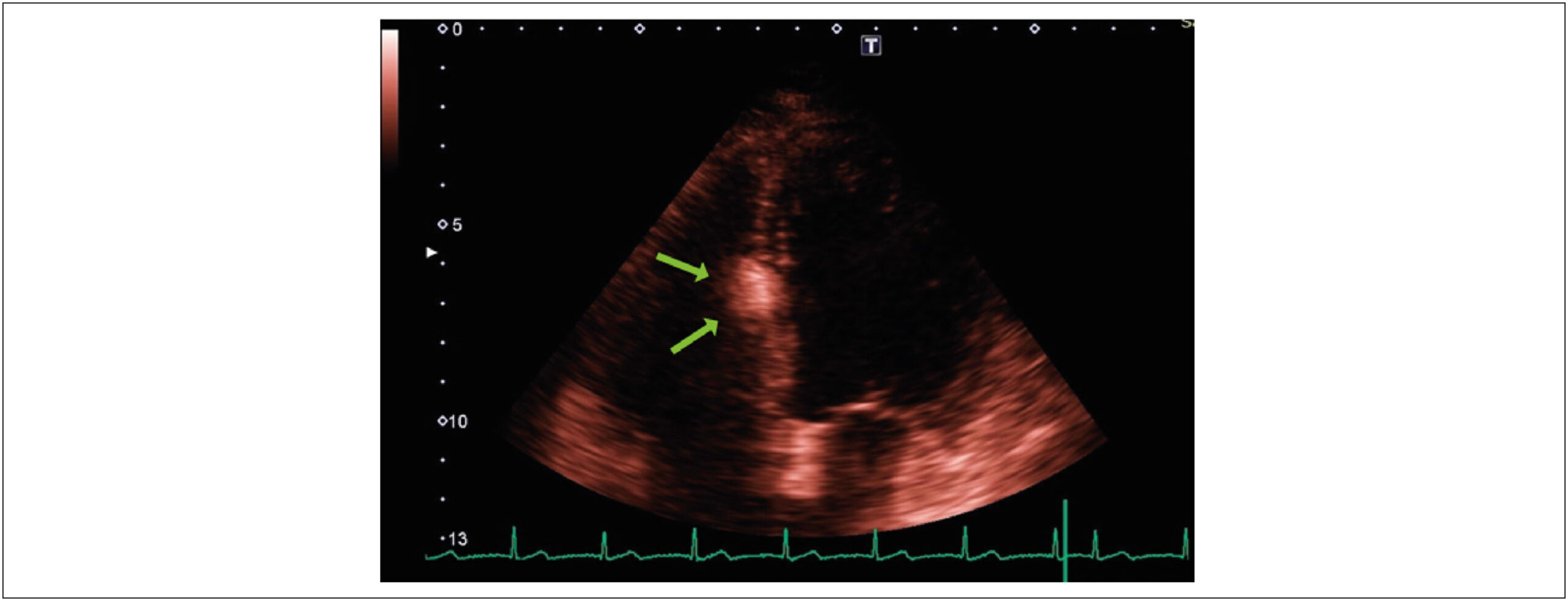
The advent of cardiac imaging has ushered in an era where incidental findings can be detected using less invasive methods, even in asymptomatic patients. The clinical presentation of cardiac masses, characterized by their varying growth rates and diverse locations within the heart, poses a formidable challenge to cardiologists. Therefore, paramount importance lies in reducing the clinical burden and costs, considering the potential of cardiac imaging to optimize the clinical pathway. The most common cardiac masses in the right atrium include thrombi, vegetations, and neoplasia. Approximately 75% of primary tumors are benign, with myxomas comprising 50% of benign cases. Other benign tumors that usually may arise in the right atrium are rhabdomyomas, fibromas, fibroelastomas, and lipomas. Cardiac lipomas are rare, accounting for 2.9% to 8% of all benign cardiac tumors. They rank third in frequency after myxomas and papillary fibroelastomas. The literature suggests that 25% of cardiac lipomas are intramyocardial; 25% are extracavitary of epicardial origin, and 50% are intracavitary of subendocardial origin. Lipomas are well-encapsulated and homogeneous masses composed of mature fat. Although the etiology of cardiac lipomas is unknown, they can originate from any of the three cardiac tissues: subendocardial (the most common), pericardial, or myocardial.

The aim of this report is to present a case of an asymptomatic right atrial lipoma and to discuss the etiology, natural history, and optimal management strategies for right atrial lipomas.

[…]
  • Cardiovascular Surgical Procedures
  • Lipoma
  • Neoplasms
Read Full Article
Creative Commons Attribution (CC BY) Creative Commons license
02/10/2023 DOI: 10.36660/abcimg.20230059i Views 2
Figure 1
ABCIC
Case Report

Lipoma in the Interventricular Septum – Case Report

Bruna Bonamigo Thomé et al. Laura Vilela Pazzini Nathalia Regina Pavan Estevan Vieira Cabeda Tobias Sato de Almeida

Abstract

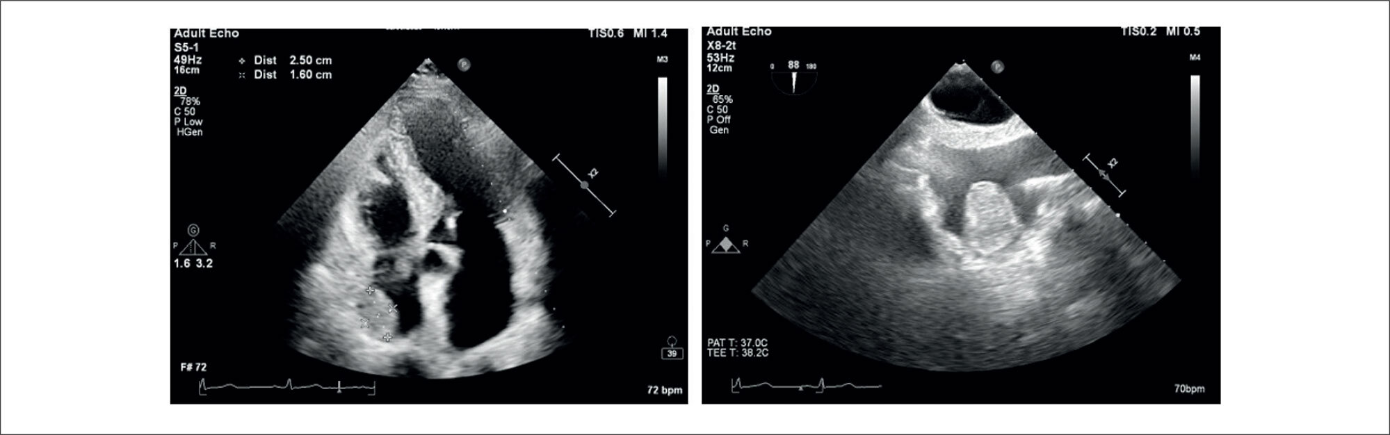
Primary cardiac tumors are a rare but mostly benign pathology found in up to 0.03% of autopsies. Among these benign tumors, with an incidence of 8.5%, lipomas are often located in the interatrial septum. When located in the interventricular septum, they are considered an even more unusual pathology with an unknown real prevalence. In most cases, the diagnosis is made incidentally on cardiovascular imaging and confirmed by histopathological examination. They are more commonly asymptomatic or feature nonspecific symptoms, but these lipomas can progress with arrhythmias, valve dysfunction, heart failure, and death, which highlights the importance of cardiovascular imaging methods in the differential diagnosis and to guide appropriate therapy. This report describes the case of a patient with an incidental echocardiographic finding of a cardiac mass located in the interventricular septum, followed by cardiac magnetic resonance imaging to better characterize the lesion and demonstrate its characteristics compatible with lipoma.

  • Diagnostic cardiovascular techniques
  • Echocardiography
  • Heart Neoplasms
  • Lipoma
Read Full Article
Creative Commons Attribution (CC BY) Creative Commons license
16/06/2021 DOI: 10.47593/2675-312X/20213401eabc152
Figure 1
ABC
Image

Fatty Left Ventricular Cardiomyopathy: An Under-Diagnosed Disease

Abdalla Elagha et al. Anthon Fuisz

During a preoperative cardiac assessment, a 57-year-old asymptomatic female, without known cardiac risk-factors, was found to have LBBB on ECG. Echocardiography revealed dilated cardiomyopathy. To rule-out coronary artery disease, coronary CT-angiography was performed and showed no significant coronary obstruction.

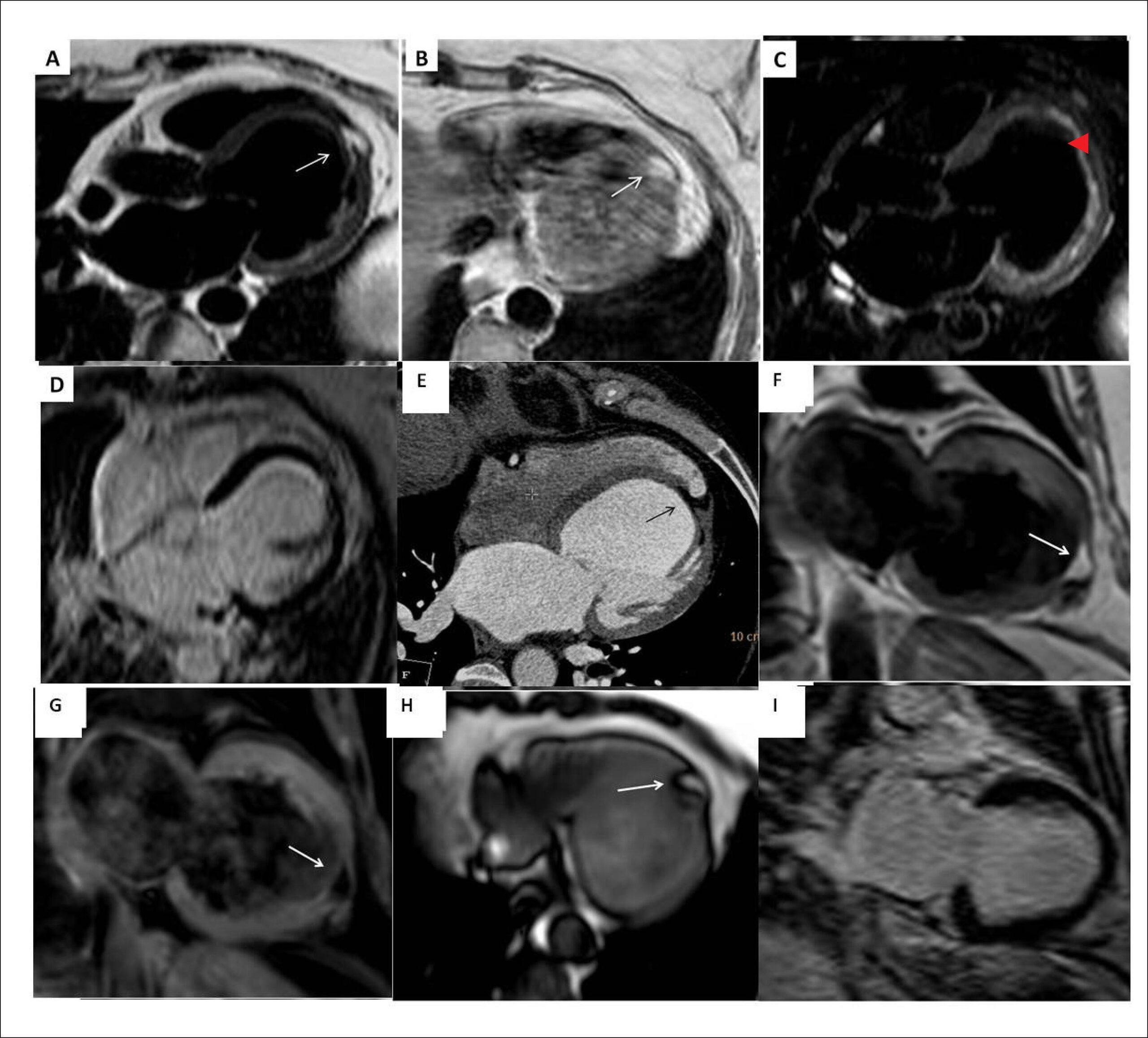
For further evaluation, a cardiac magnetic resonance (CMR) study was performed. A focal area of fatty infiltration was seen in the left ventricular (LV) apex extending from the subendocardial to the subepicardial surfaces ( and ). This area was suppressed with fat saturation pulse-sequences (), and did not show enhanced signal on delayed-hyperenhancement images (DHE) (). Both LV and LA were dilated; LV systolic function was reduced globally. Additionally, the CMR showed normal RV dimensions, function, and wall-thickness. The fatty area was also seen retrospectively on the cardiac CT ().

[…]

  • Cardiomyopathy, Dilated
  • Lipoma
  • Tomography, X Ray-Computed/methods
  • Ventricular Dysfunction, Left
Read Full Article
Creative Commons Attribution (CC BY) Creative Commons license
01/01/2017 DOI: 10.5935/abc.20160199 Views 1

How to cite this article

EndNote RIS

Electronic Document Format (Vancouver)

Taniguchi FP, Silas MG, Oliveira PM. Cardiac Lipoma as an Incidental Finding in Cardiovascular Imaging Exam. ABC Imagem Cardiovascular 2023;36(3):e20230059.

Electronic Document Format (ABNT)

Taniguchi, Fábio P.; Silas, Marcelo G.; Oliveira, Patrícia M.. Cardiac Lipoma as an Incidental Finding in Cardiovascular Imaging Exam. ABC Imagem Cardiovascular, v. 36, n. 3, e20230059, Oct. 2023.

Electronic Document Format (APA)

Taniguchi, F. P., Silas, M. G., & Oliveira, P. M. (2023). Cardiac Lipoma as an Incidental Finding in Cardiovascular Imaging Exam. ABC Imagem Cardiovascular, 36(3), e20230059.

Electronic Document Format (ISO)

Taniguchi, Fábio P. and Silas, Marcelo G. and Oliveira, Patrícia M.. Cardiac Lipoma as an Incidental Finding in Cardiovascular Imaging Exam. ABC Imagem Cardiovascular [online]. 2023, vol. 36, n. 3, [cited 2026-10-14], e20230059. Available from: <https://journals.cardiol.br/en/article/cardiac-lipoma-as-an-incidental-finding-in-cardiovascular-imaging-exam/>. ISSN 2318-8219

EndNote RIS

Electronic Document Format (Vancouver)

Thomé BB, Pazzini LV, Pavan NR, Cabeda EV, Almeida TS. Lipoma in the Interventricular Septum – Case Report. ABC Imagem Cardiovascular 2021;34(1):eabc152.

Electronic Document Format (ABNT)

Thomé, Bruna Bonamigo; Pazzini, Laura Vilela; Pavan, Nathalia Regina; Cabeda, Estevan Vieira; Almeida, Tobias Sato de. Lipoma in the Interventricular Septum – Case Report. ABC Imagem Cardiovascular, v. 34, n. 1, eabc152, Jun. 2021.

Electronic Document Format (APA)

Thomé, B. B., Pazzini, L. V., Pavan, N. R., Cabeda, E. V., & Almeida, T. S. (2021). Lipoma in the Interventricular Septum – Case Report. ABC Imagem Cardiovascular, 34(1), eabc152.

Electronic Document Format (ISO)

Thomé, Bruna Bonamigo and Pazzini, Laura Vilela and Pavan, Nathalia Regina and Cabeda, Estevan Vieira and Almeida, Tobias Sato de. Lipoma in the Interventricular Septum – Case Report. ABC Imagem Cardiovascular [online]. 2021, vol. 34, n. 1, [cited 2026-10-14], eabc152. Available from: <https://journals.cardiol.br/en/article/lipoma-in-the-interventricular-septum-case-report/>. ISSN 2318-8219

EndNote RIS

Electronic Document Format (Vancouver)

Elagha A, Fuisz A. Fatty Left Ventricular Cardiomyopathy: An Under-Diagnosed Disease. Arquivos Brasileiros de Cardiologia 2017;108(1):91-2.

Electronic Document Format (ABNT)

Elagha, Abdalla; Fuisz, Anthon. Fatty Left Ventricular Cardiomyopathy: An Under-Diagnosed Disease. Arquivos Brasileiros de Cardiologia, v. 108, n. 1, p. 91-92, Dec. 2016.

Electronic Document Format (APA)

Elagha, A., & Fuisz, A. (2017). Fatty Left Ventricular Cardiomyopathy: An Under-Diagnosed Disease. Arquivos Brasileiros de Cardiologia, 108(1), 91-92.

Electronic Document Format (ISO)

Elagha, Abdalla and Fuisz, Anthon. Fatty Left Ventricular Cardiomyopathy: An Under-Diagnosed Disease. Arquivos Brasileiros de Cardiologia [online]. 2017, vol. 108, n. 1, [cited 2026-10-14], pp. 91-92. Available from: <https://journals.cardiol.br/en/article/fatty-left-ventricular-cardiomyopathy-an-under-diagnosed-disease/>. ISSN 0066-782X

  • About ABC Family
  • Internal Regulation
  • Editorial Board
  • Brazilian Society of Cardiology (SBC)

Issues

SBC Guidelines

  • Videos
  • Podcasts
  • News