| ABCIC
ABC Family Arquivos Brasileiros de Cardiologia International Journal of Cardiovascular Sciences ABC Heart Failure & Cardiomyopathy ABC Imagem Cardiovascular
Português English
ABC Family | ABCIC
Português English
  • About
    • About the Journal
    • Internal Regulation
    • Editors Section
    • Editorial Board
    • SBC Award
  • Instructions to Authors
    • Guidelines for Publication
    • Editorial Policy
    • Ethics Policy
    • Policy on the Use of Artificial Intelligence
    • Mandatory Publication Form
  • Issues
  • SBC Guidelines
  • About
    • About the Journal
    • Internal Regulation
    • Editors Section
    • Editorial Board
    • SBC Award
  • Instructions to Authors
    • Guidelines for Publication
    • Editorial Policy
    • Ethics Policy
    • Policy on the Use of Artificial Intelligence
    • Mandatory Publication Form
  • Issues
  • SBC Guidelines

ABC Family

  • ABC Family
  • Arquivos Brasileiros de Cardiologia
  • International Journal of Cardiovascular Sciences
  • ABC Heart Failure & Cardiomyopathy
  • ABC Imagem Cardiovascular
Search in

No content found.

Journals
Ordering
Article types
Others

  1. Home
  2. ABCIC
  3. Issues
  4. 2022, Volume 35, Number 2
Figure 1
Original Article

Left Atrial Function Analysis in Patients with Functional Mitral Regurgitation Associated with Dilated Cardiomyopathy

Stephanie Macedo Andrade et al. Rodrigo Bellio de Mattos Barretto Francisco Thiago Tomaz de Sousa David Costa de Souza Le Bihan

Abstract

Background

Functional mitral regurgitation (FMR) is associated with dilated cardiomyopathy (DC), heart failure (HF) and worsening left atrial function (LAF). Patients with DC and FMR may present left atrial dysfunction resulting from both ventricular dysfunction and valve disease, but it is unknown whether the presence of valve disease will lead to greater LAF impairment.

Objective

This study aimed to evaluate the relationship between LAF parameters and FMR degree in patients with DC.

Methods

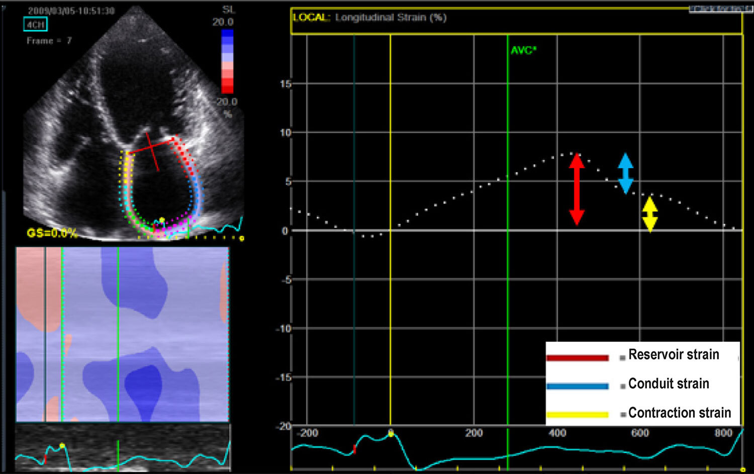
This cross-sectional observational study included 214 patients with DC, 46 without FMR (control group) and 168 with mild, moderate or severe FMR. An LAF analysis was performed by speckle tracking echocardiography (STE) and atrial volumetric variation.

Results

LAF analyzed by STE by means of reservoir strain, conduit strain and active contraction strain was reduced in the sample, with values of 14.3%, 8.49% and 5.92%, respectively. FMR degree was significantly associated with reservoir strain (0.27 ± 0.16 versus 0.15 ± 0.09; p < 0.001) and contraction strain (19.2 ± 7.3 versus 11.2 ± 2.7; p < 0.001). FMR was also associated with a reduced LAF assessed by volumetric analysis: total atrial emptying fraction of 0.51 ± 0.13 versus 0.34 ± 0.11 and active atrial emptying fraction of 0 .27 ± 0.16 versus 0.15 ± 0.09 (p < 0.001).

Conclusion

In a population with DC, FMR was associated with reduced LAF assessed by STE and atrial volume variation.
  • Atrial Function, Left
  • Cardiomyopathy, Dilated
  • Echocardiography
  • Mitral Valve Insufficiency
Read Full Article
Creative Commons Attribution (CC BY) Creative Commons license
30/06/2022 DOI: 10.47593/2675-312X/20223502eabc281 Views 1
Figure 1
Editorial

Left Atrial Appendage: Anatomy, Function, and Importance in Thrombus Formation

Taylla Mendes Silva et al. Gustavo Brandão de Oliveira Thais Lins de Souza Barros André Barbosa de Andrade … Bruno Bom Furlan Maria do Carmo de Pereira Nunes

Introduction

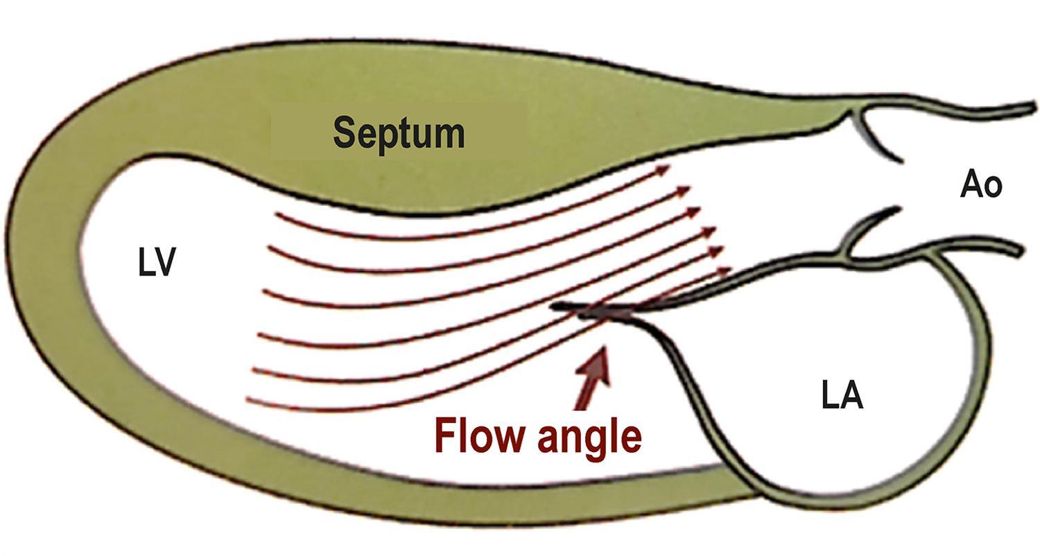
The left atrial appendage (LAA) is an extension of the left atrium (LA) and has complex anatomical structure and unique pathophysiological properties. The LAA functions as a decompression chamber during left ventricular (LV) systole and under increased left atrial pressure conditions. Despite previously being considered a relatively insignificant portion of the cardiac anatomy, the LAA has been highlighted as an important structure involved in the genesis of thrombus formation and thromboembolic events. With the recent development of percutaneous closure devices, LAA morphology assessments have become increasingly important. This article aims to describe LAA anatomy and morphology, function assessment parameters, thrombus diagnostic challenges, and the main imaging modalities, particularly transesophageal echocardiography.

[…]

Read Full Article
Creative Commons Attribution (CC BY) Creative Commons license
30/06/2022 DOI: 10.47593/2675-312X/20223502eabc301 Views 2
Editorial

Infective Endocarditis In India: A Broad Picture Of A Very Large Country

Ashish Agrawal et al. Shantanu P Sengupta

Infective endocarditis (IE) has always been challenging for the medical field, which has responded with newer innovations and persistent efforts. Habib et al. stated “Infective endocarditis is still a life-threatening disease with the frequent lethal outcome despite profound changes in its clinical, microbiological, imaging, and therapeutic profiles”.

Various factors affect the different aspects of IE diagnosis and management. As a developing country, India has a very polar healthcare delivery system. Primary health centers range from basic facilities that provide very primary level healthcare in rural areas to center of excellence hospitals that offer the latest medical innovations in metropolitan areas. The crucial point is the time it takes for a patient to reach to suitable center to receive the appropriate interventions. In particular, the diagnosis of IE by primary healthcare physicians must be obtained as soon as possible. Socio-economic factors play a pivotal role in patient outcomes. All three of these issues pose great challenges, especially in very large countries like India. Sengupta et al. concluded that socio-economic factors influence the clinical profile of patients presenting with IE worldwide, leading to delayed diagnosis and lower use of surgery. Gupta et al. reported observations about IE such as increasing patient age and increased proportions of patients without a history of cardiac ailments, better cultural positivity rates, an increasing incidence of staphylococcal infections, increased usage of transesophageal echocardiography (TEE), and increasing elective surgery rates.

[…]

Read Full Article
Creative Commons Attribution (CC BY) Creative Commons license
30/06/2022 DOI: 10.47593/2675-312X/20223502eed_15 Views 2
Figure 1
Original Article

Left Ventricular Longitudinal Linear Displacement Versus Global Longitudinal Strain on Cardiovascular Magnetic Resonance

Rafael Almeida Fonseca et al. Sergio Marrone Ribeiro Clerio Francisco de Azevedo Filho Roney Sampaio … Flávio Tarasoutchi Carlos Eduardo Rochitte

Abstract

Background

Left ventricular (LV) systolic diastolic function is prognostic in cardiovascular diseases and can be assessed via global longitudinal strain (GLS) on echocardiography and cardiac magnetic resonance (CMR). However, GLS by CMR requires the use of expensive software. Longitudinal linear displacement (LLD) may be a simple and inexpensive alternative to GLS, but the two have not been systematically compared.

Objective

To compare LLD with GLS and LV ejection fraction (LVEF) in aortic valve disease patients and controls.

Methods

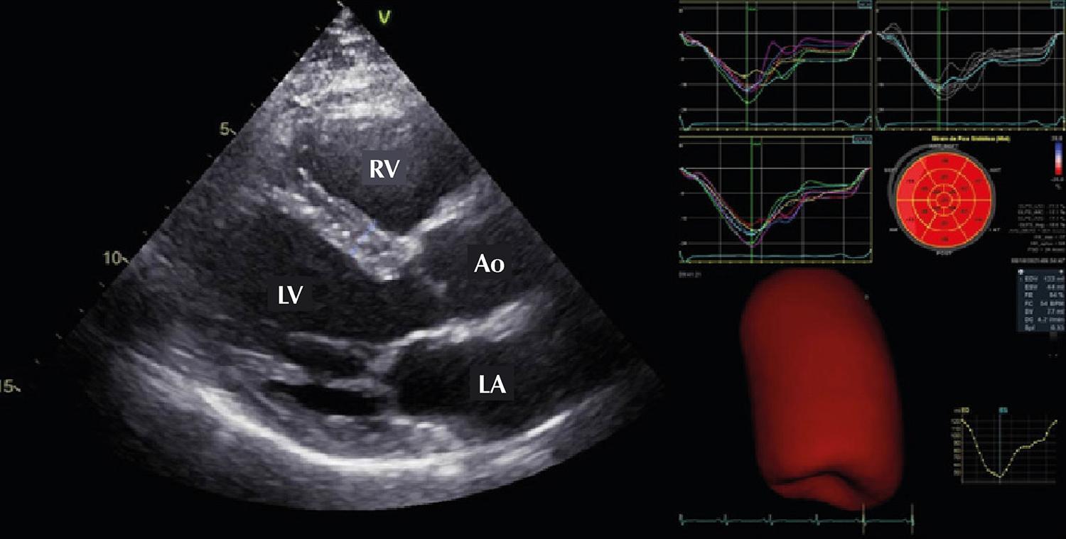
We included 44 participants (26 with aortic valve disease, 19 controls). GLS was determined using CVI42 software (Circle Cardiovascular Imaging), while the LLD linear measurements of the distance between the base/apex of the LV included maximum displacement (MD), maximum velocity in early diastole (MVED), atrioventricular junction velocity in diastasis (VDS), and VDS/MVED ratio.

Results

DM and MVED were correlated with GLS (r=0.69 and r=0.65, respectively) and LVEF (r=0.47 and r=0.57, p<0.001 for both). DM and MVED showed areas under the receiver operating characteristic curve (AUC) of 0.88 and 0.91, and at the best cut-off point (-0.13 and 0.66), sensitivities of 72.43% and 57.14% and specificities of 80.65% and 87.10%, respectively, compared to GLS. Using LVEF as a reference, we obtained AUC of 0.70 and 0.82, and at the best cut-off point (-0.11 and 0.61), sensitivities of 75.00% and 50.00% and specificities of 72.97% and 78.38%, respectively.

Conclusion

LLD demonstrated similar performance to that of GLS. MD derived from LLD was the best parameter during systole, while MVED was the best during diastole. Our findings demonstrate the routine, quick, and inexpensive assessment of diastolic function on CMR.

  • Aortic Valve Insufficiency
  • Aortic Valve Stenosis
  • Diastole
  • Magnetic Resonance
  • Myocardial Contraction
Read Full Article
Creative Commons Attribution (CC BY) Creative Commons license
30/06/2022 DOI: 10.47593/2675-312X/20223502eabc304 Views 2
Figure 1
What do Cardiologists Expect

What do Cardiologists Expect from the Echocardiogram in Heart Failure with Reduced Ejection Fraction?

Ciro Mancilha Murad et al. Fabiana Goulart Marcondes-Braga Danilo Bora Moleta

Introduction

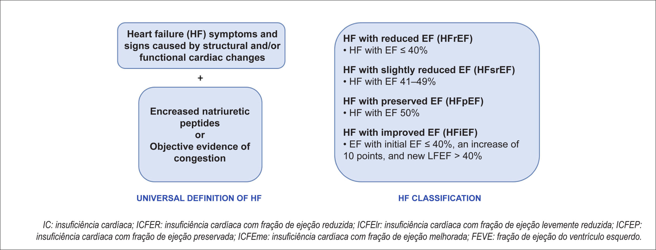
The echocardiogram plays a central role in assessments of patients with heart failure (HF), contributing to its classification, etiology definition, severity stratification, hemodynamic assessment, and clinical follow-up. HF has been historically divided according to the echocardiographic parameter left ventricular ejection fraction (LVEF). The most recent classification divided it into preserved (≥50%), reduced (≤40%), and slightly reduced LVEF (41–49%)., Different societies have recently proposed a universal definition of HF that involves the presence of symptoms and signs caused by structural and/or functional cardiac changes associated with increased natriuretic peptide levels or objective evidence of congestion. The authors also highlight the importance of classifying HF according to LVEF. () This article will describe the main parameters which the clinical cardiologist should pay attention on the echocardiogram of patients with HF with reduced ejection fraction (HFrEF) and how best to use them in clinical practice.

[…]

  • Echocardiography
  • Heart Failure
  • Stroke Volume
Read Full Article
Creative Commons Attribution (CC BY) Creative Commons license
13/06/2022 DOI: 10.47593/2675-312X/20223502ecard06 Views 2
Figure 1
Case Report

Dilated Cardimiopathy as Initial Presentation of Mucopolysacaridosis in Infant

Ludimila dos Anjos Teixeira Romão et al. Fátima Derlene da Rocha Araújo Camila Magalhães Silva Rodrigo Rezende Arantes Zilda Maria Alves Meira

Introduction

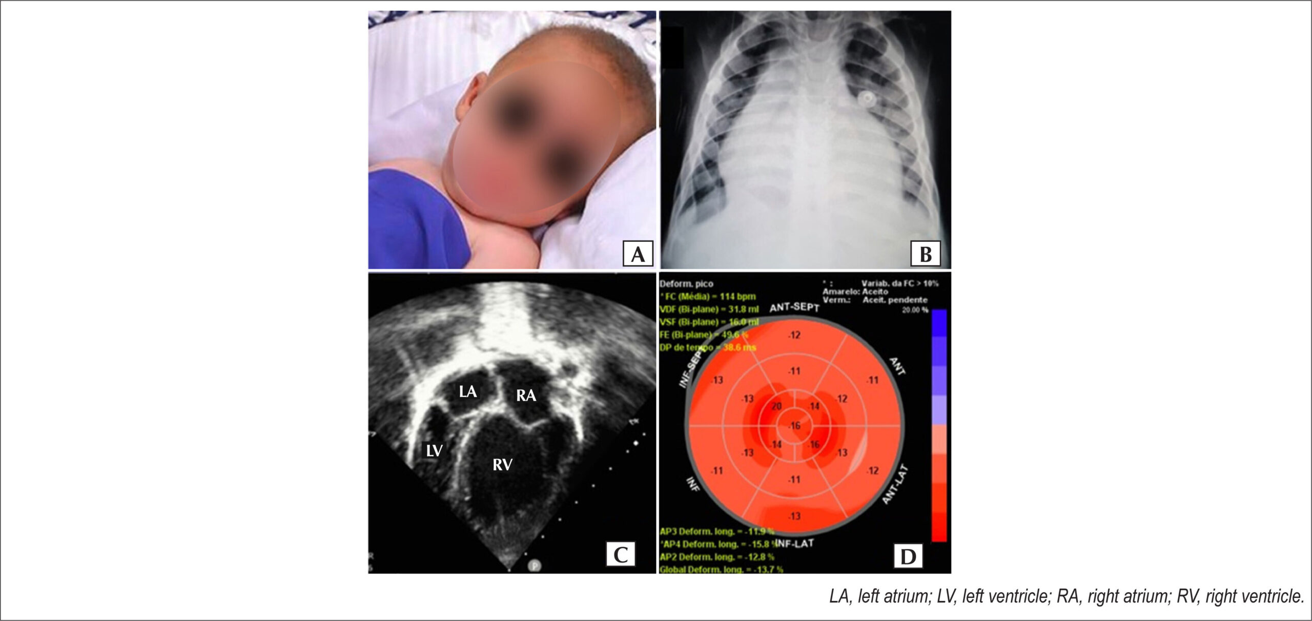
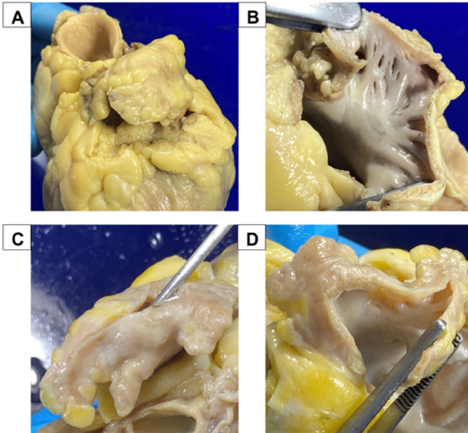
Mucopolysaccharidoses are rare genetic diseases caused by decreased lysosomal enzymes that act in the catabolism of glycosaminoglycans, connective tissue extracellular matrix polymers. Glycosaminoglycans progressively accumulate in the airways, mucous membranes, lungs, bones, joints, heart, liver, eyes, ears, and central and peripheral nervous systems. The multiple affected organs characterize its multisystemic presentation with a wide spectrum of severity and clinical presentations. The most severe phenotype is Hurler syndrome (type I mucopolysaccharidosis), with an incidence of 0.61–1.30 per 100,000 live births.

The heart is affected in more than 50% of mucopolysaccharidosis cases, with both valvular and myocardial involvement in addition to a possible association with systemic arterial hypertension (SAH).

[…]

  • cardiomyopathy
  • Child
  • Dilated
  • Mucopolysaccharidosis I
Read Full Article
Creative Commons Attribution (CC BY) Creative Commons license
13/06/2022 DOI: 10.47593/2675-312X/20223502eabc255 Views 2
Figure 1
Review Article

Assessing Myocardial Viability in Clinical Practice

Miriana Basso Gomes

Abstract

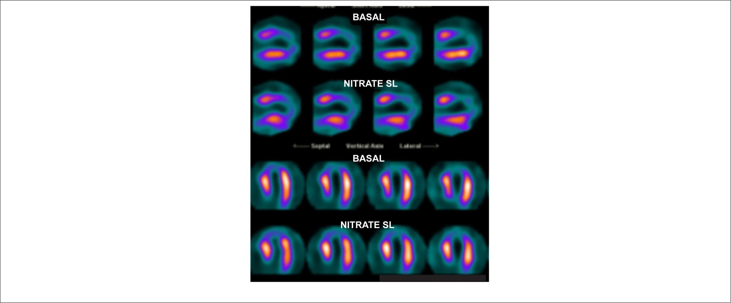
Although assessing myocardial viability is a common cardiology practice, many physicians question the results of diagnostic methods. Nuclear medicine plays an important role in viability studies, but the reports require interpretation in a clinical and pathophysiological context. this article was aimed at reviewing the origin and evolution of myocardial viability. Here we present diagnostic methods by emphasizing nuclear medicine and provide a functional explanation of each test type using example images. We also propose how to act in these cases based on clinic examination findings, the percentage of affected myocardium, and coronary lesion topography (proximal or distal).

  • Cardiology
  • Myocardial Infarction
  • Myocardial Viability
  • Nuclear Medicine
Read Full Article
Creative Commons Attribution (CC BY) Creative Commons license
13/06/2022 DOI: 10.47593/2675-312X/20223502eabc264 Views 2
Figure 1
My Approach To

My Approach to Imaging the Atrial Septal Defect by Three-Dimensional Echocardiography

Daniela Lago Kreuzig

Pre-procedural assessment

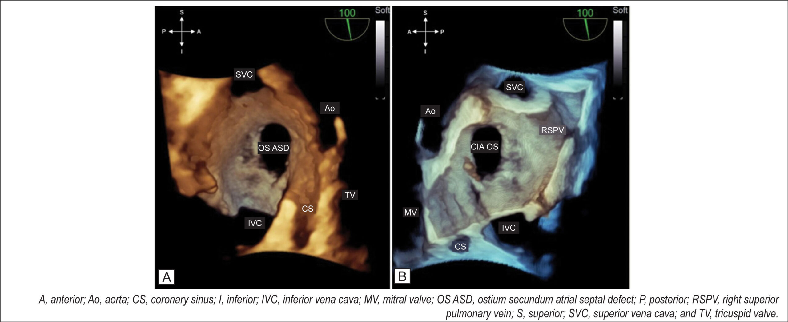
At the end of a detailed 2D TEE with a full septal analysis from the four-chamber view to bicaval section and color mapping, we commence the 3D image acquisition. The en-face projection of the ASD and the defect’s positioning in the anatomic position provide valuable information from a single image: size, shape, location in the interatrial septum, characteristics of the defect’s borders, and the presence of additional fenestration.

I usually start the 3D evaluation from the perspective of the right atrium to facilitate the identification of anatomical markers. I acquire a medium angle block (3D or 4D zoom) or a large block (full-volume using the two-section tool) starting from an initial bicaval section and proceeding with approximate 90° rotation in the z-plane so that the superior and inferior vena cava are aligned in the vertical plane (i.e., superior vena cava at the 11 o’clock position) and the aorta in the anterior and superior region. I prefer not to use the narrow-angle mode for acquisition (live 3D or bird’s eye view) or very high magnification due to a loss of anatomical references adjacent to the defect, which can interfere with this fundamental initial analysis. From this reconstruction (), I can characterize the defect as unique, the OS type, having an oval shape, being central, being medium-sized, and having adequate borders. shows the same ASD and allows assessment of the occurrence of dynamic variation in its dimension with the cardiac cycle.

[…]

  • atrial
  • Congenital
  • Echocardiography
  • Heart Defects
  • Heart septal defects
  • Three-Dimensional
Read Full Article
Creative Commons Attribution (CC BY) Creative Commons license
13/06/2022 DOI: 10.47593/2675-312X/20223502ecom32 Views 3
Figure 1
Original Article

Is Paclitaxel Less Cardiotoxic in the Treatment of Breast Cancer Before or After Doxorubicin?

André Luiz Cerqueira de Almeida et al. Edval Gomes dos Santos Júnior Aluísio José de Oliveira Monteiro Neto Thyago Monteiro do Espírito Santo … Paulo André A. Almeida Samuel Oliveira Affonseca Caroline de Souza Almeida Matheus Pamponet Freitas Cecília Lopes Viana Santos Vitória Régia Beserra Barbosa Ximenes Mariana Andrade Falcão Israel Costa Reis Suzane Pereira de Souza Maurício Gomes da Silva Serra Ana Beatriz Menezes de Oliveira Tayla Silva Santos Nilson Lima Lopes Marcelo Dantas Tavares de Melo

Abstract

Background:

The combination of doxorubicin (DOX) with paclitaxel (PTX) effectively treats breast cancer (BC). However, DOX-associated cardiotoxicity (CTX) is aggravated by the use of PTX. Consensus is lacking about which drug sequence involves the most CTX.

Objectives:

To evaluate whether DOX followed by PXT or the reverse sequence has the greatest cardiotoxic potential in the treatment of BC.

Methods:

Prospective study of women with primary BC who received four cycles of DOX and 12 infusions of PTX. Participants were divided into Group 1 (G1; PXT before DOX) and Group 2 (G2; DOX before PXT) at the discretion of the oncologist. CTX was defined as an absolute reduction in left ventricular ejection fraction (LVEF) > 10% to a value <53%. Patients underwent clinical evaluations and echocardiography before treatment (Phase 1) and one year after treatment (Phase 2).

Results:

Sixty-nine women were evaluated: 19 in G1 and 50 in G2. The groups had similar clinical characteristics. The doses of radiation, DOX, and PTX used were similar. Eight (11.6%) patients developed CTX: two (10.5%) in G1 and six (12.0%) in G2 (p=0.62). The mean LVEF was similar between groups in Phase 1 (G1=65.1±3.5%; G2=65.2±3.9%; p=0.96), with a significant reduction noted after one year in both groups: G1=61.4±8.1% (p=0.021) and G2=60.8±7.6% (p<0.001). Although lower, mean LVEF remained similar between groups after Phase 2 (p=0.79).

Conclusions:

In women with BC who underwent chemotherapy, the incidence of CTX at the end of the first year of treatment was similar regardless of whether DOX was used before or after PTX.

  • Breast neoplasm
  • Cardiotoxicity
  • Doxorubicin
  • Paclitaxel
Read Full Article
Creative Commons Attribution (CC BY) Creative Commons license
13/06/2022 DOI: 10.47593/2675-312X/20223502eabc289 Views 3
Figure 1
Case Report

Late Presentation of COVID-19-Associated Transmural Myocardial Infarction with Non-Obstructive Coronary Atherosclerosis

Jaime Paula Pessoa Linhares Filho et al. Matheus Cardoso de Aragão João Bosco Breckenfeld Bastos Filho Ricardo Paulo de Souza Rocha … Fábio Holanda Lacerda Francisco de Assis Carvalho de Santana

Introduction

There has been an increased incidence of coronavirus-associated thromboembolic events during the coronavirus disease 2019 (COVID-19) pandemic. However, most cases presented with venous thrombotic events, mainly deep vein thrombosis and pulmonary thromboembolism, whereas arterial and coronary phenomena are rare.

Several pathophysiological factors can explain the relationship between COVID-19 infection and increased embolic phenomena. The main factors are increased inflammatory mediators, the release of prothrombotic factors, and endothelial dysfunction.,

[…]

  • Biomarkers
  • Coronavirus
  • COVID-19
  • Myocardial Infarction
  • Myocarditis
Read Full Article
Creative Commons Attribution (CC BY) Creative Commons license
13/06/2022 DOI: 10.47593/2675-312X/20223502eabc290 Views 3
Figure 1
Review Article

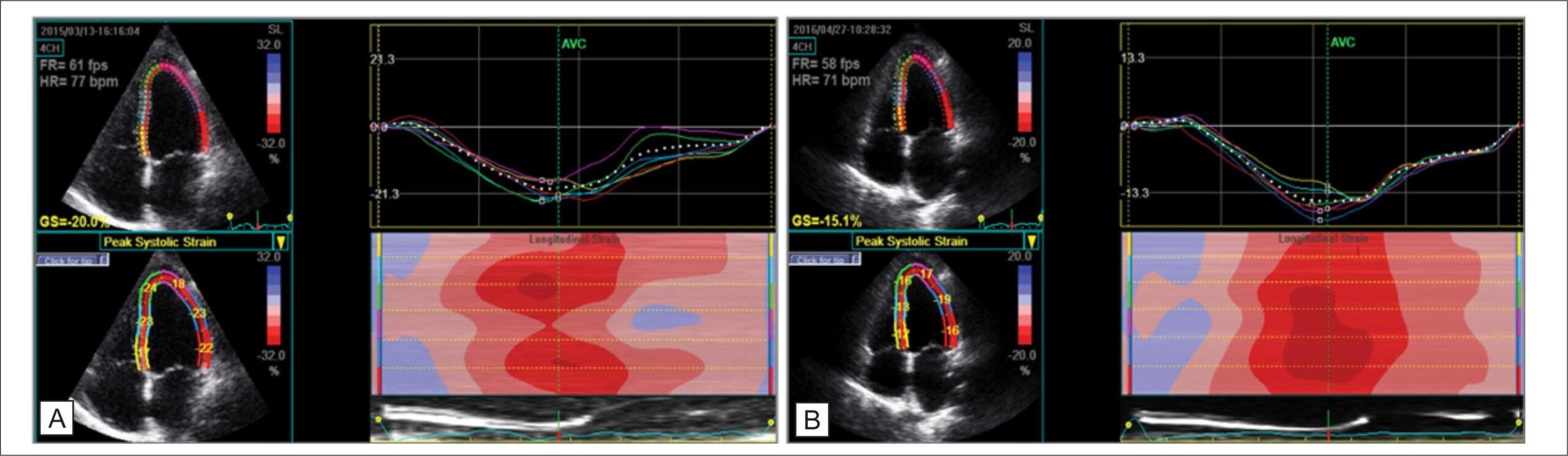
Role of Strain in the Early Diagnosis of Diabetic Cardiomyopathy

Thais Rossoni Weber da Silva et al. Roberto Léo da Silva Adriana Ferraz Martins Jefferson Luiz Brum Marques

Abstract

Background

Advanced echocardiography using two- and three-dimensional myocardial strain proposes to identify subclinical systolic dysfunction in different clinical conditions. Strain assessment plays an important role in the early diagnosis of diabetic cardiomyopathy in diabetes mellitus (DM). However, the findings of published articles are heterogeneous. Here we conducted a systematic review to analyze the current role of strain assessment in patients with DM.

Methods

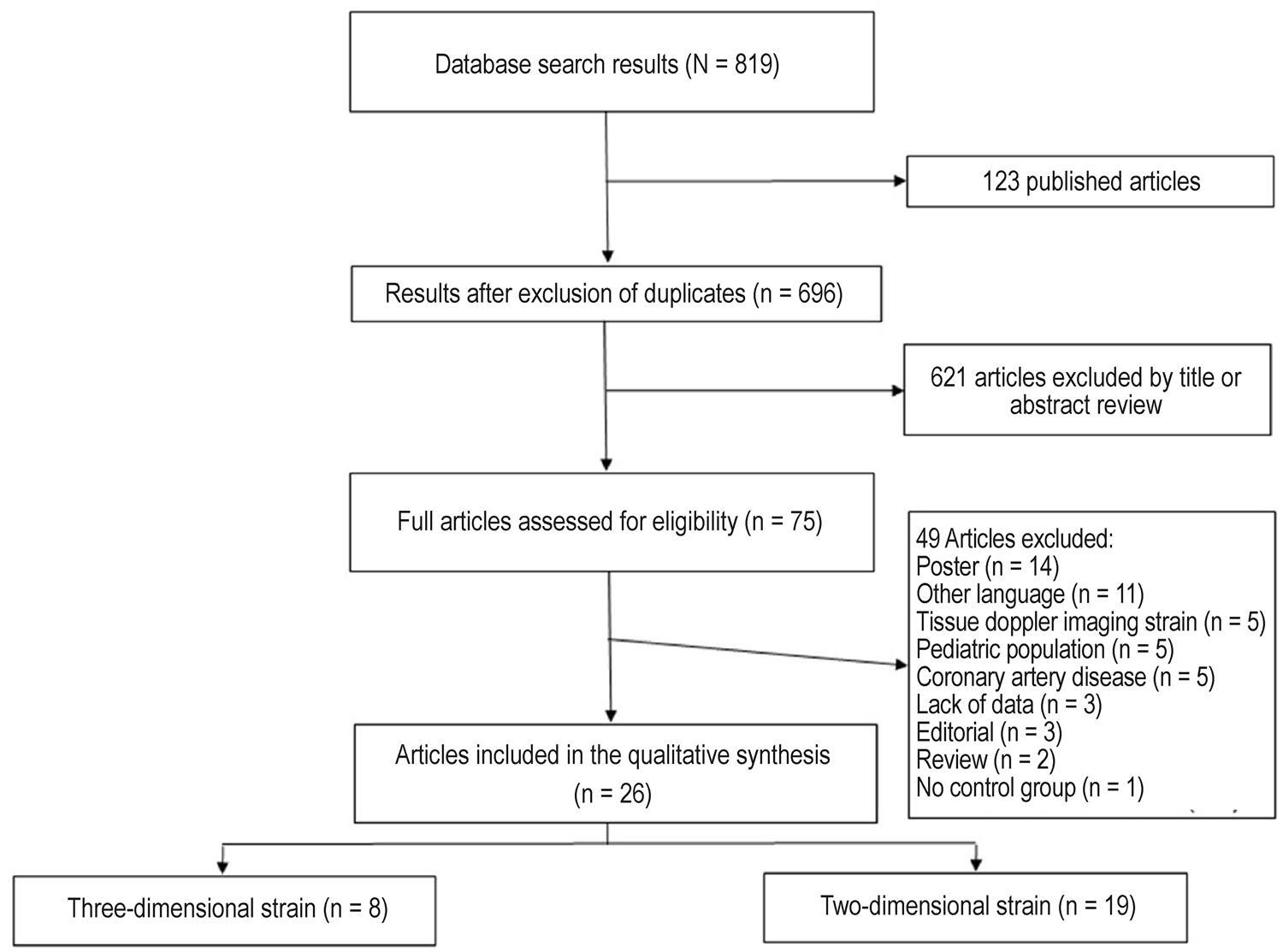
This systematic review of five databases identified 19 studies that used two-dimensional strain and 8 studies that used three-dimensional strain.

Results

The studies of two-dimensional strain included 1,774 DM patients (mean age, 57.1 years; median age, 55 years; 47.5% women), while those of three-dimensional strain included 488 DM patients (mean age, 55.7 years; median age, 63 years; 51% women). Global longitudinal strain was the myocardial deformation marker that differed most frequently between the DM and control groups.

Conclusion

Myocardial strain imaging by two- and three-dimensional speckle tracking echocardiography allows the identification of subclinical systolic dysfunction in DM patients, and differences become more marked when associated with risk factors and ventricular remodeling.

  • Diabetes Mellitus
  • Echocardiography
  • Ventricular Function
Read Full Article
Creative Commons Attribution (CC BY) Creative Commons license
28/05/2022 DOI: 10.47593/2675-312X/20223502eabc293 Views 3
Figure 1
Case Report

Primary Cardiac Lymphoma: Role of Imaging Multimodality in Diagnosis

Julia Werneck Paulino Soares de Souza et al. Luiza Telles de Andrade Alvares Maria Paula Righeti Gonçalves Ana Beatriz Aisemann Goulart Paiva … Ricardo Lopes Ferreira Marcelo Goulart Paiva

Abstract

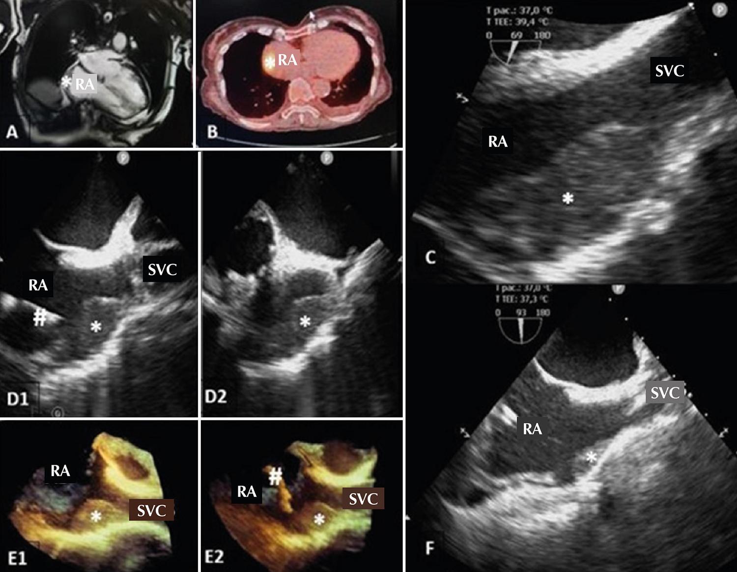
Intracardiac masses are a diagnostic challenge since their symptoms can be common to cardiovascular pathologies. Some methods, whether invasive or not, enable differential diagnosis, histological confirmation, and adequate treatment. To better understand the importance of imaging multimodality and the approach to managing cardiac tumors, we investigated a case of a primary cardiac lymphoma in which the multidisciplinary approach allowed rapid diagnosis and treatment, including of intercurrences, with a promising initial response despite fatal progression due to severe acute respiratory syndrome coronavirus 2 infection.

  • Cardiovascular Diseases
  • Echocardiography
  • Lymphoma
Read Full Article
Creative Commons Attribution (CC BY) Creative Commons license
28/05/2022 DOI: 10.47593/2675-312X/20223502eabc286 Views 3
Figure 1
My Approach To

My Approach to Echocardiography in Radiofrequency Ablation of Hypertrophic Obstructive Cardiomyopathy

Antonio Tito Paladino et al. Andrea de Andrade Vilela Jorge Eduardo Assef Bruno Pereira Valdigem

Introduction

Hypertrophic cardiomyopathy (HCM), an autosomal dominant genetic disease that affects one in 500 people, is considered the most common isolated form of hereditary heart disease. HCM is characterized by varying degrees of ventricular hypertrophy, myocardial fiber disarray, interstitial fibrosis, and microvascular disease in the absence of cardiac or systemic conditions to justify these findings.

Echocardiography, an invaluable tool for the diagnosis and follow-up of patients with HCM, is used to assess morphology, hemodynamic disorders, left ventricular (LV) function, and patient prognosis. It also has a fundamental role in cases of hypertrophic obstructive cardiomyopathy (HOCM) in the indication of invasive treatments. Radiofrequency ablation (RFA) is a possible treatment under study.

[…]

  • Ablation
  • Cardiac Hypertrophy
  • Echocardiography
  • Radio Frequency
Read Full Article
Creative Commons Attribution (CC BY) Creative Commons license
27/05/2022 DOI: 10.47593/2675-312X/20223502ecom28 Views 2
Figure 1
Case Report

Diagnosis of Coronary Anomaly of the Circumflex Artery With Retroaortic Course by Transthoracic Echocardiography (Rac Sign) Confirmed by Computed Tomography Coronary Angiography: A Case Series

Raul Serra Valério et al. Alfredo A. Eyer Rodrigues Carlos Eduardo Suaide Silva Marly Uellendahl Luciano de Figueiredo Aguiar Filho

Introduction

Congenital coronary artery anomaly (CCAA) is a relatively rare condition with an estimated population prevalence of 0.21–5.79%. CCAA presents a wide spectrum of variations and can be classified into origin and course, intrinsic, and termination anomalies. , The CCAA with the highest prevalence is the anomalous origin of the circumflex artery (CxA) from the right coronary sinus (RCS) with an estimated prevalence of 0.3–0.8%.

CCAA usually travels along a retroaortic course and may present different originations, such as from a common ostium with the right coronary artery (RCA), from separate ostia in the RCS, or from the proximal segment of the RCA.

[…]

  • Cardiovascular Abnormalities
  • Computed Tomography Angiography
  • Echocardiography
Read Full Article
Creative Commons Attribution (CC BY) Creative Commons license
27/05/2022 DOI: 10.47593/2675-312X/20223502eabc271 Views 5
My Approach to Transseptal Mitral Valve-In-Valve
My Approach To

My Approach to Transseptal Mitral Valve-In-Valve

Andrea de Andrade Vilela et al. Antonio Tito Paladino Filho Maria Estefânia Bosco Otto Vanessa Andreoli Esmanhoto

Introduction

The percutaneous treatment of heart valve disease is a subject of great interest in cardiology. Percutaneous aortic valve implantation, first performed in 2002 by Alan Cribier, paved the way for transcatheter treatment of the mitral valve.

The first valve-in-valve (VIV) procedures were performed to treat deteriorated bioprosthetic valves in the aortic position. , In 2009, the group led by Dr. John Webb published the first successful case of mitral VIV. Since then, mitral VIV has been performed in numerous centers worldwide.

[…]

  • Deart por Heart
  • Transthoracic Echocardiography
  • Treatment
Read Full Article
Creative Commons Attribution (CC BY) Creative Commons license
27/05/2022 DOI: 10.47593/2675-312X/20223502ecom24 Views 2

How to cite this article

EndNote RIS

Electronic Document Format (Vancouver)

Andrade SM, Barretto RBM, Sousa FTT, Bihan DCSL. Left Atrial Function Analysis in Patients with Functional Mitral Regurgitation Associated with Dilated Cardiomyopathy. ABC Imagem Cardiovascular 2022;35(2):eabc281.

Electronic Document Format (ABNT)

Andrade, Stephanie Macedo; Barretto, Rodrigo Bellio de Mattos; Sousa, Francisco Thiago Tomaz de; Bihan, David Costa de Souza Le. Left Atrial Function Analysis in Patients with Functional Mitral Regurgitation Associated with Dilated Cardiomyopathy. ABC Imagem Cardiovascular, v. 35, n. 2, eabc281, Jun. 2022.

Electronic Document Format (APA)

Andrade, S. M., Barretto, R. B. M., Sousa, F. T. T. , & Bihan, D. C. S. L. (2022). Left Atrial Function Analysis in Patients with Functional Mitral Regurgitation Associated with Dilated Cardiomyopathy. ABC Imagem Cardiovascular, 35(2), eabc281.

Electronic Document Format (ISO)

Andrade, Stephanie Macedo and Barretto, Rodrigo Bellio de Mattos and Sousa, Francisco Thiago Tomaz de and Bihan, David Costa de Souza Le. Left Atrial Function Analysis in Patients with Functional Mitral Regurgitation Associated with Dilated Cardiomyopathy. ABC Imagem Cardiovascular [online]. 2022, vol. 35, n. 2, [cited 2026-10-14], eabc281. Available from: <https://journals.cardiol.br/en/article/left-atrial-function-analysis-in-patients-with-functional-mitral-regurgitation-associated-with-dilated-cardiomyopathy/>. ISSN 2318-8219

EndNote RIS

Electronic Document Format (Vancouver)

Silva TM, Oliveira GB, Barros TLS, Andrade AB, Furlan BB, Nunes MCP. Left Atrial Appendage: Anatomy, Function, and Importance in Thrombus Formation. ABC Imagem Cardiovascular 2022;35(2):eabc301.

Electronic Document Format (ABNT)

Silva, Taylla Mendes; Oliveira, Gustavo Brandão de; Barros, Thais Lins de Souza; Andrade, André Barbosa de; Furlan, Bruno Bom; Nunes, Maria do Carmo de Pereira. Left Atrial Appendage: Anatomy, Function, and Importance in Thrombus Formation. ABC Imagem Cardiovascular, v. 35, n. 2, eabc301, Jun. 2022.

Electronic Document Format (APA)

Silva, T. M., Oliveira, G. B. , Barros, T. L. S., Andrade, A. B. , Furlan, B. B., & Nunes, M. C. P. (2022). Left Atrial Appendage: Anatomy, Function, and Importance in Thrombus Formation. ABC Imagem Cardiovascular, 35(2), eabc301.

Electronic Document Format (ISO)

Silva, Taylla Mendes and Oliveira, Gustavo Brandão de and Barros, Thais Lins de Souza and Andrade, André Barbosa de and Furlan, Bruno Bom and Nunes, Maria do Carmo de Pereira. Left Atrial Appendage: Anatomy, Function, and Importance in Thrombus Formation. ABC Imagem Cardiovascular [online]. 2022, vol. 35, n. 2, [cited 2026-10-14], eabc301. Available from: <https://journals.cardiol.br/en/article/left-atrial-appendage-anatomy-function-and-importance-in-thrombus-formation/>. ISSN 2318-8219

EndNote RIS

Electronic Document Format (Vancouver)

Agrawal A, Sengupta SP. Infective Endocarditis In India: A Broad Picture Of A Very Large Country. ABC Imagem Cardiovascular 2022;35(2):eed_15.

Electronic Document Format (ABNT)

Agrawal, Ashish; Sengupta, Shantanu P. Infective Endocarditis In India: A Broad Picture Of A Very Large Country. ABC Imagem Cardiovascular, v. 35, n. 2, eed_15, Jun. 2022.

Electronic Document Format (APA)

Agrawal, A., & Sengupta, S. P. (2022). Infective Endocarditis In India: A Broad Picture Of A Very Large Country. ABC Imagem Cardiovascular, 35(2), eed_15.

Electronic Document Format (ISO)

Agrawal, Ashish and Sengupta, Shantanu P. Infective Endocarditis In India: A Broad Picture Of A Very Large Country. ABC Imagem Cardiovascular [online]. 2022, vol. 35, n. 2, [cited 2026-10-14], eed_15. Available from: <https://journals.cardiol.br/en/article/infective-endocarditis-in-india-a-broad-picture-of-a-very-large-country/>. ISSN 2318-8219

EndNote RIS

Electronic Document Format (Vancouver)

Fonseca RA, Ribeiro SM, Azevedo Filho CF, Sampaio R, Tarasoutchi F, Rochitte CE. Left Ventricular Longitudinal Linear Displacement Versus Global Longitudinal Strain on Cardiovascular Magnetic Resonance. ABC Imagem Cardiovascular 2022;35(2):eabc304.

Electronic Document Format (ABNT)

Fonseca, Rafael Almeida; Ribeiro, Sergio Marrone; Azevedo Filho, Clerio Francisco de; Sampaio, Roney; Tarasoutchi, Flávio; Rochitte, Carlos Eduardo. Left Ventricular Longitudinal Linear Displacement Versus Global Longitudinal Strain on Cardiovascular Magnetic Resonance. ABC Imagem Cardiovascular, v. 35, n. 2, eabc304, Jun. 2022.

Electronic Document Format (APA)

Fonseca, R. A., Ribeiro, S. M., Azevedo Filho, C. F. , Sampaio, R., Tarasoutchi, F., & Rochitte, C. E. (2022). Left Ventricular Longitudinal Linear Displacement Versus Global Longitudinal Strain on Cardiovascular Magnetic Resonance. ABC Imagem Cardiovascular, 35(2), eabc304.

Electronic Document Format (ISO)

Fonseca, Rafael Almeida and Ribeiro, Sergio Marrone and Azevedo Filho, Clerio Francisco de and Sampaio, Roney and Tarasoutchi, Flávio and Rochitte, Carlos Eduardo. Left Ventricular Longitudinal Linear Displacement Versus Global Longitudinal Strain on Cardiovascular Magnetic Resonance. ABC Imagem Cardiovascular [online]. 2022, vol. 35, n. 2, [cited 2026-10-14], eabc304. Available from: <https://journals.cardiol.br/en/article/left-ventricular-longitudinal-linear-displacement-versus-global-longitudinal-strain-on-cardiovascular-magnetic-resonance/>. ISSN 2318-8219

EndNote RIS

Electronic Document Format (Vancouver)

Murad CM, Marcondes-Braga FG, Moleta DB. What do Cardiologists Expect from the Echocardiogram in Heart Failure with Reduced Ejection Fraction?. ABC Imagem Cardiovascular 2022;35(2):ecard06.

Electronic Document Format (ABNT)

Murad, Ciro Mancilha; Marcondes-Braga, Fabiana Goulart; Moleta, Danilo Bora. What do Cardiologists Expect from the Echocardiogram in Heart Failure with Reduced Ejection Fraction?. ABC Imagem Cardiovascular, v. 35, n. 2, ecard06, Jun. 2022.

Electronic Document Format (APA)

Murad, C. M., Marcondes-Braga, F. G., & Moleta, D. B. (2022). What do Cardiologists Expect from the Echocardiogram in Heart Failure with Reduced Ejection Fraction?. ABC Imagem Cardiovascular, 35(2), ecard06.

Electronic Document Format (ISO)

Murad, Ciro Mancilha and Marcondes-Braga, Fabiana Goulart and Moleta, Danilo Bora. What do Cardiologists Expect from the Echocardiogram in Heart Failure with Reduced Ejection Fraction?. ABC Imagem Cardiovascular [online]. 2022, vol. 35, n. 2, [cited 2026-10-14], ecard06. Available from: <https://journals.cardiol.br/en/article/what-do-cardiologists-expect-from-the-echocardiogram-in-heart-failure-with-reduced-ejection-fraction/>. ISSN 2318-8219

EndNote RIS

Electronic Document Format (Vancouver)

Romão LAT, Araújo FDR, Silva CM, Arantes RR, Meira ZMA. Dilated Cardimiopathy as Initial Presentation of Mucopolysacaridosis in Infant. ABC Imagem Cardiovascular 2022;35(2):eabc255.

Electronic Document Format (ABNT)

Romão, Ludimila dos Anjos Teixeira; Araújo, Fátima Derlene da Rocha; Silva, Camila Magalhães; Arantes, Rodrigo Rezende; Meira, Zilda Maria Alves. Dilated Cardimiopathy as Initial Presentation of Mucopolysacaridosis in Infant. ABC Imagem Cardiovascular, v. 35, n. 2, eabc255, Jun. 2022.

Electronic Document Format (APA)

Romão, L. A. T., Araújo, F. D. R., Silva, C. M., Arantes, R. R., & Meira, Z. M. A. (2022). Dilated Cardimiopathy as Initial Presentation of Mucopolysacaridosis in Infant. ABC Imagem Cardiovascular, 35(2), eabc255.

Electronic Document Format (ISO)

Romão, Ludimila dos Anjos Teixeira and Araújo, Fátima Derlene da Rocha and Silva, Camila Magalhães and Arantes, Rodrigo Rezende and Meira, Zilda Maria Alves. Dilated Cardimiopathy as Initial Presentation of Mucopolysacaridosis in Infant. ABC Imagem Cardiovascular [online]. 2022, vol. 35, n. 2, [cited 2026-10-14], eabc255. Available from: <https://journals.cardiol.br/en/article/dilated-cardimiopathy-as-initial-presentation-of-mucopolysacaridosis-in-infant/>. ISSN 2318-8219

EndNote RIS

Electronic Document Format (Vancouver)

Gomes MB. Assessing Myocardial Viability in Clinical Practice. ABC Imagem Cardiovascular 2022;35(2):eabc264.

Electronic Document Format (ABNT)

Gomes, Miriana Basso. Assessing Myocardial Viability in Clinical Practice. ABC Imagem Cardiovascular, v. 35, n. 2, eabc264, Jun. 2022.

Electronic Document Format (APA)

Gomes, M. B. (2022). Assessing Myocardial Viability in Clinical Practice. ABC Imagem Cardiovascular, 35(2), eabc264.

Electronic Document Format (ISO)

Gomes, Miriana Basso. Assessing Myocardial Viability in Clinical Practice. ABC Imagem Cardiovascular [online]. 2022, vol. 35, n. 2, [cited 2026-10-14], eabc264. Available from: <https://journals.cardiol.br/en/article/assessing-myocardial-viability-in-clinical-practice/>. ISSN 2318-8219

EndNote RIS

Electronic Document Format (Vancouver)

Kreuzig DL. My Approach to Imaging the Atrial Septal Defect by Three-Dimensional Echocardiography. ABC Imagem Cardiovascular 2022;35(2):ecom32.

Electronic Document Format (ABNT)

Kreuzig, Daniela Lago. My Approach to Imaging the Atrial Septal Defect by Three-Dimensional Echocardiography. ABC Imagem Cardiovascular, v. 35, n. 2, ecom32, Jun. 2022.

Electronic Document Format (APA)

Kreuzig, D. L. (2022). My Approach to Imaging the Atrial Septal Defect by Three-Dimensional Echocardiography. ABC Imagem Cardiovascular, 35(2), ecom32.

Electronic Document Format (ISO)

Kreuzig, Daniela Lago. My Approach to Imaging the Atrial Septal Defect by Three-Dimensional Echocardiography. ABC Imagem Cardiovascular [online]. 2022, vol. 35, n. 2, [cited 2026-10-14], ecom32. Available from: <https://journals.cardiol.br/en/article/my-approach-to-imaging-the-atrial-septal-defect-by-three-dimensional-echocardiography/>. ISSN 2318-8219

EndNote RIS

Electronic Document Format (Vancouver)

Almeida ALC, Santos Júnior EG, Monteiro Neto AJO, Santo TME, Almeida PAA, Affonseca SO, Almeida CS, et al. Is Paclitaxel Less Cardiotoxic in the Treatment of Breast Cancer Before or After Doxorubicin?. ABC Imagem Cardiovascular 2022;35(2):eabc289.

Electronic Document Format (ABNT)

Almeida, André Luiz Cerqueira de; Santos Júnior, Edval Gomes dos; Monteiro Neto, Aluísio José de Oliveira; Santo, Thyago Monteiro do Espírito; Almeida, Paulo André A.; Affonseca, Samuel Oliveira; Almeida, Caroline de Souza; Freitas, Matheus Pamponet; Santos, Cecília Lopes Viana; Ximenes, Vitória Régia Beserra Barbosa; Falcão, Mariana Andrade; Reis, Israel Costa; Souza, Suzane Pereira de; Serra, Maurício Gomes da Silva; Oliveira, Ana Beatriz Menezes de; Santos, Tayla Silva; Lopes, Nilson Lima; Melo, Marcelo Dantas Tavares de. Is Paclitaxel Less Cardiotoxic in the Treatment of Breast Cancer Before or After Doxorubicin?. ABC Imagem Cardiovascular, v. 35, n. 2, eabc289, Jun. 2022.

Electronic Document Format (APA)

Almeida, A. L. C. , Santos Júnior, E. G. , Monteiro Neto, A. J. O., Santo, T. M. E., Almeida, P. A. A., Affonseca, S. O., Almeida, C. S., Freitas, M. P., Santos, C. L. V., Ximenes, V. R. B. B., Falcão, M. A., Reis, I. C., Souza, S. P. , Serra, M. G. S., Oliveira, A. B. M. , Santos, T. S., Lopes, N. L., & Melo, M. D. T. (2022). Is Paclitaxel Less Cardiotoxic in the Treatment of Breast Cancer Before or After Doxorubicin?. ABC Imagem Cardiovascular, 35(2), eabc289.

Electronic Document Format (ISO)

Almeida, André Luiz Cerqueira de and Santos Júnior, Edval Gomes dos and Monteiro Neto, Aluísio José de Oliveira and Santo, Thyago Monteiro do Espírito and Almeida, Paulo André A. and Affonseca, Samuel Oliveira and Almeida, Caroline de Souza and Freitas, Matheus Pamponet and Santos, Cecília Lopes Viana and Ximenes, Vitória Régia Beserra Barbosa and Falcão, Mariana Andrade and Reis, Israel Costa and Souza, Suzane Pereira de and Serra, Maurício Gomes da Silva and Oliveira, Ana Beatriz Menezes de and Santos, Tayla Silva and Lopes, Nilson Lima and Melo, Marcelo Dantas Tavares de. Is Paclitaxel Less Cardiotoxic in the Treatment of Breast Cancer Before or After Doxorubicin?. ABC Imagem Cardiovascular [online]. 2022, vol. 35, n. 2, [cited 2026-10-14], eabc289. Available from: <https://journals.cardiol.br/en/article/is-paclitaxel-less-cardiotoxic-in-the-treatment-of-breast-cancer-before-or-after-doxorubicin/>. ISSN 2318-8219

EndNote RIS

Electronic Document Format (Vancouver)

Linhares Filho JPP, Aragão MC, Bastos Filho JBB, Rocha RPS, Lacerda FH, Santana FAC. Late Presentation of COVID-19-Associated Transmural Myocardial Infarction with Non-Obstructive Coronary Atherosclerosis. ABC Imagem Cardiovascular 2022;35(2):eabc290.

Electronic Document Format (ABNT)

Linhares Filho, Jaime Paula Pessoa; Aragão, Matheus Cardoso de; Bastos Filho, João Bosco Breckenfeld; Rocha, Ricardo Paulo de Souza; Lacerda, Fábio Holanda; Santana, Francisco de Assis Carvalho de. Late Presentation of COVID-19-Associated Transmural Myocardial Infarction with Non-Obstructive Coronary Atherosclerosis. ABC Imagem Cardiovascular, v. 35, n. 2, eabc290, Jun. 2022.

Electronic Document Format (APA)

Linhares Filho, J. P. P., Aragão, M. C. , Bastos Filho, J. B. B., Rocha, R. P. S., Lacerda, F. H., & Santana, F. A. C. (2022). Late Presentation of COVID-19-Associated Transmural Myocardial Infarction with Non-Obstructive Coronary Atherosclerosis. ABC Imagem Cardiovascular, 35(2), eabc290.

Electronic Document Format (ISO)

Linhares Filho, Jaime Paula Pessoa and Aragão, Matheus Cardoso de and Bastos Filho, João Bosco Breckenfeld and Rocha, Ricardo Paulo de Souza and Lacerda, Fábio Holanda and Santana, Francisco de Assis Carvalho de. Late Presentation of COVID-19-Associated Transmural Myocardial Infarction with Non-Obstructive Coronary Atherosclerosis. ABC Imagem Cardiovascular [online]. 2022, vol. 35, n. 2, [cited 2026-10-14], eabc290. Available from: <https://journals.cardiol.br/en/article/late-presentation-of-covid-19-associated-transmural-myocardial-infarction-with-non-obstructive-coronary-atherosclerosis/>. ISSN 2318-8219

EndNote RIS

Electronic Document Format (Vancouver)

Silva TRW, Silva RL, Martins AF, Marques JLB. Role of Strain in the Early Diagnosis of Diabetic Cardiomyopathy. Arquivos Brasileiros de Cardiologia Imagem cardiovascular 2022;35(2):eabc293.

Electronic Document Format (ABNT)

Silva, Thais Rossoni Weber da; Silva, Roberto Léo da; Martins, Adriana Ferraz; Marques, Jefferson Luiz Brum. Role of Strain in the Early Diagnosis of Diabetic Cardiomyopathy. Arquivos Brasileiros de Cardiologia Imagem cardiovascular, v. 35, n. 2, eabc293, May. 2022.

Electronic Document Format (APA)

Silva, T. R. W. , Silva, R. L. , Martins, A. F., & Marques, J. L. B. (2022). Role of Strain in the Early Diagnosis of Diabetic Cardiomyopathy. Arquivos Brasileiros de Cardiologia Imagem cardiovascular, 35(2), eabc293.

Electronic Document Format (ISO)

Silva, Thais Rossoni Weber da and Silva, Roberto Léo da and Martins, Adriana Ferraz and Marques, Jefferson Luiz Brum. Role of Strain in the Early Diagnosis of Diabetic Cardiomyopathy. Arquivos Brasileiros de Cardiologia Imagem cardiovascular [online]. 2022, vol. 35, n. 2, [cited 2026-10-14], eabc293. Available from: <https://journals.cardiol.br/en/article/role-of-strain-in-the-early-diagnosis-of-diabetic-cardiomyopathy/>. ISSN 2318-8219

EndNote RIS

Electronic Document Format (Vancouver)

Souza JWPS, Alvares LTA, Gonçalves MPR, Paiva ABAG, Ferreira RL, Paiva MG. Primary Cardiac Lymphoma: Role of Imaging Multimodality in Diagnosis. Arquivos Brasileiros de Cardiologia Imagem cardiovascular 2022;35(2):eabc286.

Electronic Document Format (ABNT)

Souza, Julia Werneck Paulino Soares de; Alvares, Luiza Telles de Andrade; Gonçalves, Maria Paula Righeti; Paiva, Ana Beatriz Aisemann Goulart; Ferreira, Ricardo Lopes; Paiva, Marcelo Goulart. Primary Cardiac Lymphoma: Role of Imaging Multimodality in Diagnosis. Arquivos Brasileiros de Cardiologia Imagem cardiovascular, v. 35, n. 2, eabc286, May. 2022.

Electronic Document Format (APA)

Souza, J. W. P. S. , Alvares, L. T. A., Gonçalves, M. P. R., Paiva, A. B. A. G., Ferreira, R. L., & Paiva, M. G. (2022). Primary Cardiac Lymphoma: Role of Imaging Multimodality in Diagnosis. Arquivos Brasileiros de Cardiologia Imagem cardiovascular, 35(2), eabc286.

Electronic Document Format (ISO)

Souza, Julia Werneck Paulino Soares de and Alvares, Luiza Telles de Andrade and Gonçalves, Maria Paula Righeti and Paiva, Ana Beatriz Aisemann Goulart and Ferreira, Ricardo Lopes and Paiva, Marcelo Goulart. Primary Cardiac Lymphoma: Role of Imaging Multimodality in Diagnosis. Arquivos Brasileiros de Cardiologia Imagem cardiovascular [online]. 2022, vol. 35, n. 2, [cited 2026-10-14], eabc286. Available from: <https://journals.cardiol.br/en/article/primary-cardiac-lymphoma-role-of-imaging-multimodality-in-diagnosis/>. ISSN 2318-8219

EndNote RIS

Electronic Document Format (Vancouver)

Paladino AT, Vilela AA, Assef JE, Valdigem BP. My Approach to Echocardiography in Radiofrequency Ablation of Hypertrophic Obstructive Cardiomyopathy. Arquivos Brasileiros de Cardiologia Imagem cardiovascular 2022;35(2):ecom28.

Electronic Document Format (ABNT)

Paladino, Antonio Tito; Vilela, Andrea de Andrade; Assef, Jorge Eduardo; Valdigem, Bruno Pereira. My Approach to Echocardiography in Radiofrequency Ablation of Hypertrophic Obstructive Cardiomyopathy. Arquivos Brasileiros de Cardiologia Imagem cardiovascular, v. 35, n. 2, ecom28, May. 2022.

Electronic Document Format (APA)

Paladino, A. T., Vilela, A. A., Assef, J. E., & Valdigem, B. P. (2022). My Approach to Echocardiography in Radiofrequency Ablation of Hypertrophic Obstructive Cardiomyopathy. Arquivos Brasileiros de Cardiologia Imagem cardiovascular, 35(2), ecom28.

Electronic Document Format (ISO)

Paladino, Antonio Tito and Vilela, Andrea de Andrade and Assef, Jorge Eduardo and Valdigem, Bruno Pereira. My Approach to Echocardiography in Radiofrequency Ablation of Hypertrophic Obstructive Cardiomyopathy. Arquivos Brasileiros de Cardiologia Imagem cardiovascular [online]. 2022, vol. 35, n. 2, [cited 2026-10-14], ecom28. Available from: <https://journals.cardiol.br/en/article/my-approach-to-echocardiography-in-radiofrequency-ablation-of-hypertrophic-obstructive-cardiomyopathy/>. ISSN 2318-8219

EndNote RIS

Electronic Document Format (Vancouver)

Valério RS, Rodrigues AAE, Silva CES, Uellendahl M, Aguiar Filho LF. Diagnosis of Coronary Anomaly of the Circumflex Artery With Retroaortic Course by Transthoracic Echocardiography (Rac Sign) Confirmed by Computed Tomography Coronary Angiography: A Case Series. Arquivos Brasileiros de Cardiologia Imagem cardiovascular 2022;35(2):eabc271.

Electronic Document Format (ABNT)

Valério, Raul Serra; Rodrigues, Alfredo A. Eyer; Silva, Carlos Eduardo Suaide; Uellendahl, Marly; Aguiar Filho, Luciano de Figueiredo. Diagnosis of Coronary Anomaly of the Circumflex Artery With Retroaortic Course by Transthoracic Echocardiography (Rac Sign) Confirmed by Computed Tomography Coronary Angiography: A Case Series. Arquivos Brasileiros de Cardiologia Imagem cardiovascular, v. 35, n. 2, eabc271, May. 2022.

Electronic Document Format (APA)

Valério, R. S., Rodrigues, A. A. E., Silva, C. E. S., Uellendahl, M., & Aguiar Filho, L. F. (2022). Diagnosis of Coronary Anomaly of the Circumflex Artery With Retroaortic Course by Transthoracic Echocardiography (Rac Sign) Confirmed by Computed Tomography Coronary Angiography: A Case Series. Arquivos Brasileiros de Cardiologia Imagem cardiovascular, 35(2), eabc271.

Electronic Document Format (ISO)

Valério, Raul Serra and Rodrigues, Alfredo A. Eyer and Silva, Carlos Eduardo Suaide and Uellendahl, Marly and Aguiar Filho, Luciano de Figueiredo. Diagnosis of Coronary Anomaly of the Circumflex Artery With Retroaortic Course by Transthoracic Echocardiography (Rac Sign) Confirmed by Computed Tomography Coronary Angiography: A Case Series. Arquivos Brasileiros de Cardiologia Imagem cardiovascular [online]. 2022, vol. 35, n. 2, [cited 2026-10-14], eabc271. Available from: <https://journals.cardiol.br/en/article/diagnosis-of-coronary-anomaly-of-the-circumflex-artery-with-retroaortic-course-by-transthoracic-echocardiography-rac-sign-confirmed-by-computed-tomography-coronary-angiography-a-case-series/>. ISSN 2318-8219

EndNote RIS

Electronic Document Format (Vancouver)

Vilela AA, Paladino Filho AT, Otto MEB, Esmanhoto VA. My Approach to Transseptal Mitral Valve-In-Valve. Arquivos Brasileiros de Cardiologia Imagem cardiovascular 2022;35(2):ecom24.

Electronic Document Format (ABNT)

Vilela, Andrea de Andrade; Paladino Filho, Antonio Tito; Otto, Maria Estefânia Bosco; Esmanhoto, Vanessa Andreoli. My Approach to Transseptal Mitral Valve-In-Valve. Arquivos Brasileiros de Cardiologia Imagem cardiovascular, v. 35, n. 2, ecom24, May. 2022.

Electronic Document Format (APA)

Vilela, A. A., Paladino Filho, A. T., Otto, M. E. B., & Esmanhoto, V. A. (2022). My Approach to Transseptal Mitral Valve-In-Valve. Arquivos Brasileiros de Cardiologia Imagem cardiovascular, 35(2), ecom24.

Electronic Document Format (ISO)

Vilela, Andrea de Andrade and Paladino Filho, Antonio Tito and Otto, Maria Estefânia Bosco and Esmanhoto, Vanessa Andreoli. My Approach to Transseptal Mitral Valve-In-Valve. Arquivos Brasileiros de Cardiologia Imagem cardiovascular [online]. 2022, vol. 35, n. 2, [cited 2026-10-14], ecom24. Available from: <https://journals.cardiol.br/en/article/my-approach-to-transseptal-mitral-valve-in-valve/>. ISSN 2318-8219

Sede – São Paulo
Rua Barata Ribeiro, 380 cj 54 Bela Vista
São Paulo, Brasil – CEP: 01308-000
Tel.: +55 (11) 3120-3363
Telefax: +55 (11) 3259-2988
E-mail: depeco@cardiol.br

Sede – Rio de Janeiro
Av. Marechal Câmara, 160
3º andar – Sala: 330 – Centro
Rio de Janeiro – CEP: 20020-907
Tel.: +55 (21) 3478-2700
ou: +55 (21) 99386-3961
E-mail: abcimaging@cardiol.br

  • About the Journal
  • Internal Regulation
  • Editors Section
  • Editorial Board
  • SBC Award

  • Guidelines for Publication
  • Editorial Policy
  • Ethics Policy
  • Policy on the Use of Artificial Intelligence
  • Mandatory Publication Form

Issues

SBC Guidelines