ABC Family Arquivos Brasileiros de Cardiologia International Journal of Cardiovascular Sciences ABC Heart Failure & Cardiomyopathy ABC Imagem Cardiovascular
Português English
ABC Family
Português English
  • About
    • About ABC Family
    • Internal Regulation
    • Editorial Board
    • Brazilian Society of Cardiology (SBC)
  • Issues
  • SBC Guidelines
  • Content
    • Videos
    • Podcasts
    • News
  • About
    • About ABC Family
    • Internal Regulation
    • Editorial Board
    • Brazilian Society of Cardiology (SBC)
  • Issues
  • SBC Guidelines
  • Content
    • Videos
    • Podcasts
    • News

ABC Family

  • ABC Family
  • Arquivos Brasileiros de Cardiologia
  • International Journal of Cardiovascular Sciences
  • ABC Heart Failure & Cardiomyopathy
  • ABC Imagem Cardiovascular
Search in

No content found.

Journals
Ordering
Article types
Others

  1. Home
  2. Keyword: Heart septal defects
Figure 1
ABCIC
My Approach To

My Approach to Imaging the Atrial Septal Defect by Three-Dimensional Echocardiography

Daniela Lago Kreuzig

Pre-procedural assessment

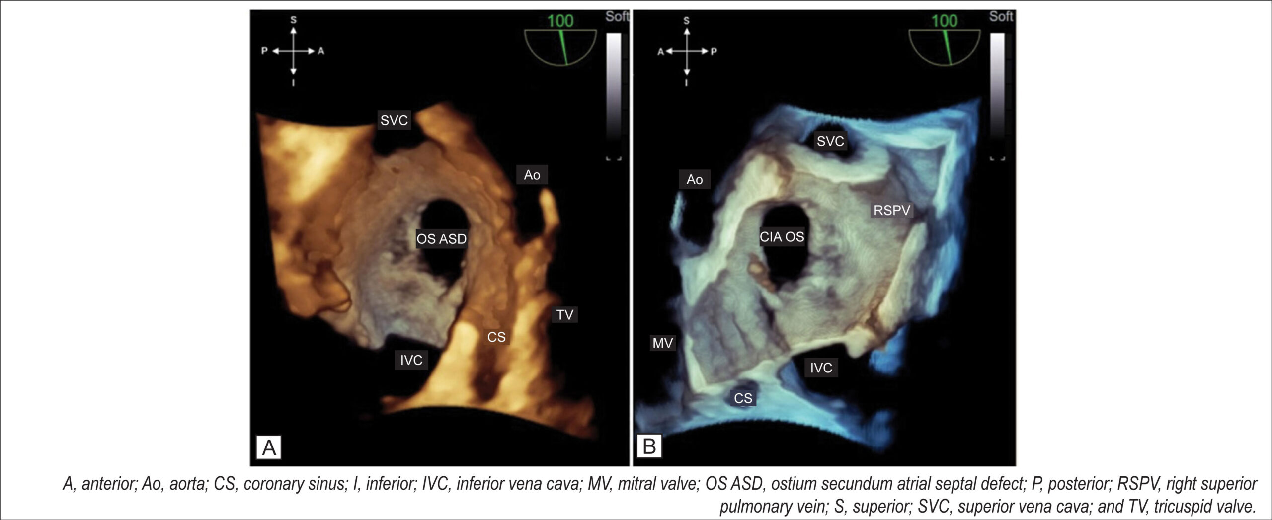
At the end of a detailed 2D TEE with a full septal analysis from the four-chamber view to bicaval section and color mapping, we commence the 3D image acquisition. The en-face projection of the ASD and the defect’s positioning in the anatomic position provide valuable information from a single image: size, shape, location in the interatrial septum, characteristics of the defect’s borders, and the presence of additional fenestration.

I usually start the 3D evaluation from the perspective of the right atrium to facilitate the identification of anatomical markers. I acquire a medium angle block (3D or 4D zoom) or a large block (full-volume using the two-section tool) starting from an initial bicaval section and proceeding with approximate 90° rotation in the z-plane so that the superior and inferior vena cava are aligned in the vertical plane (i.e., superior vena cava at the 11 o’clock position) and the aorta in the anterior and superior region. I prefer not to use the narrow-angle mode for acquisition (live 3D or bird’s eye view) or very high magnification due to a loss of anatomical references adjacent to the defect, which can interfere with this fundamental initial analysis. From this reconstruction (), I can characterize the defect as unique, the OS type, having an oval shape, being central, being medium-sized, and having adequate borders. shows the same ASD and allows assessment of the occurrence of dynamic variation in its dimension with the cardiac cycle.

[…]

  • atrial
  • Congenital
  • Echocardiography
  • Heart Defects
  • Heart septal defects
  • Three-Dimensional
Read Full Article
Creative Commons Attribution (CC BY) Creative Commons license
13/06/2022 DOI: 10.47593/2675-312X/20223502ecom32 Views 3
ABC
Editorial

Coronary Arteries after Jatene Operation for Transposition of Great Arteries: The Role of CT Coronary Angiography on Follow-up

Antonio Joaquim Marinho-da-Silva

The correction of transposition of the great arteries (TGA) by the Jatene technique was one of the greatest achievements of cardiac surgery in congenital heart disease. By replacing the transposed arteries in their proper anatomical arrangement, this technique — atrial switch operation (ASO) – achieves a normal spatial arterial relationship. However, this new arrangement requires excision of the coronary artery buttons and their implantation in the new aorta. The difficulty of this step relates to the type of coronary origin and distance to the new insertion site.

Once this anatomical normalization step has been achieved, cardiac functional normalization is sought. The most common post-surgical complications are related to the complexity of associated anomalies (ventricular septal defect (VSD), coarctation of the aorta, valve stenosis, and others), suture sites of the neovessels, pulmonary ramification and dilation of the neoaorta, Not neglectable and object of concern, late complications of either congenital or acquired coronary circulation are well known. The actual incidence of such complications ranges from 0,8% to 27,5% according to reporting series and time of follow-up. ,

[…]

  • Aortic Stenosis, Subvalvular
  • Arterial Switch Operation
  • coronary circulation
  • Heart Defects, Congenital/surgery
  • Heart septal defects
  • Ventricular
Read Full Article
Creative Commons Attribution (CC BY) Creative Commons license
14/06/2021 DOI: 10.36660/abc.20210403 Views 1
Figure 1
ABC
Case Report

Pseudo-pulmonary Embolism – A Case of Hypoxemia Associated with Right-to-left Shunt

Mariana Faustino et al. Ana Oliveira Soares Fernando Rodrigues Rui Anjos … António Freitas Victor M. Gil

Introduction

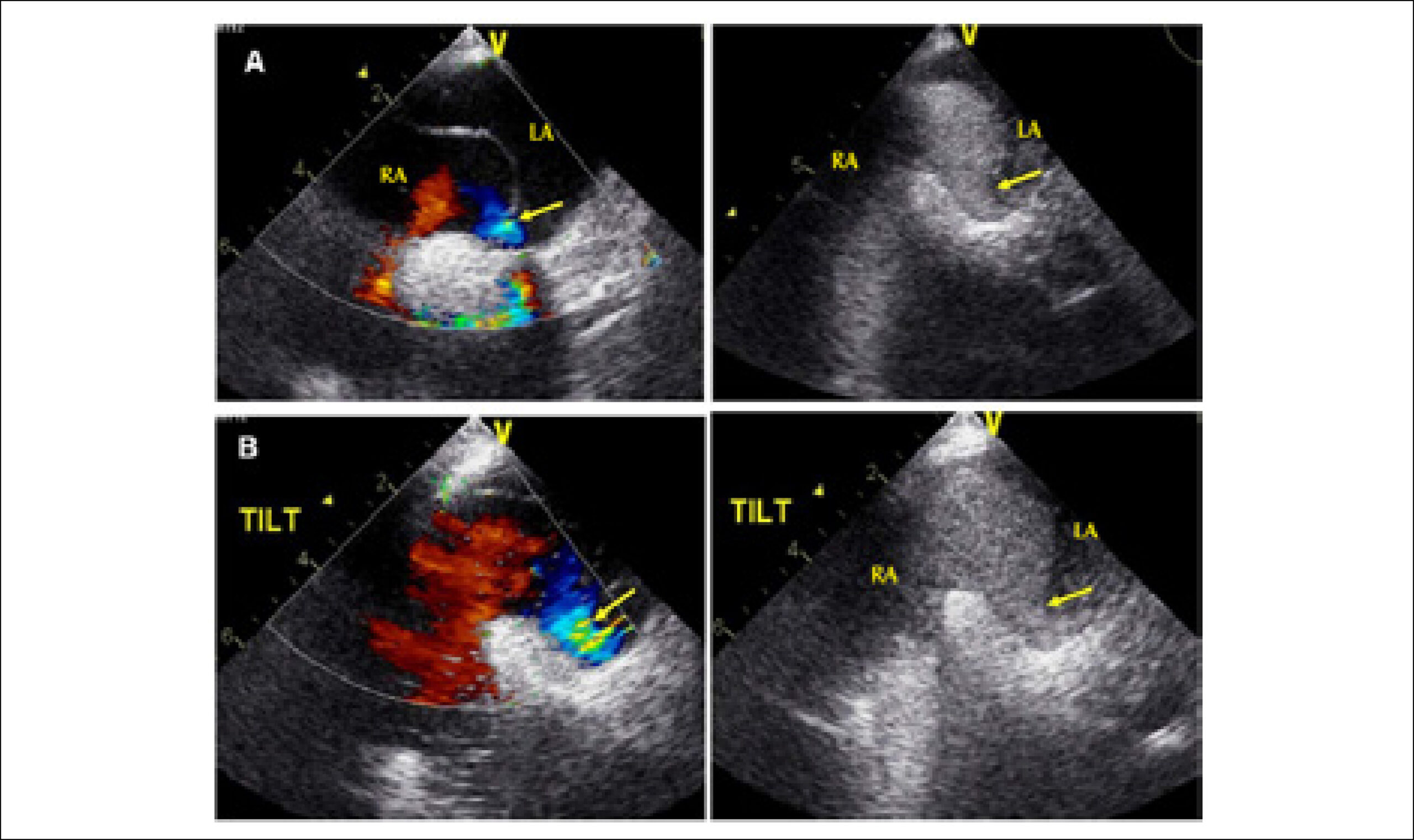
Platypnea orthodeoxia is a rare syndrome characterized by dyspnea and decreased arterial oxygen saturation when passing from the dorsal decubitus position to the sitting or upright position. It usually results from an intracardiac right-to-left shunt; however, the pathophysiological mechanism responsible for the positional nature of the shunt has not been clarified,. We report a case of dyspnea and orthostatic hypoxemia, which was attributed to a right-to-left shunt due to atrial septal defect (ASD).

[…]

  • atrial
  • Dyspnea
  • Echocardiography
  • Heart septal defects
  • Transesophageal
Read Full Article
Creative Commons Attribution (CC BY) Creative Commons license
01/01/2015 DOI: 10.5935/abc.20140208
Case 1/2009: 21-month-old infant with partial atrioventricular septal defect, consisting of atrial septal defect and tricuspid insufficiency
ABC
Correlação Clínico-Radiográfica

Case 1/2009: 21-month-old infant with partial atrioventricular septal defect, consisting of atrial septal defect and tricuspid insufficiency

Edmar Atik

CLINICORADIOLOGICAL SESSION

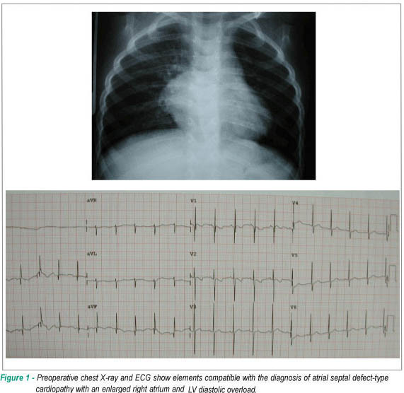
Case 1/2009 – 21-month-old infant with partial atrioventricular septal defect, consisting of atrial septal defect and tricuspid insufficiency

[…]

  • Heart Defects
  • Heart septal defects
  • Tricuspid Valve Insufficiency
Read Full Article
Creative Commons Attribution (CC BY) Creative Commons license
04/02/2009 DOI: 10.1590/S0066-782X2009000100012 Views 2
Anatomy of a dextrocardia case with situs solitus
ABC
Case Report

Anatomy of a dextrocardia case with situs solitus

Fabíola Sawaguchi Faig-Leite et al. Horácio Faig-Leite

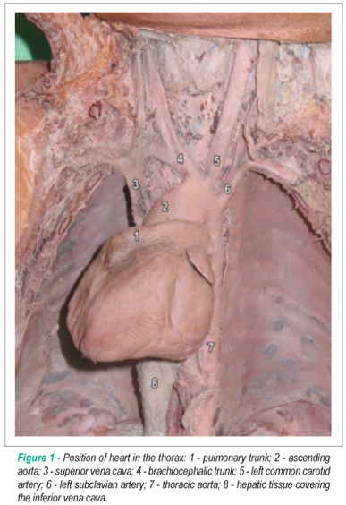
We present a rare case of dextrocardia in the corpse of a female child, aged approximately one year old, presenting situs solitus. The cases of dextrocardia reported in the literature normally describe multiple associated cardiac malformations. In the present study, it is noteworthy the fact that a complete specular image of the heart and the vessels of the base of the heart was not found. There was no atrioventricular discordance or other intra and extracardiac malformations, which are commonly seen in cases of dextrocardia. A large ductus arteriosus was found, as well as the anomalous position of the right portion of the heart.

  • Dextrocardia
  • Dissection
  • Heart septal defects
  • thoracic duct
Read Full Article
Creative Commons Attribution (CC BY) Creative Commons license
05/01/2009 DOI: 10.1590/S0066-782X2008001800013 Views 1

How to cite this article

EndNote RIS

Electronic Document Format (Vancouver)

Kreuzig DL. My Approach to Imaging the Atrial Septal Defect by Three-Dimensional Echocardiography. ABC Imagem Cardiovascular 2022;35(2):ecom32.

Electronic Document Format (ABNT)

Kreuzig, Daniela Lago. My Approach to Imaging the Atrial Septal Defect by Three-Dimensional Echocardiography. ABC Imagem Cardiovascular, v. 35, n. 2, ecom32, Jun. 2022.

Electronic Document Format (APA)

Kreuzig, D. L. (2022). My Approach to Imaging the Atrial Septal Defect by Three-Dimensional Echocardiography. ABC Imagem Cardiovascular, 35(2), ecom32.

Electronic Document Format (ISO)

Kreuzig, Daniela Lago. My Approach to Imaging the Atrial Septal Defect by Three-Dimensional Echocardiography. ABC Imagem Cardiovascular [online]. 2022, vol. 35, n. 2, [cited 2026-10-14], ecom32. Available from: <https://journals.cardiol.br/en/article/my-approach-to-imaging-the-atrial-septal-defect-by-three-dimensional-echocardiography/>. ISSN 2318-8219

EndNote RIS

Electronic Document Format (Vancouver)

Marinho-da-Silva AJ. Coronary Arteries after Jatene Operation for Transposition of Great Arteries: The Role of CT Coronary Angiography on Follow-up. Arquivos Brasileiros de Cardiologia 2021;116(6):1117-8.

Electronic Document Format (ABNT)

Marinho-da-Silva, Antonio Joaquim. Coronary Arteries after Jatene Operation for Transposition of Great Arteries: The Role of CT Coronary Angiography on Follow-up. Arquivos Brasileiros de Cardiologia, v. 116, n. 6, p. 1117-1118, Jun. 2021.

Electronic Document Format (APA)

Marinho-da-Silva, A. J. (2021). Coronary Arteries after Jatene Operation for Transposition of Great Arteries: The Role of CT Coronary Angiography on Follow-up. Arquivos Brasileiros de Cardiologia, 116(6), 1117-1118.

Electronic Document Format (ISO)

Marinho-da-Silva, Antonio Joaquim. Coronary Arteries after Jatene Operation for Transposition of Great Arteries: The Role of CT Coronary Angiography on Follow-up. Arquivos Brasileiros de Cardiologia [online]. 2021, vol. 116, n. 6, [cited 2026-10-14], pp. 1117-1118. Available from: <https://journals.cardiol.br/en/article/coronary-arteries-after-jatene-operation-for-transposition-of-great-arteries-the-role-of-ct-coronary-angiography-on-follow-up/>. ISSN 0066-782X

EndNote RIS

Electronic Document Format (Vancouver)

Faustino M, Soares AO, Rodrigues F, Anjos R, Freitas A, Gil VM. Pseudo-pulmonary Embolism – A Case of Hypoxemia Associated with Right-to-left Shunt. Arquivos Brasileiros de Cardiologia 2015;104(1):e4-9.

Electronic Document Format (ABNT)

Faustino, Mariana; Soares, Ana Oliveira; Rodrigues, Fernando; Anjos, Rui; Freitas, António; Gil, Victor M.. Pseudo-pulmonary Embolism – A Case of Hypoxemia Associated with Right-to-left Shunt. Arquivos Brasileiros de Cardiologia, v. 104, n. 1, p. e4-e9, Dec. 2014.

Electronic Document Format (APA)

Faustino, M., Soares, A. O., Rodrigues, F., Anjos, R., Freitas, A., & Gil, V. M. (2015). Pseudo-pulmonary Embolism – A Case of Hypoxemia Associated with Right-to-left Shunt. Arquivos Brasileiros de Cardiologia, 104(1), e4-e9.

Electronic Document Format (ISO)

Faustino, Mariana and Soares, Ana Oliveira and Rodrigues, Fernando and Anjos, Rui and Freitas, António and Gil, Victor M.. Pseudo-pulmonary Embolism – A Case of Hypoxemia Associated with Right-to-left Shunt. Arquivos Brasileiros de Cardiologia [online]. 2015, vol. 104, n. 1, [cited 2026-10-14], pp. e4-e9. Available from: <https://journals.cardiol.br/en/article/pseudo-pulmonary-embolism-a-case-of-hypoxemia-associated-with-right-to-left-shunt/>. ISSN 0066-782X

EndNote RIS

Electronic Document Format (Vancouver)

Atik E. Case 1/2009: 21-month-old infant with partial atrioventricular septal defect, consisting of atrial septal defect and tricuspid insufficiency. Arquivos Brasileiros de Cardiologia 2009;92(1):77-8.

Electronic Document Format (ABNT)

Atik, Edmar. Case 1/2009: 21-month-old infant with partial atrioventricular septal defect, consisting of atrial septal defect and tricuspid insufficiency. Arquivos Brasileiros de Cardiologia, v. 92, n. 1, p. 77-78, Feb. 2009.

Electronic Document Format (APA)

Atik, E. (2009). Case 1/2009: 21-month-old infant with partial atrioventricular septal defect, consisting of atrial septal defect and tricuspid insufficiency. Arquivos Brasileiros de Cardiologia, 92(1), 77-78.

Electronic Document Format (ISO)

Atik, Edmar. Case 1/2009: 21-month-old infant with partial atrioventricular septal defect, consisting of atrial septal defect and tricuspid insufficiency. Arquivos Brasileiros de Cardiologia [online]. 2009, vol. 92, n. 1, [cited 2026-10-14], pp. 77-78. Available from: <https://journals.cardiol.br/en/article/case-1-2009-21-month-old-infant-with-partial-atrioventricular-septal-defect-consisting-of-atrial-septal-defect-and-tricuspid-insufficiency/>. ISSN 0066-782X

EndNote RIS

Electronic Document Format (Vancouver)

Faig-Leite FS, Faig-Leite H. Anatomy of a dextrocardia case with situs solitus. Arquivos Brasileiros de Cardiologia 2009;91(6):e56-8.

Electronic Document Format (ABNT)

Faig-Leite, Fabíola Sawaguchi; Faig-Leite, Horácio. Anatomy of a dextrocardia case with situs solitus. Arquivos Brasileiros de Cardiologia, v. 91, n. 6, p. e56-e58, Jan. 2009.

Electronic Document Format (APA)

Faig-Leite, F. S., & Faig-Leite, H. (2009). Anatomy of a dextrocardia case with situs solitus. Arquivos Brasileiros de Cardiologia, 91(6), e56-e58.

Electronic Document Format (ISO)

Faig-Leite, Fabíola Sawaguchi and Faig-Leite, Horácio. Anatomy of a dextrocardia case with situs solitus. Arquivos Brasileiros de Cardiologia [online]. 2009, vol. 91, n. 6, [cited 2026-10-14], pp. e56-e58. Available from: <https://journals.cardiol.br/en/article/anatomy-of-a-dextrocardia-case-with-situs-solitus/>. ISSN 0066-782X

  • About ABC Family
  • Internal Regulation
  • Editorial Board
  • Brazilian Society of Cardiology (SBC)

Issues

SBC Guidelines

  • Videos
  • Podcasts
  • News