ABC Family Arquivos Brasileiros de Cardiologia International Journal of Cardiovascular Sciences ABC Heart Failure & Cardiomyopathy ABC Imagem Cardiovascular
Português English
ABC Family
Português English
  • About
    • About ABC Family
    • Internal Regulation
    • Editorial Board
    • Brazilian Society of Cardiology (SBC)
  • Issues
  • SBC Guidelines
  • Content
    • Videos
    • Podcasts
    • News
  • About
    • About ABC Family
    • Internal Regulation
    • Editorial Board
    • Brazilian Society of Cardiology (SBC)
  • Issues
  • SBC Guidelines
  • Content
    • Videos
    • Podcasts
    • News

ABC Family

  • ABC Family
  • Arquivos Brasileiros de Cardiologia
  • International Journal of Cardiovascular Sciences
  • ABC Heart Failure & Cardiomyopathy
  • ABC Imagem Cardiovascular
Search in

No content found.

Journals
Ordering
Article types
Others

  1. Home
  2. Keyword: Tomography, X Ray-Computed/methods
Figure 1
ABC
Image

Intracavitary Right Coronary Artery: An Incidental Finding with Potential Implications for Invasive Cardiac Procedures

Sara Cristina da Silva Borges et al. Catarina Isabel Ribeiro Carvalho Miguel Eduardo Teixeira Moz Gonçalves Ana Isabel Santos Baptista José Ilídio Moreira

A 66-year-old man with a history of palpitations suggestive of paroxysmal supraventricular tachycardia was referred for CT angiography (CTA) for exertional dyspnea etiology investigation. ECG-gated cardiac CT was performed using the 64-slices dual-source Somatom Go Scanner.

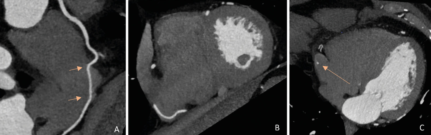
CTA showed the normal origin of the right and left main coronary arteries, and there was no evidence of obstructive coronary artery disease. The proximal right coronary artery (RCA) had a normal epicardial course, but mid-RCA was noted to penetrate the right atrial wall for a 30 mm course within the right atrium, exiting to its usual course in the posterior atrioventricular groove, as demonstrated via the multiplanar reconstruction CT images at maximum intensity projection ( ) as well as 3-dimensional reconstructions ( ).

[…]
  • Anomalous Intracaitary /diagnosis
  • Cardiovascular Abnormalities
  • Coronary Angiography/methods
  • Coronary Vessel Anomalies
  • Tomography, X Ray-Computed/methods
Read Full Article
Creative Commons Attribution (CC BY) Creative Commons license
13/05/2022 DOI: 10.36660/abc.20210819 Views 1
Figure 1
ABC
Original Article

Diagnostic Performance of Coronary Tomography Angiography and Serial Measurements of Sensitive Cardiac Troponin in Patients With Chest Pain and Intermediate Risk for Cardiovascular Events

Alexandre de Matos Soeiro et al. Bruno Biselli Tatiana C.A.T. Leal Aline Siqueira Bossa … Maria Cristina César Sérgio Jallad Priscila Gherardi Goldstein Patrícia Oliveira Guimarães Carlos Vicente Serrano Jr Cesar Higa Nomura Débora Nakamura Carlos Eduardo Rochitte Paulo Rogério Soares Múcio Tavares de Oliveira Jr.

Abstract

Background

Coronary tomography angiography (CTA) has been mainly used for chest pain evaluation in low-risk patients, and few data exist regarding patients at intermediate risk.

Objective

To evaluate the performance of serial measures of sensitive troponin and CTA in intermediate-risk patients.

Methods

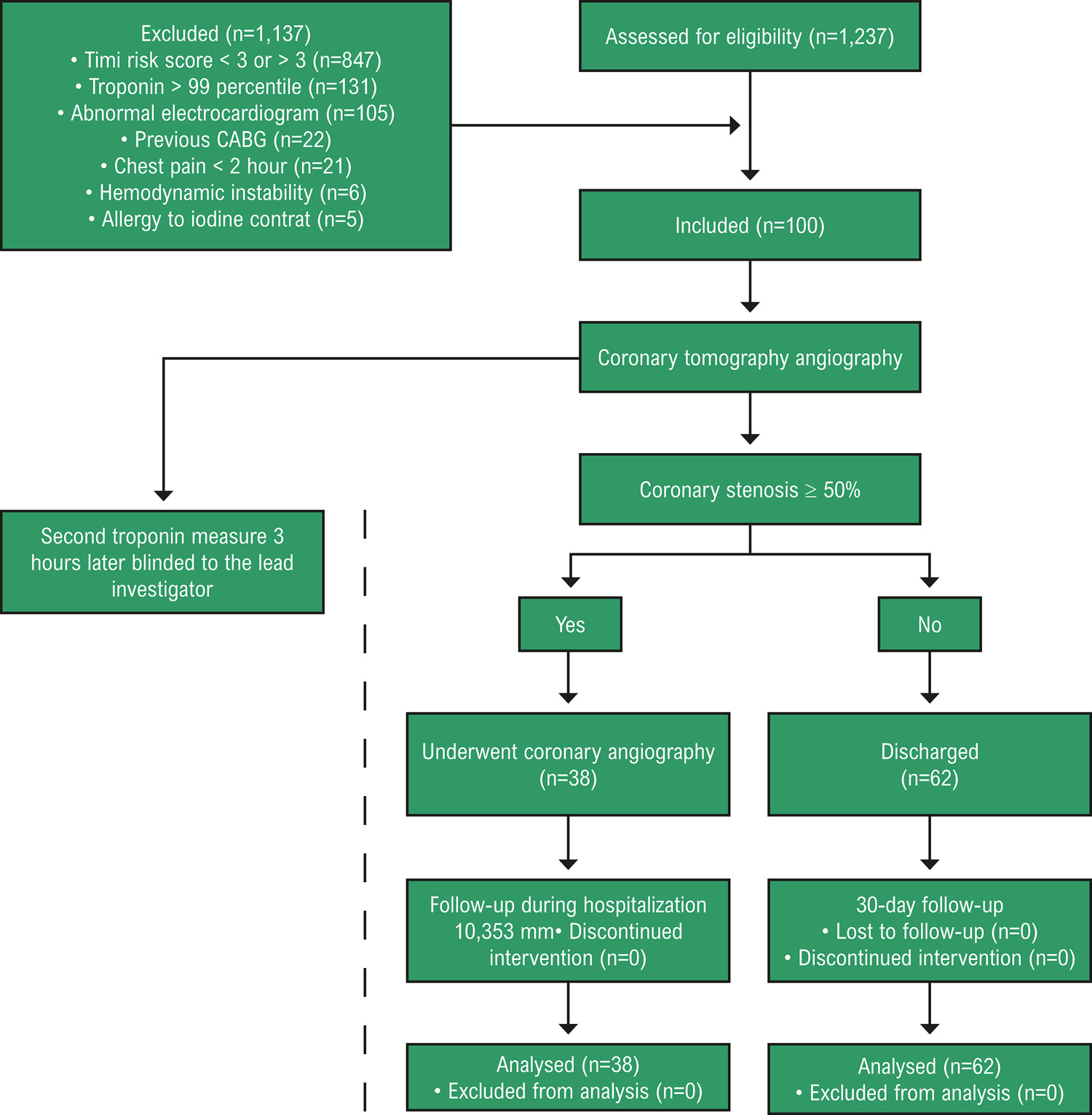
A total of 100 patients with chest pain, TIMI risk scores of 3 or 4, and negative troponin were prospectively included. All patients underwent CTA and those with coronary stenosis ≥ 50% were referred to invasive coronary angiography. Patients with coronary lesions <50% were discharged and contacted 30 days later by a telephone call to assess clinical outcomes. Outcomes were hospitalization, death, and myocardial infarction at 30 days. The comparison between methods was performed by Kappa agreement test. The performance of troponin measures and CTA for detecting significant coronary lesions and clinical outcomes was calculated. Results were considered statistically significant when p < 0.05.

Results

Coronary stenosis ≥ 50% on CTA was found in 38% of patients and significant coronary lesions on coronary angiography were found in 31 patients. Two clinical events were observed. Kappa agreement analysis showed low agreement between troponin measures and CTA in the detection of significant coronary lesions (kappa = 0.022, p = 0.78). The performance of CTA for detecting significant coronary lesions on coronary angiography or for predicting clinical events at 30 days was better than sensitive troponin measures (accuracy of 91% versus 60%).

Conclusion

CTA performed better than sensitive troponin measures in the detection of significant coronary disease in patients with chest pain and intermediate risk for cardiovascular events.

  • Angiotomography Coronary/methods
  • Cardiovascular Diseases
  • Chest Pain
  • Risk Factors
  • Risk Management
  • Tomography, X Ray-Computed/methods
  • Troponin I
  • Troponin T
Read Full Article
Creative Commons Attribution (CC BY) Creative Commons license
13/05/2022 DOI: 10.36660/abc.20210006
Figure 1
IJCS
Review Article

Translational Approach for Percutaneous Interventions for the Treatment of Cardiac Arrhythmias

Angelo Amato Vincenzo de Paola

Abstract

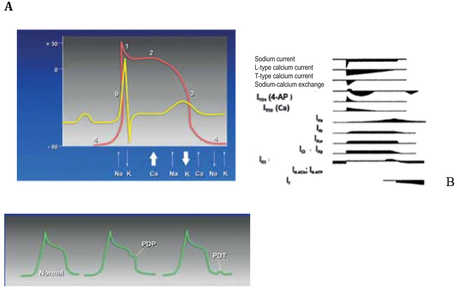
New translational concepts on cellular and tissue substrate of cardiac arrhythmias have been responsible for the development of non-pharmacological interventions, with important achievements compared to the conventional approach with antiarrhythmic drugs. In addition, the increasing knowledge of anatomical and electrophysiological studies, sophisticated mapping methods, special catheters, and controlled clinical trials have favored the progression of ablation of tachyarrhythmias, particularly of ventricular tachyarrhythmias and atrial fibrillation.

  • Anti-Arrhythmia Agents
  • Arrhythmias Cardiac/physiopathology
  • atrioventricular node
  • Catheter Ablation
  • Cryosurgery / methods
  • Echocardiography/methods
  • Tomography, X Ray-Computed/methods
  • Translational Medical Research
Read Full Article
Creative Commons Attribution (CC BY) Creative Commons license
09/10/2020 DOI: 10.36660/ijcs.20200152 Views 2
Figure 1
IJCS
Original Article

Lung Ultrasound as a Triage Tool in an Emergency Setting during the Covid-19 Outbreak: comparison with CT Findings

Monica Luiza de Alcantara et al. Marcos Paulo Lacerda Bernardo Tatiana Bagrichevsky Autran Rodolfo de Paula Lustosa … Marcelo Tayah Lucia Antunes Chagas Dequitier Carvalho Machado

Abstract

Background

Lung ultrasound (LUS) can detect interstitial alveolar changes confined to the subpleural region, like those described in Covid-19.

Objetive

To evaluate how LUS findings correlate with chest computed tomography (CT) in patients admitted to the emergency department (ED) with suspicion of Covid-19.

Methods

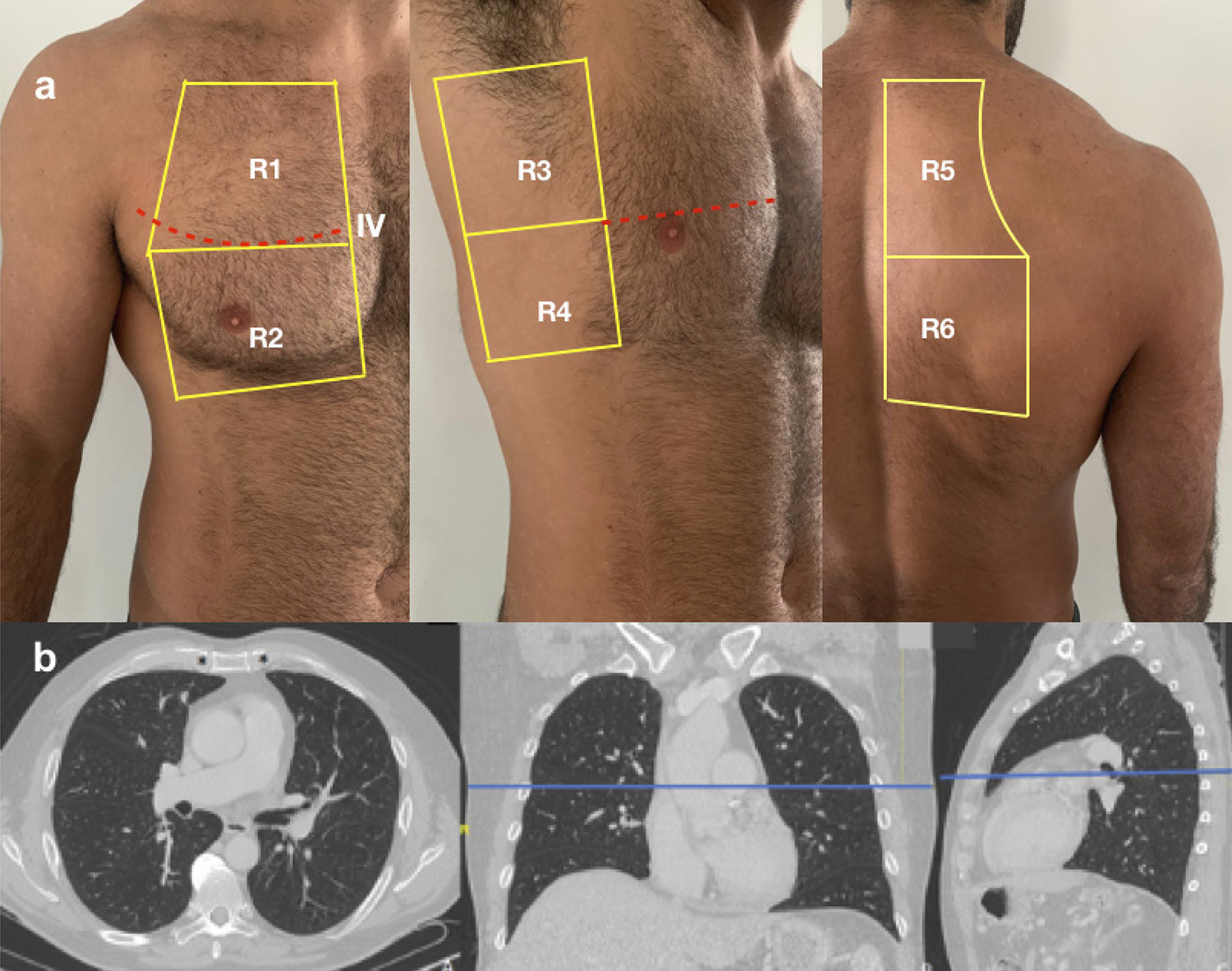
Cross-sectional study of 20 patients (median age 43 years; interquartile range, 37–63 years; 50% male). All patients underwent LUS and chest CT on the day of ED admission. Each hemithorax was divided into 6 segments with similar landmarks, and equivalent scores (sc) of lesion severity were defined for both methods. The number of affected segments on LUS (LUSseg) was divided into tertiles (0-1, 2-5, and ≥6), and compared with number of affected segments on CT (CTseg), LUSsc, CTsc, and percentage of affected lung parenchyma through visual analysis (CTvis). ANOVA or Kruskal–Wallis test for continuous variables, chi-square test for categorical variables, and receiver operating characteristic (ROC) curve analysis to define optimal cutoff points were performed. P<0.05 was considered statistically significant.

Results

Median LUSsc, CTsc, CTseg, and CTvis were significantly different between groups. A clear separation between groups was demonstrated; patients with 7 LUSsc and >3 LUSseg, respectively.

Conclusion

LUS findings correlate with chest CT, and can help identify patients with normal lung or minor pulmonary involvement secondary to Covid-19. Int J Cardiovasc Sci. 2020; [online].ahead print, PP.0-0

  • COVID-19
  • Lung
  • Pandemics
  • Pleura
  • Pulmonary Alveoli
  • Tomography, X Ray-Computed/methods
  • Triage
  • Ultrasonography/methods
Read Full Article
Creative Commons Attribution (CC BY) Creative Commons license
28/08/2020 DOI: 10.36660/ijcs.20200133 Views 1
Figure 1
ABC
Case Report

Novel Coronavirus Pneumonia and Cardiomyopathy: A Case Report

Mustafa Ahmet Huyut

Case report

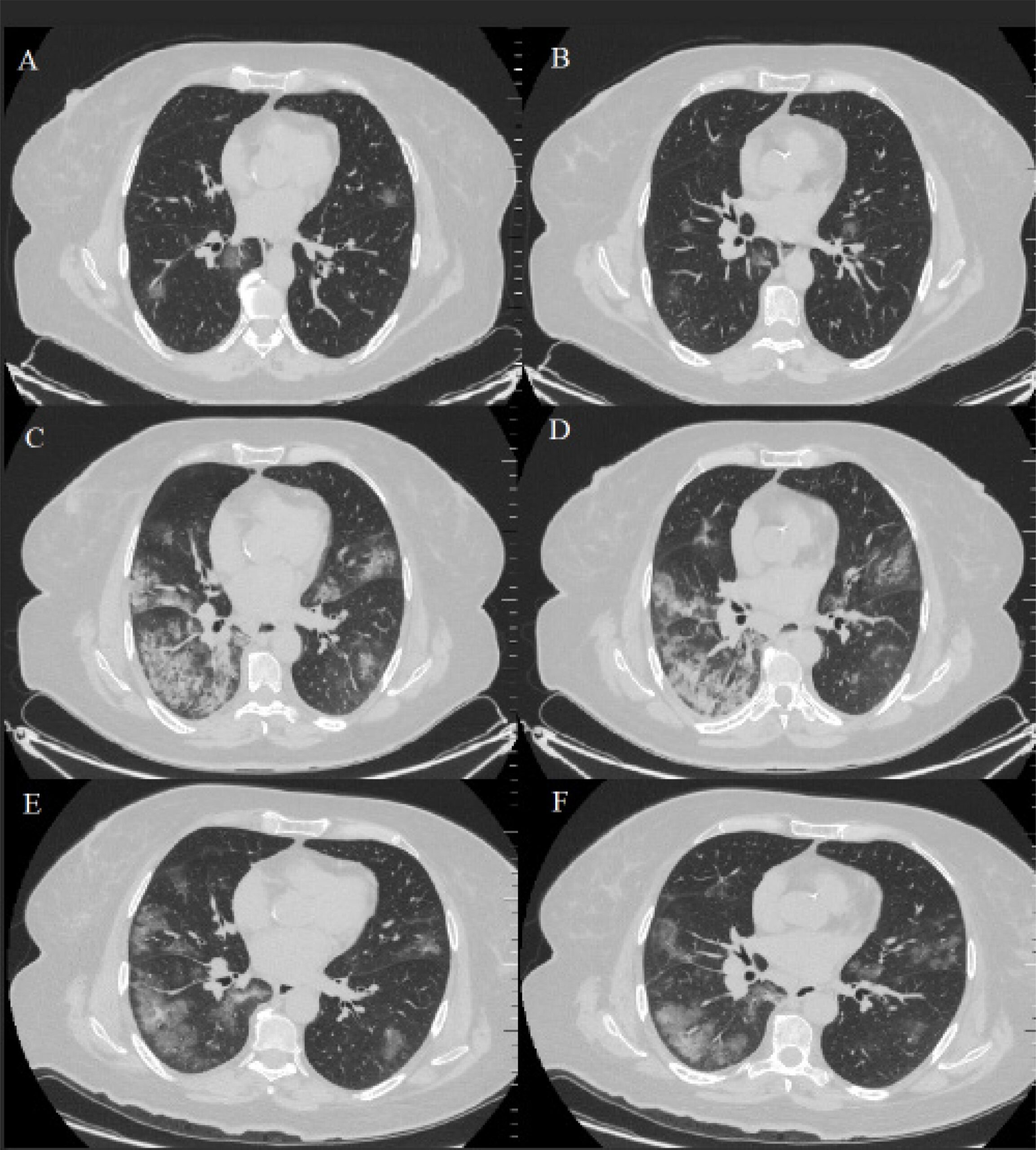
A 59-year-old female had had fever for 4 days, after catching a cold. One day before visiting the hospital, she had fever and cough, but no chest tightness, chest pain, chills, nausea and vomiting or diarrhea. She did not feel better after receiving antifebrile agents. Then, she was admitted to our outpatient clinic in BHT Clinic Tema Hospital. Four days before, the patient had had contact with her relative who had traveled from Europe. In her previous medical history, bariatric surgery had been performed 3 years before and she had still type II diabetes mellitus, hyperlipidemia, and hypertension as pre-existing conditions. She was hospitalized in our hospital on March/20/2020, and still had fever after admission, with the highest temperature of 39.5 °C, heart rate of 119 beats per minute; the electrocardiography was consistent with sinus tachycardia and QTc was calculated at 0.398 seconds, blood pressure at 94/60 mmHg, and conspicuous tachypnea with a respiratory rate of 24/min with sufficient oxygenation (95% ambient air saturation). The identification of 2019 novel coronavirus (2019-nCoV) in RT-PCR was positive from a throat swab. The risk of simultaneous contamination with other respiratory viruses and other pathogens were negative for the throat swab. The CT characteristics of the patient were similar to the case series reported by Pan et al. (). Laboratory results showed leukopenia, with 4.1×10/L, lymphopenia with 0.8×10/L, an only slightly increased CRP level, with 18.4 mg/L and a low procalcitonin level, with 0.01 ng/mL. The patient underwent a TTE examination with a 3.5-MHz transducer (Vivid-7 GE Medical System, Horten, Norway). Examinations and measurements were performed according to the recommendations of the American Echocardiography Unit. Simpson’s method was used to calculate LVEF. At the admission, LVEF was calculated at 65%, with normal TTE findings.

[…]

  • Acute Respiratory Syndrome
  • Cardiomyopathies
  • Cardiovascular Diseases/complications
  • Coronavirus
  • COVID-19
  • Echocardiography/methods
  • Hospitalization
  • Infectious Diseases
  • Myocarditis
  • Pneumonia
  • Tomography, X Ray-Computed/methods
Read Full Article
Creative Commons Attribution (CC BY) Creative Commons license
01/06/2020 DOI: 10.36660/abc.20200268 Views 1
Figure 1
ABC
Image

Abdominal Pain: an Uncommon Presentation of Myocardial Rupture

Daniel Seabra et al. Ana Neto Inês Oliveira Rui Pontes dos Santos … João Azevedo Paula Pinto

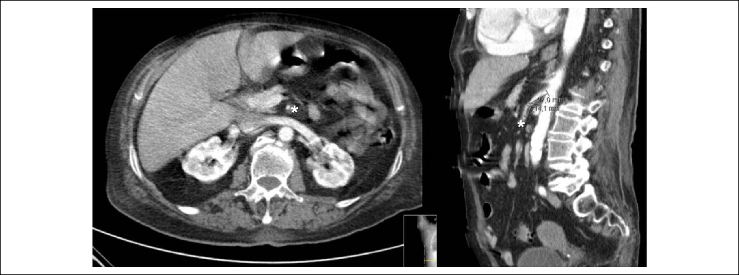
An 84-years-old woman with hypertension and dyslipidemia was admitted in the emergency room with acute abdominal pain; patient complained of chest pain three weeks before hospital admission. On physical examination, she was hypotensive, tachycardic (with arrhythmic pulse), tachypneic and with diffuse abdominal pain. Electrocardiogram showed atrial fibrillation with rapid ventricular response, complete left bundle branch block and inferior Q waves. Abdominal computed tomography (CT) revealed a thrombus in the superior mesenteric artery (, white asterisk). The patient showed a clinical course with congestive heart failure and low cardiac output. Transthoracic echocardiogram (-) showed mild left ventricular dilatation with a mild dysfunction and a pseudoaneurysm of the basal half of the posterior and inferior walls with left-to-right shunt, confirmed by color Doppler imaging (-). Cardiac CT () revealed contained myocardial rupture, located at the basal segments of the inferior and posterior septal walls, extending to the free wall of the right ventricle, forming a pseudo-cavity, which communicates with the true cavity of the right ventricle (). Despite vasopressor and inotropic support and proposal for cardiac surgery, the patient had an unfavorable course.

[…]

  • Abdominal Pain
  • Dyslipidemias
  • Echocardiography, Doppler/methods
  • Hypertension
  • Myocardial Infarction/complications
  • Thrombosis/surgery
  • Tomography, X Ray-Computed/methods
Read Full Article
Creative Commons Attribution (CC BY) Creative Commons license
20/03/2020 DOI: 10.36660/abc.20190495
Figure 1
ABC
Image

Fatty Left Ventricular Cardiomyopathy: An Under-Diagnosed Disease

Abdalla Elagha et al. Anthon Fuisz

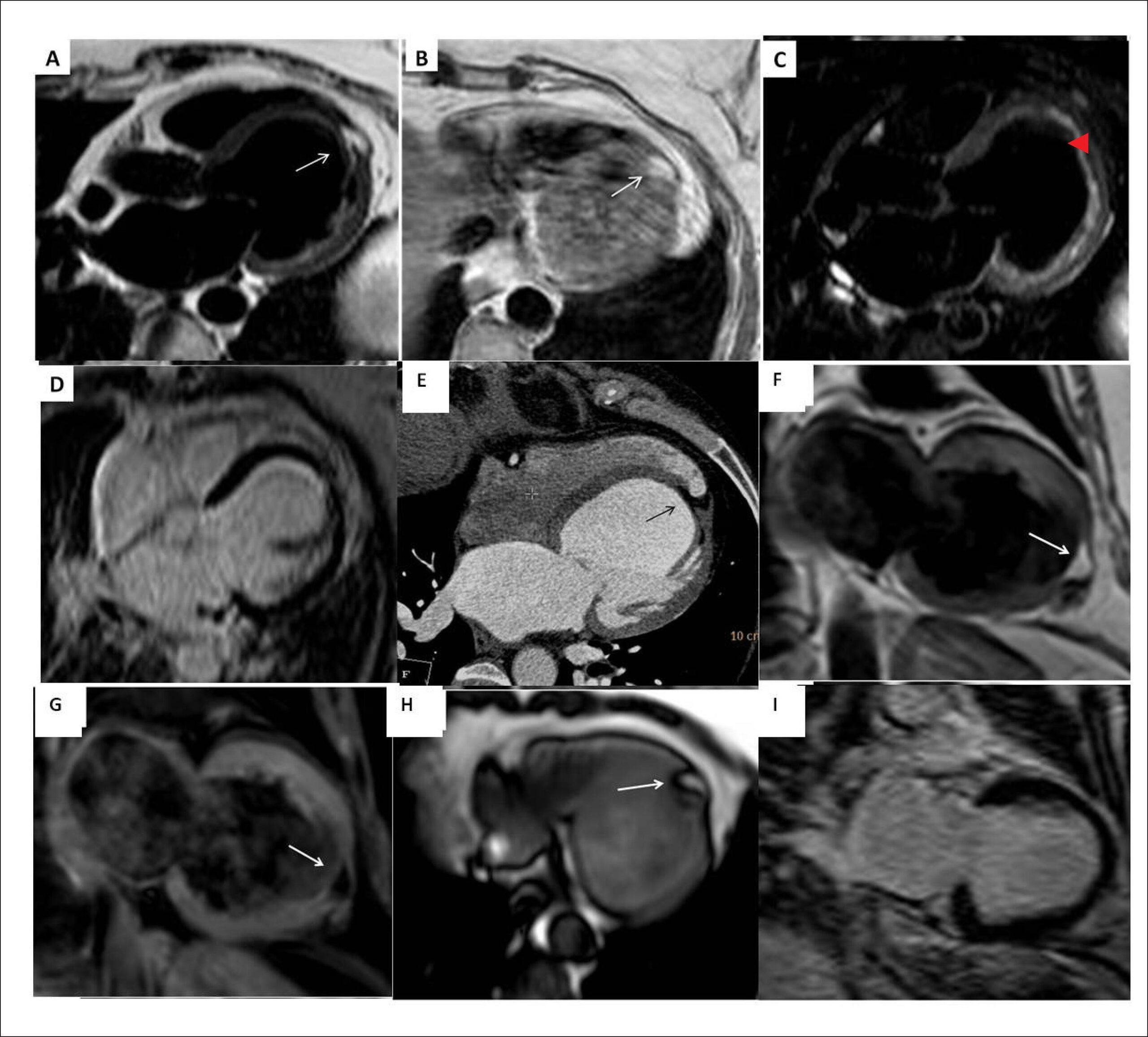
During a preoperative cardiac assessment, a 57-year-old asymptomatic female, without known cardiac risk-factors, was found to have LBBB on ECG. Echocardiography revealed dilated cardiomyopathy. To rule-out coronary artery disease, coronary CT-angiography was performed and showed no significant coronary obstruction.

For further evaluation, a cardiac magnetic resonance (CMR) study was performed. A focal area of fatty infiltration was seen in the left ventricular (LV) apex extending from the subendocardial to the subepicardial surfaces ( and ). This area was suppressed with fat saturation pulse-sequences (), and did not show enhanced signal on delayed-hyperenhancement images (DHE) (). Both LV and LA were dilated; LV systolic function was reduced globally. Additionally, the CMR showed normal RV dimensions, function, and wall-thickness. The fatty area was also seen retrospectively on the cardiac CT ().

[…]

  • Cardiomyopathy, Dilated
  • Lipoma
  • Tomography, X Ray-Computed/methods
  • Ventricular Dysfunction, Left
Read Full Article
Creative Commons Attribution (CC BY) Creative Commons license
01/01/2017 DOI: 10.5935/abc.20160199 Views 1

How to cite this article

EndNote RIS

Electronic Document Format (Vancouver)

Borges SCS, Carvalho CIR, Gonçalves METM, Baptista AIS, Moreira JI. Intracavitary Right Coronary Artery: An Incidental Finding with Potential Implications for Invasive Cardiac Procedures. Arquivos Brasileiros de Cardiologia 2022;118(5):1000-1.

Electronic Document Format (ABNT)

Borges, Sara Cristina da Silva; Carvalho, Catarina Isabel Ribeiro; Gonçalves, Miguel Eduardo Teixeira Moz; Baptista, Ana Isabel Santos; Moreira, José Ilídio. Intracavitary Right Coronary Artery: An Incidental Finding with Potential Implications for Invasive Cardiac Procedures. Arquivos Brasileiros de Cardiologia, v. 118, n. 5, p. 1000-1001, May. 2022.

Electronic Document Format (APA)

Borges, S. C. S., Carvalho, C. I. R., Gonçalves, M. E. T. M., Baptista, A. I. S., & Moreira, J. I. (2022). Intracavitary Right Coronary Artery: An Incidental Finding with Potential Implications for Invasive Cardiac Procedures. Arquivos Brasileiros de Cardiologia, 118(5), 1000-1001.

Electronic Document Format (ISO)

Borges, Sara Cristina da Silva and Carvalho, Catarina Isabel Ribeiro and Gonçalves, Miguel Eduardo Teixeira Moz and Baptista, Ana Isabel Santos and Moreira, José Ilídio. Intracavitary Right Coronary Artery: An Incidental Finding with Potential Implications for Invasive Cardiac Procedures. Arquivos Brasileiros de Cardiologia [online]. 2022, vol. 118, n. 5, [cited 2026-10-15], pp. 1000-1001. Available from: <https://journals.cardiol.br/en/article/intracavitary-right-coronary-artery-an-incidental-finding-with-potential-implications-for-invasive-cardiac-procedures/>. ISSN 0066-782X

EndNote RIS

Electronic Document Format (Vancouver)

Soeiro AM, Biselli B, Leal TC, Bossa AS, César MC, Jallad S, Goldstein PG, et al. Diagnostic Performance of Coronary Tomography Angiography and Serial Measurements of Sensitive Cardiac Troponin in Patients With Chest Pain and Intermediate Risk for Cardiovascular Events. Arquivos Brasileiros de Cardiologia 2022;118(5):894-902.

Electronic Document Format (ABNT)

Soeiro, Alexandre de Matos; Biselli, Bruno; Leal, Tatiana C.A.T.; Bossa, Aline Siqueira; César, Maria Cristina; Jallad, Sérgio; Goldstein, Priscila Gherardi; Guimarães, Patrícia Oliveira; Serrano Jr, Carlos Vicente; Nomura, Cesar Higa; Nakamura, Débora; Rochitte, Carlos Eduardo; Soares, Paulo Rogério; Oliveira Jr., Múcio Tavares de. Diagnostic Performance of Coronary Tomography Angiography and Serial Measurements of Sensitive Cardiac Troponin in Patients With Chest Pain and Intermediate Risk for Cardiovascular Events. Arquivos Brasileiros de Cardiologia, v. 118, n. 5, p. 894-902, May. 2022.

Electronic Document Format (APA)

Soeiro, A. M., Biselli, B., Leal, T. C., Bossa, A. S., César, M. C., Jallad, S., Goldstein, P. G., Guimarães, P. O., Serrano Jr, C. V., Nomura, C. H., Nakamura, D., Rochitte, C. E., Soares, P. R., & Oliveira Jr., M. T. (2022). Diagnostic Performance of Coronary Tomography Angiography and Serial Measurements of Sensitive Cardiac Troponin in Patients With Chest Pain and Intermediate Risk for Cardiovascular Events. Arquivos Brasileiros de Cardiologia, 118(5), 894-902.

Electronic Document Format (ISO)

Soeiro, Alexandre de Matos and Biselli, Bruno and Leal, Tatiana C.A.T. and Bossa, Aline Siqueira and César, Maria Cristina and Jallad, Sérgio and Goldstein, Priscila Gherardi and Guimarães, Patrícia Oliveira and Serrano Jr, Carlos Vicente and Nomura, Cesar Higa and Nakamura, Débora and Rochitte, Carlos Eduardo and Soares, Paulo Rogério and Oliveira Jr., Múcio Tavares de. Diagnostic Performance of Coronary Tomography Angiography and Serial Measurements of Sensitive Cardiac Troponin in Patients With Chest Pain and Intermediate Risk for Cardiovascular Events. Arquivos Brasileiros de Cardiologia [online]. 2022, vol. 118, n. 5, [cited 2026-10-15], pp. 894-902. Available from: <https://journals.cardiol.br/en/article/diagnostic-performance-of-coronary-tomography-angiography-and-serial-measurements-of-sensitive-cardiac-troponin-in-patients-with-chest-pain-and-intermediate-risk-for-cardiovascular-events/>. ISSN 0066-782X

EndNote RIS

Electronic Document Format (Vancouver)

Paola AAV. Translational Approach for Percutaneous Interventions for the Treatment of Cardiac Arrhythmias. International Journal of Cardiovascular Sciences 2020;33(5):550-64.

Electronic Document Format (ABNT)

Paola, Angelo Amato Vincenzo de. Translational Approach for Percutaneous Interventions for the Treatment of Cardiac Arrhythmias. International Journal of Cardiovascular Sciences, v. 33, n. 5, p. 550-564, Oct. 2020.

Electronic Document Format (APA)

Paola, A. A. V. (2020). Translational Approach for Percutaneous Interventions for the Treatment of Cardiac Arrhythmias. International Journal of Cardiovascular Sciences, 33(5), 550-564.

Electronic Document Format (ISO)

Paola, Angelo Amato Vincenzo de. Translational Approach for Percutaneous Interventions for the Treatment of Cardiac Arrhythmias. International Journal of Cardiovascular Sciences [online]. 2020, vol. 33, n. 5, [cited 2026-10-15], pp. 550-564. Available from: <https://journals.cardiol.br/en/article/translational-approach-for-percutaneous-interventions-for-the-treatment-of-cardiac-arrhythmias/>. ISSN 2359-4802

EndNote RIS

Electronic Document Format (Vancouver)

Alcantara ML, Bernardo MPL, Autran TB, Lustosa RP, Tayah M, Chagas LA, Machado DC, et al. Lung Ultrasound as a Triage Tool in an Emergency Setting during the Covid-19 Outbreak: comparison with CT Findings. International Journal of Cardiovascular Sciences 2020;33(5):479-87.

Electronic Document Format (ABNT)

Alcantara, Monica Luiza de; Bernardo, Marcos Paulo Lacerda; Autran, Tatiana Bagrichevsky; Lustosa, Rodolfo de Paula; Tayah, Marcelo; Chagas, Lucia Antunes; Machado, Dequitier Carvalho. Lung Ultrasound as a Triage Tool in an Emergency Setting during the Covid-19 Outbreak: comparison with CT Findings. International Journal of Cardiovascular Sciences, v. 33, n. 5, p. 479-487, Aug. 2020.

Electronic Document Format (APA)

Alcantara, M. L. , Bernardo, M. P. L., Autran, T. B., Lustosa, R. P., Tayah, M., Chagas, L. A., & Machado, D. C. (2020). Lung Ultrasound as a Triage Tool in an Emergency Setting during the Covid-19 Outbreak: comparison with CT Findings. International Journal of Cardiovascular Sciences, 33(5), 479-487.

Electronic Document Format (ISO)

Alcantara, Monica Luiza de and Bernardo, Marcos Paulo Lacerda and Autran, Tatiana Bagrichevsky and Lustosa, Rodolfo de Paula and Tayah, Marcelo and Chagas, Lucia Antunes and Machado, Dequitier Carvalho. Lung Ultrasound as a Triage Tool in an Emergency Setting during the Covid-19 Outbreak: comparison with CT Findings. International Journal of Cardiovascular Sciences [online]. 2020, vol. 33, n. 5, [cited 2026-10-15], pp. 479-487. Available from: <https://journals.cardiol.br/en/article/lung-ultrasound-as-a-triage-tool-in-an-emergency-setting-during-the-covid-19-outbreak-comparison-with-ct-findings/>. ISSN 2359-4802

EndNote RIS

Electronic Document Format (Vancouver)

Huyut MA. Novel Coronavirus Pneumonia and Cardiomyopathy: A Case Report. Arquivos Brasileiros de Cardiologia 2020;114(5):843-5.

Electronic Document Format (ABNT)

Huyut, Mustafa Ahmet. Novel Coronavirus Pneumonia and Cardiomyopathy: A Case Report. Arquivos Brasileiros de Cardiologia, v. 114, n. 5, p. 843-845, May. 2020.

Electronic Document Format (APA)

Huyut, M. A. (2020). Novel Coronavirus Pneumonia and Cardiomyopathy: A Case Report. Arquivos Brasileiros de Cardiologia, 114(5), 843-845.

Electronic Document Format (ISO)

Huyut, Mustafa Ahmet. Novel Coronavirus Pneumonia and Cardiomyopathy: A Case Report. Arquivos Brasileiros de Cardiologia [online]. 2020, vol. 114, n. 5, [cited 2026-10-15], pp. 843-845. Available from: <https://journals.cardiol.br/en/article/novel-coronavirus-pneumonia-and-cardiomyopathy-a-case-report/>. ISSN 0066-782X

EndNote RIS

Electronic Document Format (Vancouver)

Seabra D, Ana Neto , Oliveira I, Santos RP, Azevedo J, Pinto P. Abdominal Pain: an Uncommon Presentation of Myocardial Rupture. Arquivos Brasileiros de Cardiologia 2020;114(2):319-22.

Electronic Document Format (ABNT)

Seabra, Daniel; Ana Neto, ; Oliveira, Inês; Santos, Rui Pontes dos; Azevedo, João; Pinto, Paula. Abdominal Pain: an Uncommon Presentation of Myocardial Rupture. Arquivos Brasileiros de Cardiologia, v. 114, n. 2, p. 319-322, Mar. 2020.

Electronic Document Format (APA)

Seabra, D., Ana Neto, , Oliveira, I., Santos, R. P. , Azevedo, J., & Pinto, P. (2020). Abdominal Pain: an Uncommon Presentation of Myocardial Rupture. Arquivos Brasileiros de Cardiologia, 114(2), 319-322.

Electronic Document Format (ISO)

Seabra, Daniel and Ana Neto and Oliveira, Inês and Santos, Rui Pontes dos and Azevedo, João and Pinto, Paula. Abdominal Pain: an Uncommon Presentation of Myocardial Rupture. Arquivos Brasileiros de Cardiologia [online]. 2020, vol. 114, n. 2, [cited 2026-10-15], pp. 319-322. Available from: <https://journals.cardiol.br/en/article/abdominal-pain-an-uncommon-presentation-of-myocardial-rupture/>. ISSN 0066-782X

EndNote RIS

Electronic Document Format (Vancouver)

Elagha A, Fuisz A. Fatty Left Ventricular Cardiomyopathy: An Under-Diagnosed Disease. Arquivos Brasileiros de Cardiologia 2017;108(1):91-2.

Electronic Document Format (ABNT)

Elagha, Abdalla; Fuisz, Anthon. Fatty Left Ventricular Cardiomyopathy: An Under-Diagnosed Disease. Arquivos Brasileiros de Cardiologia, v. 108, n. 1, p. 91-92, Dec. 2016.

Electronic Document Format (APA)

Elagha, A., & Fuisz, A. (2017). Fatty Left Ventricular Cardiomyopathy: An Under-Diagnosed Disease. Arquivos Brasileiros de Cardiologia, 108(1), 91-92.

Electronic Document Format (ISO)

Elagha, Abdalla and Fuisz, Anthon. Fatty Left Ventricular Cardiomyopathy: An Under-Diagnosed Disease. Arquivos Brasileiros de Cardiologia [online]. 2017, vol. 108, n. 1, [cited 2026-10-15], pp. 91-92. Available from: <https://journals.cardiol.br/en/article/fatty-left-ventricular-cardiomyopathy-an-under-diagnosed-disease/>. ISSN 0066-782X

  • About ABC Family
  • Internal Regulation
  • Editorial Board
  • Brazilian Society of Cardiology (SBC)

Issues

SBC Guidelines

  • Videos
  • Podcasts
  • News