Fatty Left Ventricular Cardiomyopathy: An Under-Diagnosed Disease
During a preoperative cardiac assessment, a 57-year-old asymptomatic female, without known cardiac risk-factors, was found to have LBBB on ECG. Echocardiography revealed dilated cardiomyopathy. To rule-out coronary artery disease, coronary CT-angiography was performed and showed no significant coronary obstruction.
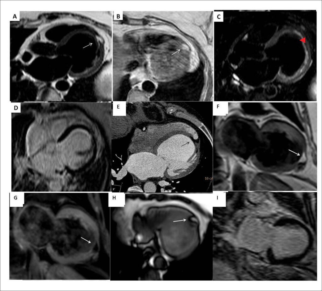
For further evaluation, a cardiac magnetic resonance (CMR) study was performed. A focal area of fatty infiltration was seen in the left ventricular (LV) apex extending from the subendocardial to the subepicardial surfaces ( and ). This area was suppressed with fat saturation pulse-sequences (), and did not show enhanced signal on delayed-hyperenhancement images (DHE) (). Both LV and LA were dilated; LV systolic function was reduced globally. Additionally, the CMR showed normal RV dimensions, function, and wall-thickness. The fatty area was also seen retrospectively on the cardiac CT ().
[…]
Images
Image gallery fullscreen viewer
Comments are moderated by the editorial team before publication. Discussions should be evidence-based and use scientific language. Conflicts of interest should be declared.