Abdominal Pain: an Uncommon Presentation of Myocardial Rupture
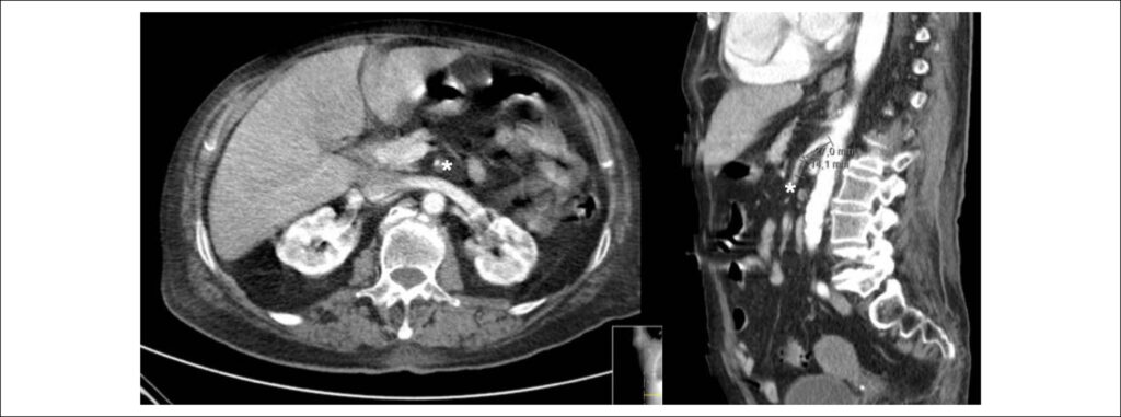
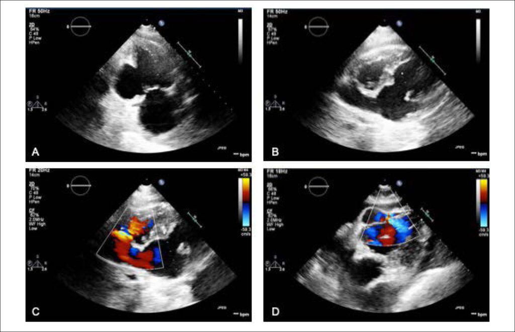
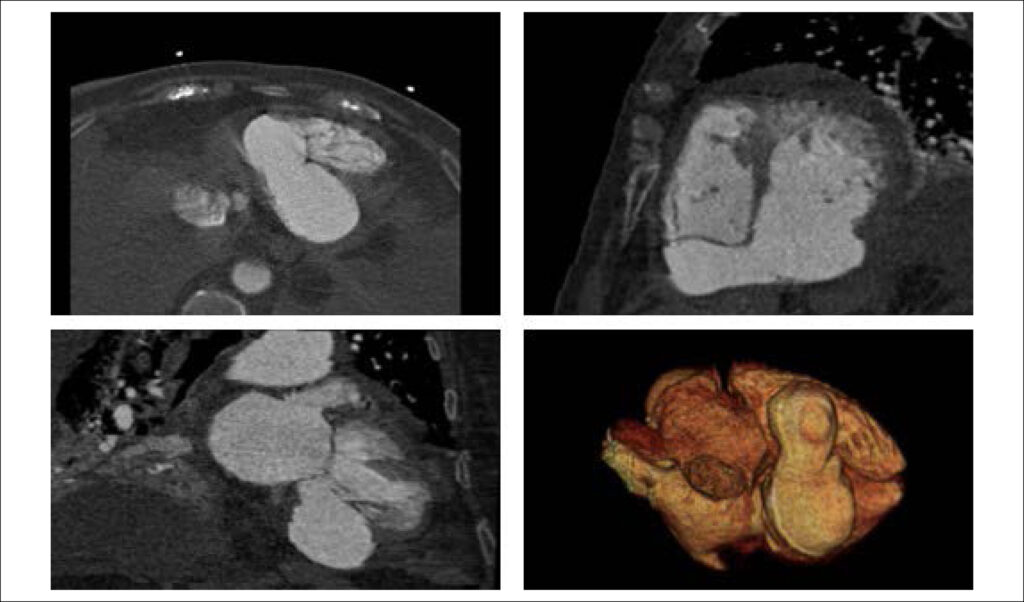
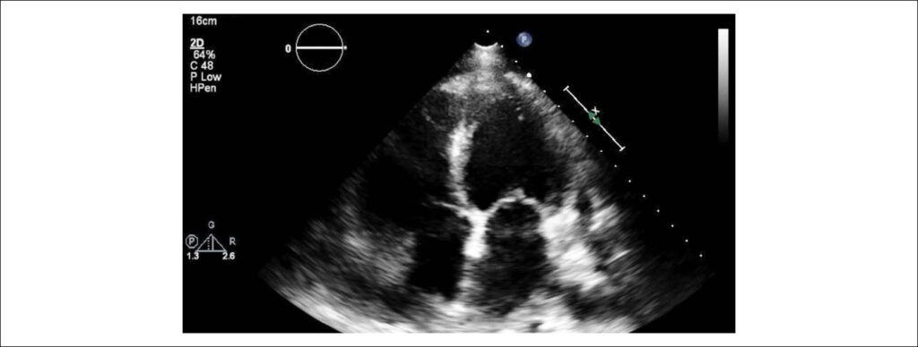
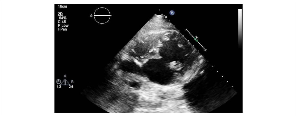
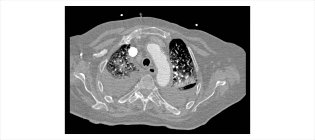
An 84-years-old woman with hypertension and dyslipidemia was admitted in the emergency room with acute abdominal pain; patient complained of chest pain three weeks before hospital admission. On physical examination, she was hypotensive, tachycardic (with arrhythmic pulse), tachypneic and with diffuse abdominal pain. Electrocardiogram showed atrial fibrillation with rapid ventricular response, complete left bundle branch block and inferior Q waves. Abdominal computed tomography (CT) revealed a thrombus in the superior mesenteric artery (, white asterisk). The patient showed a clinical course with congestive heart failure and low cardiac output. Transthoracic echocardiogram (-) showed mild left ventricular dilatation with a mild dysfunction and a pseudoaneurysm of the basal half of the posterior and inferior walls with left-to-right shunt, confirmed by color Doppler imaging (-). Cardiac CT () revealed contained myocardial rupture, located at the basal segments of the inferior and posterior septal walls, extending to the free wall of the right ventricle, forming a pseudo-cavity, which communicates with the true cavity of the right ventricle (). Despite vasopressor and inotropic support and proposal for cardiac surgery, the patient had an unfavorable course.
[…]
Images
Image gallery fullscreen viewer
Comments are moderated by the editorial team before publication. Discussions should be evidence-based and use scientific language. Conflicts of interest should be declared.