ABC Family Arquivos Brasileiros de Cardiologia International Journal of Cardiovascular Sciences ABC Heart Failure & Cardiomyopathy ABC Imagem Cardiovascular
Português English
ABC Family
Português English
  • About
    • About ABC Family
    • Internal Regulation
    • Editorial Board
    • Brazilian Society of Cardiology (SBC)
  • Issues
  • SBC Guidelines
  • Content
    • Videos
    • Podcasts
    • News
  • About
    • About ABC Family
    • Internal Regulation
    • Editorial Board
    • Brazilian Society of Cardiology (SBC)
  • Issues
  • SBC Guidelines
  • Content
    • Videos
    • Podcasts
    • News

ABC Family

  • ABC Family
  • Arquivos Brasileiros de Cardiologia
  • International Journal of Cardiovascular Sciences
  • ABC Heart Failure & Cardiomyopathy
  • ABC Imagem Cardiovascular
Search in

No content found.

Journals
Ordering
Article types
Others

  1. Home
  2. Keyword: myocardial perfusion
ABC
Editorial

One-Stop Shop for Non-Invasive Cardiovascular Imagers?

Rodrigo Julio Cerci et al. Afonso Akio Shiosaki

Over the past fifteen years, coronary computed tomography angiography (CCTA) has witnessed rapid technological and scientific advances in the detection of anatomical coronary artery disease (CAD), leading to an improvement in patient care. Visual assessment of stenosis severity using CCTA has a high sensitivity and negative predictive value when compared to invasive angiography, making it an ideal test to exclude obstructive CAD. With its high diagnostic performance associated with an important prognostic impact in the management of CAD, CCTA has finally established itself as a Class I recommendation in international guidelines (European Society of Cardiology – ESC).

However, CCTA is limited by modest diagnostic specificity and only provides anatomical assessment, which does not inform hemodynamic significance of specific lesions. CCTA combined with stress tomography evaluation of myocardial perfusion (CTP) is an accurate modality to determine regional myocardial flow repercussions of coronary stenosis, though it usually requires additional acquisition and is still underused. Derived flow fractional reserve – computed tomography (FFR-CT) is another “physiologic” CT approach in which computational fluid dynamics is applied to standard CCTA data and has emerged as a promising tool for the functional assessment of coronary stenosis. The diagnostic value of remotely performed FFR-CT has been prospectively validated in several large multicenter studies, but requires the use of offsite supercomputers, which can be time-consuming and cost-intensive, limiting its widespread clinical utility. –

[…]

  • Cardiovascular Diseases/diagnostic, imaging
  • Coronary Artery Disease
  • Coronary, Tomography
  • Diagnostic Imaging/trends
  • myocardial perfusion
Read Full Article
Creative Commons Attribution (CC BY) Creative Commons license
14/06/2021 DOI: 10.36660/abc.20210245 Views 2
ABC
Editorial

In Search of the Perfect Coronary Perfusion

Carlos Eduardo Lucena Montenegro

Since medical school, we always heard that “time is muscle” and that the faster we reperfuse a culprit artery in acute coronary syndrome (ACS), the better for the patient. Over time, evidence-based cardiology has taught us that not every patent artery is the same. The fact that we have an artery with a “TIMI 3” flow seemed enough to define the patient’s prognosis, but after the concept of microvascular ischemia, we also started to care about small-vessel perfusion.,

Based on this concern, came the concept of ‘no reflow’, which means that even after the recanalization of a culprit artery, the tissue flow related to that myocardial territory might not be reestablished. Going back in time, the phenomenon of slow flow has been described since 1972, defined as delayed coronary opacification in the absence of epicardial obstructive coronary disease, while maintaining myocardial perfusion. Slow flow seems to be more common in patients with metabolic syndrome, in the male gender and smokers. Both the phenomenon of no reflow and slow flow are associated with significant cardiovascular outcomes, the first being related to ventricular dysfunction and cardiac remodeling, and the latter, to cases of ventricular arrhythmias or sudden death,, in addition to refractory angina.

[…]

  • Acute coronary syndrome
  • Coronary Thrombosis
  • Heart Rate
  • Kidney Injury Prognosis
  • Metabolic Syndrome
  • Myocardial Infarction
  • myocardial perfusion
  • Stroke
Read Full Article
Creative Commons Attribution (CC BY) Creative Commons license
17/05/2021 DOI: 10.36660/abc.20210228
Figure 1
IJCS
Review Article

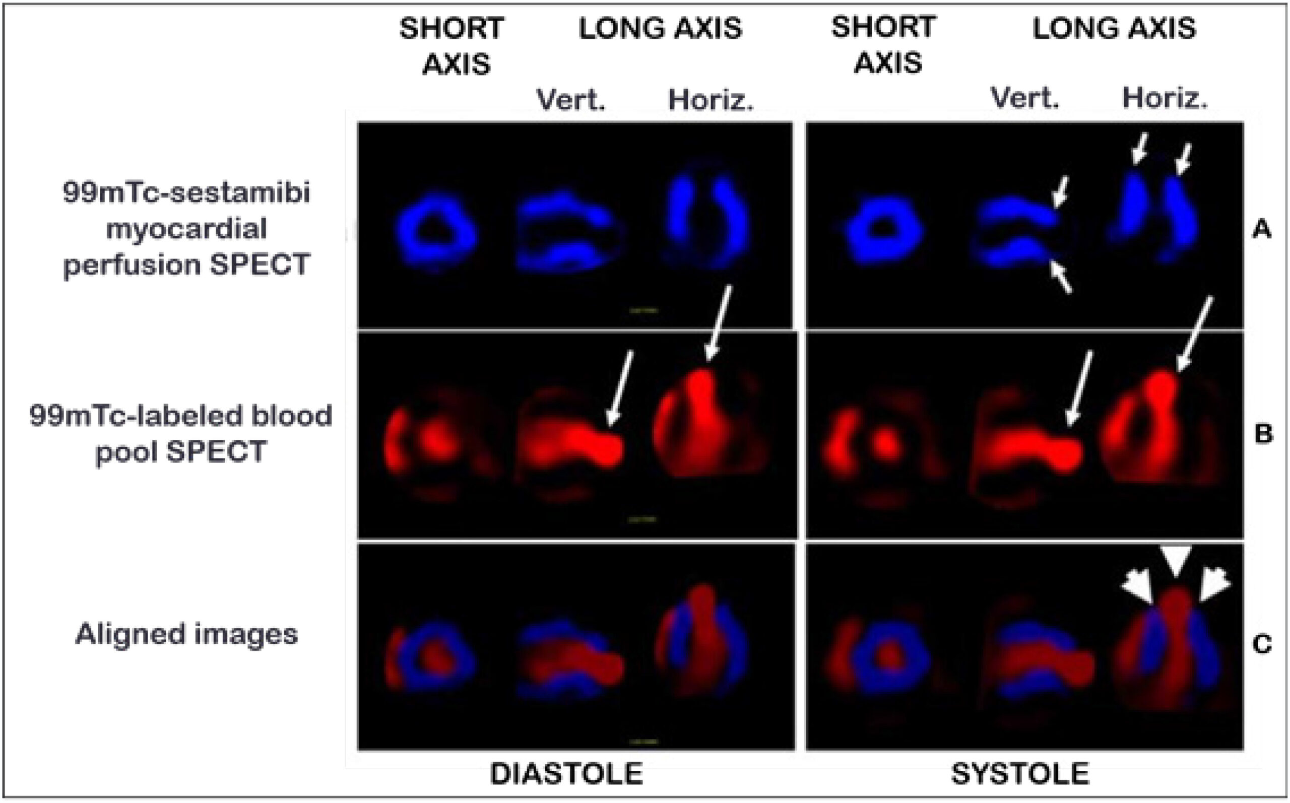
Nuclear Medicine Methods for Assessment of Chronic Chagas Heart Disease

Marcus Vinicius Simões et al. Denise Mayumi Tanaka José Antonio Marin-Neto

Abstract

Several different imaging methods can be used to evaluate patients with Chagas heart disease (CHD) for diagnostic and prognostic purposes, including plain chest radiography; echocardiography; myocardial perfusion scintigraphy, for detection of ischemia and fibrosis; radionuclide gated-angiography, for evaluation of biventricular function; 123I-MIBG labeling of sympathetic myocardial innervation; MRI, for detection and quantitation of myocardial fibrosis; and coronary angiography. This study aims to review the contributions of these nuclear medicine methods to understanding of the pathophysiology of chronic Chagas cardiomyopathy (CCC). Careful analysis and integration of findings provided by these imaging methods in patients with CCC at different stages has contributed significantly to improving understanding of several peculiarities of the disease. Clinical and experimental studies in animal models show that perfusion abnormalities detected in association with dysfunctional but viable myocardium are a common finding in CCC patients and correspond to areas of cardiac sympathetic denervation, as assessed by 123I-MIBG imaging. Furthermore, recent reports have demonstrated a close relationship between coronary microvascular disturbances and myocardial inflammation. Thus, ongoing research, mainly focused on refinements of 18F-FDF -PET techniques and further exploration of nuclear methods, such as SPECT, have the potential to contribute to detection and monitoring of early subclinical myocardial damage thereby enabling evaluation of therapeutic strategies targeting inflammation and microvascular ischemia that could result in better prognostic stratification of patients with CHD.

  • Chagas Cardiomyopathy
  • Chagas disease
  • Diagnostic Imaging
  • Echocardiography
  • myocardial perfusion
  • Prognosis
Read Full Article
Creative Commons Attribution (CC BY) Creative Commons license
25/11/2020 DOI: 10.36660/ijcs.20200153 Views 2
Figure 1
ABC
Original Article

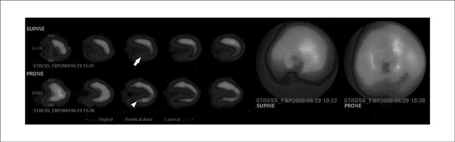
The Benefits of Prone SPECT Myocardial Perfusion Imaging in Reducing Both Artifact Defects and Patient Radiation Exposure

Maria Stathaki et al. Sophia Koukouraki Emmanouela Papadaki Angeliki Tsaroucha Nikolaos Karkavitsas

Abstract

Background:

Prone imaging has been demonstrated to minimize diaphragmatic and breast tissue attenuation.

Objectives:

To determine the role of prone imaging on the reduction of unnecessary rest perfusion studies and coronary angiographies performed, thus decreasing investigation time and radiation exposure.

Methods:

We examined 139 patients, 120 with an inferior wall and 19 with an anterior wall perfusion defect that might represented attenuation artifact. Post-stress images were acquired in both the supine and prone position. Coronary angiography was used as the “gold standard” for evaluating coronary artery patency. The study was terminated and rest imaging was obviated in the presence of complete improvement of the defect in the prone position. Quantitative interpretation was performed. Results were compared with clinical data and coronary angiographic findings.

Results:

Prone acquisition correctly revealed defect improvement in 89 patients (89/120) with inferior wall and 12 patients (12/19) with anterior wall attenuation artifact. Quantitative analysis demonstrated statistically significant difference in the mean summed stress scores (SSS) of supine and mean SSS of prone studies in patients with disappearing inferior wall defect in the prone position and patent right coronary artery (true negative results). The mean difference between SSS in supine and in prone position was higher with disappearing than with remaining defects.

Conclusion:

Technetium-99m (Tc-99m) tetrofosmin myocardial perfusion imaging with the patient in the prone position overcomes soft tissue attenuation; moreover it provides an inexpensive, accurate approach to limit the number of unnecessary rest perfusion studies and coronary angiographies performed.

  • myocardial perfusion
  • Prone Position
  • Radioactive Emission
  • SPECT instead of Tomography emission-computed single-photon
  • Technetium (Tc-99m) Tetrofosmin
Read Full Article
Creative Commons Attribution (CC BY) Creative Commons license
25/09/2015 DOI: 10.5935/abc.20150122
Late enhancement and myocardial perfusion in hypertrophic cardiomyopathy (comparison betw een groups)
ABC
Artigo Original

Late enhancement and myocardial perfusion in hypertrophic cardiomyopathy (comparison betw een groups)

Clarissa Almeida Sarmento Barbosa et al. Cláudio Campi de Castro Luiz Francisco Rodrigues de Ávila José Rodrigues Parga Filho … Domingos Mohanad Hattem Edmundo Arteaga Fernandez

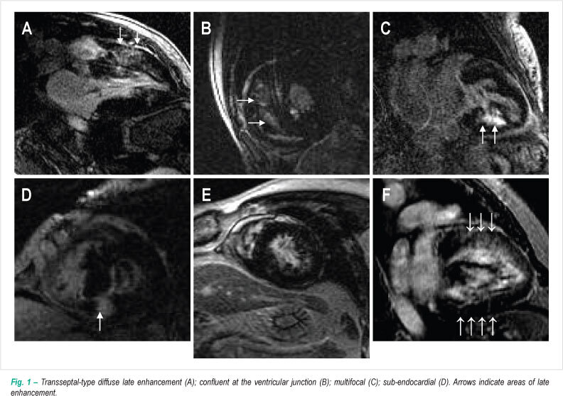
BACKGROUND: The magnetic resonance imaging (MRI) is an effective method to study hypertrophic cardiomyopathy (HCM). OBJECTIVE: To evaluate, using MRI, the parameters of systolic function, perfusion and myocardial viability in patients with HCM, comparing the groups with and without obstruction of the left ventricular outflow tract. METHODS: Twenty-one patients with a diagnosis of HCM underwent the assessment of myocardial function, viability and perfusion under stress and at rest through MRI. RESULTS: The ventricular segments most severely impaired by hypertrophy were those of the septal region. The obstructive group presented segmental myocardial thickening distribution similar to the non-obstructive group, but with higher means than the first group. The mean ejection fraction of the patients in the obstructive group was higher than in the non-obstructive group, whereas the means of the end systolic and diastolic volumes were lower in the obstructive group. There was a positive correlation between the ventricular segmental thickening and the late enhancement segmental mass. The stress induction resulted in an increase in the number of segments with perfusion alterations and this alteration was more evident in the obstructive group. CONCLUSION: The thickest ventricular segments are the septal ones. The hypertrophic regions are associated to a greater extension of late enhancement. There was a positive correlation between the areas of ventricular hypertrophy and altered myocardial perfusion and these findings were more evident in the obstructive group.

  • Comparative Study
  • Hypertrophic Cardiomyopathy
  • myocardial perfusion
  • resonance imaging
Read Full Article
Creative Commons Attribution (CC BY) Creative Commons license
24/11/2009 DOI: 10.1590/S0066-782X2009001000017
Cardiac resynchronization therapy improves the uptake of MIBI-99mTc and cardiac function
ABC
Relato de Caso

Cardiac resynchronization therapy improves the uptake of MIBI-99mTc and cardiac function

Simone Cristina Soares Brandão et al. Maria Clementina Giorgi Silvana D'Orio Nishioka Martino Martinelli Filho … José Soares Jr. José Cláudio Meneghetti

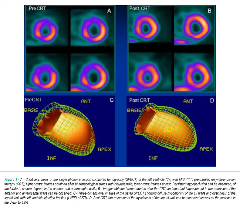
This case shows the improvement promoted by cardiac resynchronization therapy (CRT) on myocardial perfusion and left ventricular (LV) performance assessed by gated myocardial perfusion scintigraphy. The patient had idiopathic dilated cardiomyopathy, left bundle branch block and severe heart failure despite optimized medical treatment. After CRT, clinical improvement, QRS reduction and improvement of previously hypoperfused anterior and septal walls were observed. There was also decrease in LV end-diastolic and systolic volumes and increase in LV ejection fraction.

  • Dilated Cardiomyopathy
  • Left Ventricular Function
  • myocardial perfusion
  • Ventricular Function
Read Full Article
Creative Commons Attribution (CC BY) Creative Commons license
01/10/2008 DOI: 10.1590/S0066-782X2008001500013
Erectile dysfunction: a marker for myocardial perfusion impairment?
ABC
Artigo Original

Erectile dysfunction: a marker for myocardial perfusion impairment?

Juarez Ortiz et al. Sergio Tranchesi Ortiz Claudia Gianini Monaco Cristina Harumi Yamashita … Miriam Cássia Mendes Moreira Carlos Alberto F. Monaco

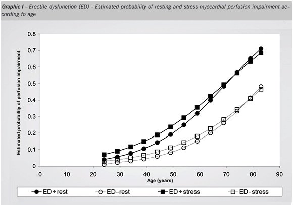
OBJECTIVE: To study the correlation between erectile dysfunction (ED) and myocardial perfusion impairment in men with suspected or diagnosed coronary artery disease (CAD). METHODS: In this prospective study a self-administered IIEF-5 questionnaire was answered by 287 patients that underwent myocardial perfusion imaging under both resting and stress condition with technetium-99m sestamibi, through gated SPECT nuclear scintigraphy technique, before and after physical or pharmacological stress. RESULTS: Some degree of erectile dysfunction (group ED+) was found in 137 (47.8%) patients and in these, age was significantly higher (60.60±9.84 vs 50.67±9.94 – p<0.001)) than in those without erectile dysfunction (group ED-). In the ED+ group, it was observed a higher prevalence of hypertension, diabetes, myocardial infarction (MI) and percutaneous coronary angioplasty (PCTA). Regardless of the age factor, ED+ patients also presented higher occurrence of myocardial perfusion impairment (necrosis and/or ischemia) and left ventricular systolic wall motion and thickening abnormalities. CONCLUSION: Patients with ED have higher estimated probability of presenting segmental myocardial perfusion and functional contraction impairment and, therefore, CAD, independent of the aging factor. The authors conclude that ED may be considered not only a marker for CAD but also a condition related to the occurrence of major coronary events such as MI and PCTA.

  • Atherosclerosis
  • Coronary Artery Disease
  • erectile dysfunction
  • myocardial perfusion
  • Nuclear Medicine
  • Risk Factors
Read Full Article
Creative Commons Attribution (CC BY) Creative Commons license
07/11/2005 DOI: 10.1590/S0066-782X2005001700003 Views 2
Myocardial contrast echocardiography in patients with suspected or known coronary artery disease: comparison with myocardial nuclear scintigraphy
ABC
Artigo Original

Myocardial contrast echocardiography in patients with suspected or known coronary artery disease: comparison with myocardial nuclear scintigraphy

Marcia Carrinho et al. Alvaro Moraes Fernando Morcerf Caio Medeiros … Márcia Castier Luis José Martins Romeo

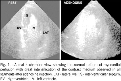
OBJECTIVE: To compare myocardial contrast echocardiography (MCE) using PESDA and adenosine in bolus (ADN) with myocardial nuclear scintigraphy (NS) in patients (pts) undergoing routine investigation with a high probability of having coronary artery disease. METHODS: This study comprised 125 pts (85 men) with 58.4 ± 10.6 years, who underwent MCE and NS within 4 weeks. MCE was performed with PESDA in a continuous infusion at rest and after administration of an adenosine bolus. The LV walls was divided into 3 territories related to the coronary arteries, in a total of 375 territories. MCE was normal when an increase in contrast intensity occurred after ADN. The reduction in contrast intensity at rest or after ADN was defined as an abnormal MCE result. NS was performed according to classical protocols. When compared per patient, both examinations were considered concordant when they were normal or abnormal, independent of its location. The comparison by territory was considered concordant when perfusion defects existed or not in the same territory. The chi-square test was used to determine the significance of concordance. RESULTS: In 106/125 pts, MCE and NS were concordant (84.8% – P < 0.001). Concordance occurred in 342/375 territories (91.2% – P < 0.001). For the LAD territory, concordance was 87.2%; for the RCA, 93.6%; and for the CX, 92.8% (P < 0.001). CONCLUSION: An excellent concordance exists between MCE and NS in assessing pts for coronary artery disease; therefore, MCE may represent a good alternative for assessing myocardial perfusion.

  • Adenosine
  • Coronary Artery Disease
  • echocardiographic contrast medium
  • Echocardiography
  • myocardial nuclear scintigraphy
  • myocardial perfusion
Read Full Article
Creative Commons Attribution (CC BY) Creative Commons license
11/11/2004 DOI: 10.1590/S0066-782X2004001700008
Study of Coronary Flow Reserve with Intravenous Use of Microbubbles (Contrast Echocardiography) and Adenosine: Protocol for Clinical Application in Patients Suspected of Having Coronary Heart Disease
ABC
Original Article

Study of Coronary Flow Reserve with Intravenous Use of Microbubbles (Contrast Echocardiography) and Adenosine: Protocol for Clinical Application in Patients Suspected of Having Coronary Heart Disease

Fernando Morcerf et al. Alvaro Moraes Marcia Carrinho Hans J. F. Dohmann

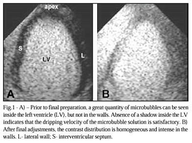
OBJECTIVE: To test the feasibility, safety and accuracy of the adenosine protocol in the study of myocardial perfusion with microbubbles contrast echocardiography. METHODS: 81 pts (64 male, 60+11 years) were submitted to contrast echocardiography with PESDA (sonicated solution of albumin 20%-1ml, dextrose 5%-12ml and deca-fluorobutane gas-8ml) to study the myocardial perfusion at rest and after bolus injection of adenosine (6 to 18mg) and to coronary angiography within 1 month each other. For each patient 3 left ventricle perfusion beds were considered (total of 243 territories). 208 territories were analyzed and 35 territories were excluded. PESDA was continuously infused (1-2ml/min), titrated for best myocardial contrast. Triggered (1:1) second harmonic imaging was used. RESULTS: Coronary angiography showed 70 flow limiting (> 75%) lesions and 138 no flow limiting lesions. At rest an obvious myocardium contrast enhancement was seen in at least 1 segment of a territory in all patients. After adenosine injection an unquestionable further increase in myocardial contrast was observed in 136 territories (99%) related to no flow limiting lesions, lasting < 10 s, and a myocardial perfusion defect was detected in 68 territories (97%) related to flow limiting lesions. It was observed only 4 false results. There were no serious complications. CONCLUSION: Myocardial perfusion study with PESDA and adenosine protocol is a practical, safe and accurate method to analyze the coronary flow reserve.

  • Coronary Disease
  • echocardiographic contrast
  • myocardial perfusion
Read Full Article
Creative Commons Attribution (CC BY) Creative Commons license
19/04/2002 DOI: 10.1590/S0066-782X2002000300004 Views 1
Myocardial perfusion by contrast echocardiography. Establishment of normal pattern of intracoronary injection and safety in humans
ABC
Original Article

Myocardial perfusion by contrast echocardiography. Establishment of normal pattern of intracoronary injection and safety in humans

Fernando Morcerf et al. Armando Luiz Cantisano Flavia Salek Carlos Alberto Mendonça … Guilherme Paulino Flavio Cure Palheiro Antonio Carlos Nogueira Wanderley Quarte Pereira Álvaro Moraes

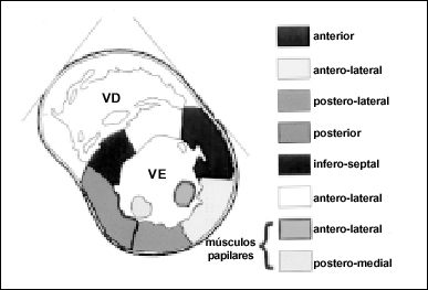
OBJECTIVE: To establish the normal pattern and safety of echocardiographic contrast in patients with no significant obstruction of epicardial coronary arteries. METHODS: 67 patients with normal coronary arteries or obstructions < 50% were selected from 277 patients who underwent coronary angiography (CA). Mean age was 56 ± 11years and 36 were males. At the end CA, echocardiographic contrast was selectively injected into each coronary artery. The parasternal short axis of the left ventricle (LV) was divided into six segments: anterior (A), antero-lateral (AL), postero-lateral (PL), posterior (P), infero-septal (IS) and antero-septal (AS). Anterolateral (ALPM) and posteromedial papillary muscles (PMPM) were also considered. The pattern and intensity of the appearance of the myocardial contrast was visually analyzed. RESULTS: The right coronary artery (RCA) was dominant in 60 patients. Contrast appearance was sudden and simultaneous in the 3 muscle layers. All segments could be contrasted after the injection in both coronary arteries. 100% of the AS, A and AL segments, 97% of the PL and 98% of the ALPM were perfused by the left coronary artery (LCA). P and IS segments were perfused by the RCA in 85% and 82%, respectively, and by a dominant LCA in 71% of the cases. The PMPM was perfused by a dominant RCA in 77% and by a dominant LCA in 86%. There were no symptoms. CONCLUSION: Intracoronary injection of the sonicated solution is a safe procedure that allows for an excellent opacification of the myocardium and can potentially be used during routine CA.

  • Echocardiography
  • Microbubbles
  • myocardial contrast echocardiography
  • myocardial perfusion
Read Full Article
Creative Commons Attribution (CC BY) Creative Commons license
08/01/2002 DOI: 10.1590/S0066-782X1999000600003 Views 2

How to cite this article

EndNote RIS

Electronic Document Format (Vancouver)

Cerci RJ, Shiosaki AA. One-Stop Shop for Non-Invasive Cardiovascular Imagers?. Arquivos Brasileiros de Cardiologia 2021;116(6):1099-100.

Electronic Document Format (ABNT)

Cerci, Rodrigo Julio; Shiosaki, Afonso Akio. One-Stop Shop for Non-Invasive Cardiovascular Imagers?. Arquivos Brasileiros de Cardiologia, v. 116, n. 6, p. 1099-1100, Jun. 2021.

Electronic Document Format (APA)

Cerci, R. J., & Shiosaki, A. A. (2021). One-Stop Shop for Non-Invasive Cardiovascular Imagers?. Arquivos Brasileiros de Cardiologia, 116(6), 1099-1100.

Electronic Document Format (ISO)

Cerci, Rodrigo Julio and Shiosaki, Afonso Akio. One-Stop Shop for Non-Invasive Cardiovascular Imagers?. Arquivos Brasileiros de Cardiologia [online]. 2021, vol. 116, n. 6, [cited 2026-10-14], pp. 1099-1100. Available from: <https://journals.cardiol.br/en/article/one-stop-shop-for-non-invasive-cardiovascular-imagers/>. ISSN 0066-782X

EndNote RIS

Electronic Document Format (Vancouver)

Montenegro CEL. In Search of the Perfect Coronary Perfusion. Arquivos Brasileiros de Cardiologia 2021;116(5):865-6.

Electronic Document Format (ABNT)

Montenegro, Carlos Eduardo Lucena. In Search of the Perfect Coronary Perfusion. Arquivos Brasileiros de Cardiologia, v. 116, n. 5, p. 865-866, May. 2021.

Electronic Document Format (APA)

Montenegro, C. E. L. (2021). In Search of the Perfect Coronary Perfusion. Arquivos Brasileiros de Cardiologia, 116(5), 865-866.

Electronic Document Format (ISO)

Montenegro, Carlos Eduardo Lucena. In Search of the Perfect Coronary Perfusion. Arquivos Brasileiros de Cardiologia [online]. 2021, vol. 116, n. 5, [cited 2026-10-14], pp. 865-866. Available from: <https://journals.cardiol.br/en/article/in-search-of-the-perfect-coronary-perfusion/>. ISSN 0066-782X

EndNote RIS

Electronic Document Format (Vancouver)

Simões MV, Tanaka DM, Marin-Neto JA. Nuclear Medicine Methods for Assessment of Chronic Chagas Heart Disease. International Journal of Cardiovascular Sciences 2020;33(6):686-9.

Electronic Document Format (ABNT)

Simões, Marcus Vinicius; Tanaka, Denise Mayumi; Marin-Neto, José Antonio. Nuclear Medicine Methods for Assessment of Chronic Chagas Heart Disease. International Journal of Cardiovascular Sciences, v. 33, n. 6, p. 686-696, Nov. 2020.

Electronic Document Format (APA)

Simões, M. V., Tanaka, D. M., & Marin-Neto, J. A. (2020). Nuclear Medicine Methods for Assessment of Chronic Chagas Heart Disease. International Journal of Cardiovascular Sciences, 33(6), 686-696.

Electronic Document Format (ISO)

Simões, Marcus Vinicius and Tanaka, Denise Mayumi and Marin-Neto, José Antonio. Nuclear Medicine Methods for Assessment of Chronic Chagas Heart Disease. International Journal of Cardiovascular Sciences [online]. 2020, vol. 33, n. 6, [cited 2026-10-14], pp. 686-696. Available from: <https://journals.cardiol.br/en/article/nuclear-medicine-methods-for-assessment-of-chronic-chagas-heart-disease/>. ISSN 2359-4802

EndNote RIS

Electronic Document Format (Vancouver)

Stathaki M, Koukouraki S, Papadaki E, Tsaroucha A, Karkavitsas N. The Benefits of Prone SPECT Myocardial Perfusion Imaging in Reducing Both Artifact Defects and Patient Radiation Exposure. Arquivos Brasileiros de Cardiologia 2015;105(4):345-52.

Electronic Document Format (ABNT)

Stathaki, Maria; Koukouraki, Sophia; Papadaki, Emmanouela; Tsaroucha, Angeliki; Karkavitsas, Nikolaos. The Benefits of Prone SPECT Myocardial Perfusion Imaging in Reducing Both Artifact Defects and Patient Radiation Exposure. Arquivos Brasileiros de Cardiologia, v. 105, n. 4, p. 345-352, Sep. 2015.

Electronic Document Format (APA)

Stathaki, M., Koukouraki, S., Papadaki, E., Tsaroucha, A., & Karkavitsas, N. (2015). The Benefits of Prone SPECT Myocardial Perfusion Imaging in Reducing Both Artifact Defects and Patient Radiation Exposure. Arquivos Brasileiros de Cardiologia, 105(4), 345-352.

Electronic Document Format (ISO)

Stathaki, Maria and Koukouraki, Sophia and Papadaki, Emmanouela and Tsaroucha, Angeliki and Karkavitsas, Nikolaos. The Benefits of Prone SPECT Myocardial Perfusion Imaging in Reducing Both Artifact Defects and Patient Radiation Exposure. Arquivos Brasileiros de Cardiologia [online]. 2015, vol. 105, n. 4, [cited 2026-10-14], pp. 345-352. Available from: <https://journals.cardiol.br/en/article/the-benefits-of-prone-spect-myocardial-perfusion-imaging-in-reducing-both-artifact-defects-and-patient-radiation-exposure/>. ISSN 0066-782X

EndNote RIS

Electronic Document Format (Vancouver)

Barbosa CAS, Castro CC, Ávila LFR, Parga Filho JR, Hattem DM, Fernandez EA. Late enhancement and myocardial perfusion in hypertrophic cardiomyopathy (comparison betw een groups). Arquivos Brasileiros de Cardiologia 2009;93(4):426-33.

Electronic Document Format (ABNT)

Barbosa, Clarissa Almeida Sarmento; Castro, Cláudio Campi de; Ávila, Luiz Francisco Rodrigues de; Parga Filho, José Rodrigues; Hattem, Domingos Mohanad; Fernandez, Edmundo Arteaga. Late enhancement and myocardial perfusion in hypertrophic cardiomyopathy (comparison betw een groups). Arquivos Brasileiros de Cardiologia, v. 93, n. 4, p. 426-433, Nov. 2009.

Electronic Document Format (APA)

Barbosa, C. A. S., Castro, C. C. , Ávila, L. F. R. , Parga Filho, J. R., Hattem, D. M., & Fernandez, E. A. (2009). Late enhancement and myocardial perfusion in hypertrophic cardiomyopathy (comparison betw een groups). Arquivos Brasileiros de Cardiologia, 93(4), 426-433.

Electronic Document Format (ISO)

Barbosa, Clarissa Almeida Sarmento and Castro, Cláudio Campi de and Ávila, Luiz Francisco Rodrigues de and Parga Filho, José Rodrigues and Hattem, Domingos Mohanad and Fernandez, Edmundo Arteaga. Late enhancement and myocardial perfusion in hypertrophic cardiomyopathy (comparison betw een groups). Arquivos Brasileiros de Cardiologia [online]. 2009, vol. 93, n. 4, [cited 2026-10-14], pp. 426-433. Available from: <https://journals.cardiol.br/en/article/late-enhancement-and-myocardial-perfusion-in-hypertrophic-cardiomyopathy-comparison-betw-een-groups/>. ISSN 0066-782X

EndNote RIS

Electronic Document Format (Vancouver)

Brandão SCS, Giorgi MC, Nishioka SD, Martinelli Filho M, Soares Jr. J, Meneghetti JC. Cardiac resynchronization therapy improves the uptake of MIBI-99mTc and cardiac function. Arquivos Brasileiros de Cardiologia 2008;91(3):e25-8.

Electronic Document Format (ABNT)

Brandão, Simone Cristina Soares; Giorgi, Maria Clementina; Nishioka, Silvana D’Orio; Martinelli Filho, Martino; Soares Jr., José; Meneghetti, José Cláudio. Cardiac resynchronization therapy improves the uptake of MIBI-99mTc and cardiac function. Arquivos Brasileiros de Cardiologia, v. 91, n. 3, p. e25-e28, Sep. 2008.

Electronic Document Format (APA)

Brandão, S. C. S., Giorgi, M. C., Nishioka, S. D., Martinelli Filho, M., Soares Jr., J., & Meneghetti, J. C. (2008). Cardiac resynchronization therapy improves the uptake of MIBI-99mTc and cardiac function. Arquivos Brasileiros de Cardiologia, 91(3), e25-e28.

Electronic Document Format (ISO)

Brandão, Simone Cristina Soares and Giorgi, Maria Clementina and Nishioka, Silvana D’Orio and Martinelli Filho, Martino and Soares Jr., José and Meneghetti, José Cláudio. Cardiac resynchronization therapy improves the uptake of MIBI-99mTc and cardiac function. Arquivos Brasileiros de Cardiologia [online]. 2008, vol. 91, n. 3, [cited 2026-10-14], pp. e25-e28. Available from: <https://journals.cardiol.br/en/article/cardiac-resynchronization-therapy-improves-the-uptake-of-mibi-99mtc-and-cardiac-function/>. ISSN 0066-782X

EndNote RIS

Electronic Document Format (Vancouver)

Ortiz J, Ortiz ST, Monaco CG, Yamashita CH, Moreira MCM, Monaco CAF. Erectile dysfunction: a marker for myocardial perfusion impairment?. Arquivos Brasileiros de Cardiologia 2005;85(4):241-6.

Electronic Document Format (ABNT)

Ortiz, Juarez; Ortiz, Sergio Tranchesi; Monaco, Claudia Gianini; Yamashita, Cristina Harumi; Moreira, Miriam Cássia Mendes; Monaco, Carlos Alberto F.. Erectile dysfunction: a marker for myocardial perfusion impairment?. Arquivos Brasileiros de Cardiologia, v. 85, n. 4, p. 241-246, Nov. 2005.

Electronic Document Format (APA)

Ortiz, J., Ortiz, S. T., Monaco, C. G., Yamashita, C. H., Moreira, M. C. M., & Monaco, C. A. F. (2005). Erectile dysfunction: a marker for myocardial perfusion impairment?. Arquivos Brasileiros de Cardiologia, 85(4), 241-246.

Electronic Document Format (ISO)

Ortiz, Juarez and Ortiz, Sergio Tranchesi and Monaco, Claudia Gianini and Yamashita, Cristina Harumi and Moreira, Miriam Cássia Mendes and Monaco, Carlos Alberto F.. Erectile dysfunction: a marker for myocardial perfusion impairment?. Arquivos Brasileiros de Cardiologia [online]. 2005, vol. 85, n. 4, [cited 2026-10-14], pp. 241-246. Available from: <https://journals.cardiol.br/en/article/erectile-dysfunction-a-marker-for-myocardial-perfusion-impairment/>. ISSN 0066-782X

EndNote RIS

Electronic Document Format (Vancouver)

Carrinho M, Moraes A, Morcerf F, Medeiros C, Castier M, Romeo LJM. Myocardial contrast echocardiography in patients with suspected or known coronary artery disease: comparison with myocardial nuclear scintigraphy. Arquivos Brasileiros de Cardiologia 2004;83(5):414-8.

Electronic Document Format (ABNT)

Carrinho, Marcia; Moraes, Alvaro; Morcerf, Fernando; Medeiros, Caio; Castier, Márcia; Romeo, Luis José Martins. Myocardial contrast echocardiography in patients with suspected or known coronary artery disease: comparison with myocardial nuclear scintigraphy. Arquivos Brasileiros de Cardiologia, v. 83, n. 5, p. 414-418, Nov. 2004.

Electronic Document Format (APA)

Carrinho, M., Moraes, A., Morcerf, F., Medeiros, C., Castier, M., & Romeo, L. J. M. (2004). Myocardial contrast echocardiography in patients with suspected or known coronary artery disease: comparison with myocardial nuclear scintigraphy. Arquivos Brasileiros de Cardiologia, 83(5), 414-418.

Electronic Document Format (ISO)

Carrinho, Marcia and Moraes, Alvaro and Morcerf, Fernando and Medeiros, Caio and Castier, Márcia and Romeo, Luis José Martins. Myocardial contrast echocardiography in patients with suspected or known coronary artery disease: comparison with myocardial nuclear scintigraphy. Arquivos Brasileiros de Cardiologia [online]. 2004, vol. 83, n. 5, [cited 2026-10-14], pp. 414-418. Available from: <https://journals.cardiol.br/en/article/myocardial-contrast-echocardiography-in-patients-with-suspected-or-known-coronary-artery-disease-comparison-with-myocardial-nuclear-scintigraphy/>. ISSN 0066-782X

EndNote RIS

Electronic Document Format (Vancouver)

Morcerf F, Moraes A, Carrinho M, Dohmann HJF. Study of Coronary Flow Reserve with Intravenous Use of Microbubbles (Contrast Echocardiography) and Adenosine: Protocol for Clinical Application in Patients Suspected of Having Coronary Heart Disease. Arquivos Brasileiros de Cardiologia 2002;78(3):290-8.

Electronic Document Format (ABNT)

Morcerf, Fernando; Moraes, Alvaro; Carrinho, Marcia; Dohmann, Hans J. F.. Study of Coronary Flow Reserve with Intravenous Use of Microbubbles (Contrast Echocardiography) and Adenosine: Protocol for Clinical Application in Patients Suspected of Having Coronary Heart Disease. Arquivos Brasileiros de Cardiologia, v. 78, n. 3, p. 290-298, Apr. 2002.

Electronic Document Format (APA)

Morcerf, F., Moraes, A., Carrinho, M., & Dohmann, H. J. F. (2002). Study of Coronary Flow Reserve with Intravenous Use of Microbubbles (Contrast Echocardiography) and Adenosine: Protocol for Clinical Application in Patients Suspected of Having Coronary Heart Disease. Arquivos Brasileiros de Cardiologia, 78(3), 290-298.

Electronic Document Format (ISO)

Morcerf, Fernando and Moraes, Alvaro and Carrinho, Marcia and Dohmann, Hans J. F.. Study of Coronary Flow Reserve with Intravenous Use of Microbubbles (Contrast Echocardiography) and Adenosine: Protocol for Clinical Application in Patients Suspected of Having Coronary Heart Disease. Arquivos Brasileiros de Cardiologia [online]. 2002, vol. 78, n. 3, [cited 2026-10-14], pp. 290-298. Available from: <https://journals.cardiol.br/en/article/study-of-coronary-flow-reserve-with-intravenous-use-of-microbubbles-contrast-echocardiography-and-adenosine-protocol-for-clinical-application-in-patients-suspected-of-having-coronary-heart-disease/>. ISSN 0066-782X

EndNote RIS

Electronic Document Format (Vancouver)

Morcerf F, Cantisano AL, Salek F, Mendonça CA, Paulino G, Palheiro FC, Nogueira AC, et al. Myocardial perfusion by contrast echocardiography. Establishment of normal pattern of intracoronary injection and safety in humans. Arquivos Brasileiros de Cardiologia 2002;72(6):682-6.

Electronic Document Format (ABNT)

Morcerf, Fernando; Cantisano, Armando Luiz; Salek, Flavia; Mendonça, Carlos Alberto; Paulino, Guilherme; Palheiro, Flavio Cure; Nogueira, Antonio Carlos; Pereira, Wanderley Quarte; Moraes, Álvaro. Myocardial perfusion by contrast echocardiography. Establishment of normal pattern of intracoronary injection and safety in humans. Arquivos Brasileiros de Cardiologia, v. 72, n. 6, p. 682-686, Jan. 2002.

Electronic Document Format (APA)

Morcerf, F., Cantisano, A. L., Salek, F., Mendonça, C. A., Paulino, G., Palheiro, F. C., Nogueira, A. C., Pereira, W. Q., & Moraes, (2002). Myocardial perfusion by contrast echocardiography. Establishment of normal pattern of intracoronary injection and safety in humans. Arquivos Brasileiros de Cardiologia, 72(6), 682-686.

Electronic Document Format (ISO)

Morcerf, Fernando and Cantisano, Armando Luiz and Salek, Flavia and Mendonça, Carlos Alberto and Paulino, Guilherme and Palheiro, Flavio Cure and Nogueira, Antonio Carlos and Pereira, Wanderley Quarte and Moraes, Álvaro. Myocardial perfusion by contrast echocardiography. Establishment of normal pattern of intracoronary injection and safety in humans. Arquivos Brasileiros de Cardiologia [online]. 2002, vol. 72, n. 6, [cited 2026-10-14], pp. 682-686. Available from: <https://journals.cardiol.br/en/article/myocardial-perfusion-by-contrast-echocardiography-establishment-of-normal-pattern-of-intracoronary-injection-and-safety-in-humans/>. ISSN 0066-782X

  • About ABC Family
  • Internal Regulation
  • Editorial Board
  • Brazilian Society of Cardiology (SBC)

Issues

SBC Guidelines

  • Videos
  • Podcasts
  • News