
| ABCIC
ABC Family Arquivos Brasileiros de Cardiologia International Journal of Cardiovascular Sciences ABC Heart Failure & Cardiomyopathy ABC Imagem Cardiovascular
Português English
ABC Family | ABCIC
Português English
  • About
    • About the Journal
    • Internal Regulation
    • Editors Section
    • Editorial Board
    • SBC Award
  • Instructions to Authors
    • Guidelines for Publication
    • Editorial Policy
    • Ethics Policy
    • Policy on the Use of Artificial Intelligence
    • Mandatory Publication Form
  • Issues
  • SBC Guidelines
  • About
    • About the Journal
    • Internal Regulation
    • Editors Section
    • Editorial Board
    • SBC Award
  • Instructions to Authors
    • Guidelines for Publication
    • Editorial Policy
    • Ethics Policy
    • Policy on the Use of Artificial Intelligence
    • Mandatory Publication Form
  • Issues
  • SBC Guidelines

ABC Family

  • ABC Family
  • Arquivos Brasileiros de Cardiologia
  • International Journal of Cardiovascular Sciences
  • ABC Heart Failure & Cardiomyopathy
  • ABC Imagem Cardiovascular
Search in

No content found.

Journals
Ordering
Article types
Others

  1. Home
  2. ABCIC
  3. Issues
  4. 2022, Volume 35, Number 3
Figure 1
Editorial

What does the navigator system in interventional procedures in the hybrid room?

Denisse Guzman Ramirez et al. Jose Maria Hernandez Hernandez

What does the navigator system add to interventional procedures in the hybrid room?

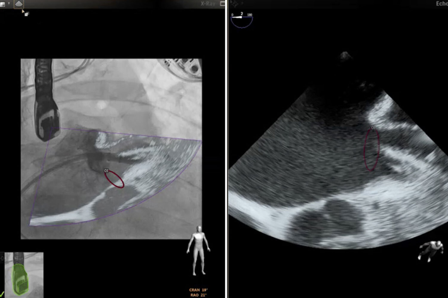
The usefulness of two-dimensional fixed projection fluoroscopic imaging in the characterization of soft tissues and complex cardiac anatomy is limited. Moreover, it is currently insufficient for cardiac structural interventionism. Including recent three-dimensional cardiac ultrasound imaging techniques is necessary to improve anatomical images and spatial resolution.

Fusion or hybrid imaging using two-dimensional fluoroscopy in combination with static or dynamic imaging obtained from cardiac computed tomography angiography (CCT), cardiac magnetic resonance (CMR), and transesophageal echocardiography (TEE) have been successfully used for structural cardiac interventions. A fusion image is the superimposition of images acquired from different modalities within the same spatial coordinate. This image mapping process is called co-registration or image registration.

[…]

  • Cardiac Surgical Procedures
  • Echocardiography
  • Fluoroscopy
Read Full Article
Creative Commons Attribution (CC BY) Creative Commons license
23/09/2022 DOI: 10.47593/2675-312X/20223503eabc332
Figure 1
Original Article

Correlation Between Echocardiographic and Hemodynamic Measurements in Right Atrium and Right Ventricle Assessments of Patients with Pulmonary Hypertension

Ana Tereza Azeredo Bastos Brito et al. João Batista Masson Silva Daniela do Carmo Rassi Daniela Graner Schuwartz Tannus Silva … Sandra Araújo Costa Diogo Sampaio de Oliveira Flavio de Souza Veiga Jardim Leandro Zacarias F. de Freitas Aguinaldo F. de Freitas Júnior Salvador Rassi

Abstract

Introduction

Pulmonary hypertension (PH), a serious clinical condition, can lead to right ventricular systolic dysfunction (RVSD) with prognostic implications. Patients with suspected PH should undergo transthoracic echocardiography (TTE) for diagnosis and evaluation as the main screening and follow-up exam.

Objective

To verify the associations of and agreement between measurements of mean pressure in the right atrium (RA) and RVSD with TTE

Method

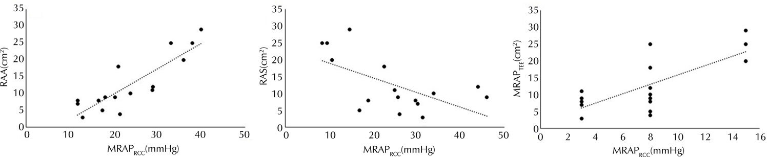
Individuals diagnosed with PH were included. All patients underwent TTE and RCC. The following were evaluated by TTE: right atrial area (RAA), mean right atrial pressure through the diameter and collapsibility of the inferior vena cava (RMAPTTE), RA strain (RAS), tricuspid annular plane systolic excursion, fractional area change, RV free wall strain, and tricuspid s’ wave. Mean RA pressure (RMAPRCC) and cardiac index (CI) were evaluated through the RCC.

Results

Of the 16 patients, 13 were female. The mean patient age was 44.4 (±14.9) years. An association was found between RMAPRCC and AAD, RMAPTTE, and RAS (r=0.845, r=0.621, and r=-0.523, respectively; p<0.05). There was an association between the mortality risk categories measured by the RAA and RMAPRCC measures (X2=10.42; p=0.003), with moderate agreement (k=0.44; p=0.012). RVSDJ was present in 10 patients. There was an association between RVSD (present or absent) and CI (r=0.522; p=0.04) with moderate agreement (k=0.43; p=0.037).

Conclusion

The TTE and RCC measurements showed an association in the assessment of mean right atrial pressure, especially between RAA and RMAPRCC. An association with RVSD and moderate agreement between methods were also noted.

  • Catheterization
  • Echocardiography
  • Hypertension
  • Pulmonary
  • Right
  • Ventricular Dysfunction
Read Full Article
Creative Commons Attribution (CC BY) Creative Commons license
23/09/2022 DOI: 10.47593/2675-312X/20223503eabc308 Views 1
What do Cardiologists Expect

What do cardiologists expect from Echocardiography in the evaluation of non-compacted myocardium

Luciano Nastari

Recommendation level IIb; evidence level B

[…]

  • Echocardiography
  • Isolated Noncompaction of the Ventricular Myocardium
  • Nuclear magnetic resonance
Read Full Article
Creative Commons Attribution (CC BY) Creative Commons license
23/09/2022 DOI: 10.47593/2675-312X/20223503ecard07
Figure 1
Original Article

Impairment of Right Ventricular Function in Patients with Systemic Sclerosis and Interstitial Lung Disease:Tissue Doppler Evaluation

Ana Clara Tude Rodrigues et al. Fernando U. Kay Marina Roque Thais Emerick Henriques Gripp … Derly Becker Ana Lucia Arruda Giovanni Guido Cerri Percival D. Sampaio-Barros

Abstract

Introduction

Systemic sclerosis (SSc) is an autoimmune tissue connective disease that courses with fibrosis and microvascular dysfunction. Involvement of the visceral organs, including the lungs and heart, is the main cause of death among patients with SSc. In this context, here we analyzed the relationship between right ventricle (RV) parameters assessed by tissue Doppler echocardiography and lung involvement in patients with SSc.

Methods

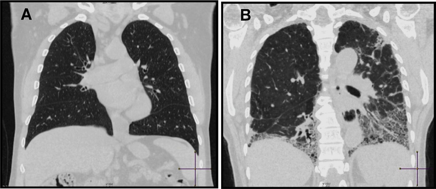
Patients fulfilling the 2013 SSc Classification Criteria underwent tissue Doppler echocardiography for the assessment of left ventricular (LV) systolic function (ejection fraction) and RV fractional area change (FAC), tissue Doppler s’ (systolic) velocity, myocardial performance index, and tricuspid annular plane systolic excursion for the assessment of RV systolic function. Pulmonary systolic pressure was estimated using tricuspid regurgitation. Chest high-resolution computed tomography was used to evaluate the presence of pulmonary fibrosis. The patients were divided into two subgroups accordingly: Group I, patients with pulmonary fibrosis (n=26); and Group II, those without fibrosis (n=17).

Results

Among the 43 patients with SSc, most were female (86%), and the mean age was 51 ± 12 years. All patients had normal systolic ventricular function as evidenced by an LV ejection fraction > 55% and an RV FAC > 35%. No significant intergroup difference was noted in age or disease duration. Except for a decreased tissue Doppler s’ velocity in patients with lung fibrosis, all indexes of RV performance were similar. Conclusion: In patients with SSc and pulmonary fibrosis, tissue Doppler identified early RV longitudinal myocardial involvement despite preserved RV radial systolic performance.

  • Pulmonary Fibrosis
  • Systemic Sclerosis
  • Tissue Doppler
Read Full Article
Creative Commons Attribution (CC BY) Creative Commons license
23/09/2022 DOI: 10.47593/2675-312X/20223503eabc331 Views 1
Editorial

Role of Echocardiography in Assessment of Patients with Suspected Ph: Beyond the Peak Tricuspid Regurgitant Velocity

Viviane Tiemi Hotta

Pulmonary hypertension (PH) causes great clinical and functional limitations and significantly impacts quality of life and survival., Current pharmacological treatment options for PH include phosphodiesterase-5 inhibitors, endothelin receptor antagonists, and prostanoids. However, an early diagnosis and etiological definition are essential to improve prognosis.

Transthoracic echocardiography (TTE) is the first-line test for suspected PH, as it is widely available, noninvasive, relatively low cost, does not involve radiation exposure, and carries minimal risk to patients. However, TTE can only suggest the diagnosis of PH since the definitive diagnosis requires invasive hemodynamic measurements via right heart catheterization.

[…]

  • Echocardiography
  • Pulmonary Hypretension
  • Right Atrium
  • Right ventricle
Read Full Article
Creative Commons Attribution (CC BY) Creative Commons license
23/09/2022 DOI: 10.47593/2675-312X/20223503eed24
Figure 1
My Approach To

My Approach to Managing Left Ventricular Diastolic Function of Children and in Congenital Heart Disease

Laura Mercer Rosa et al. Samira Saady Morhy

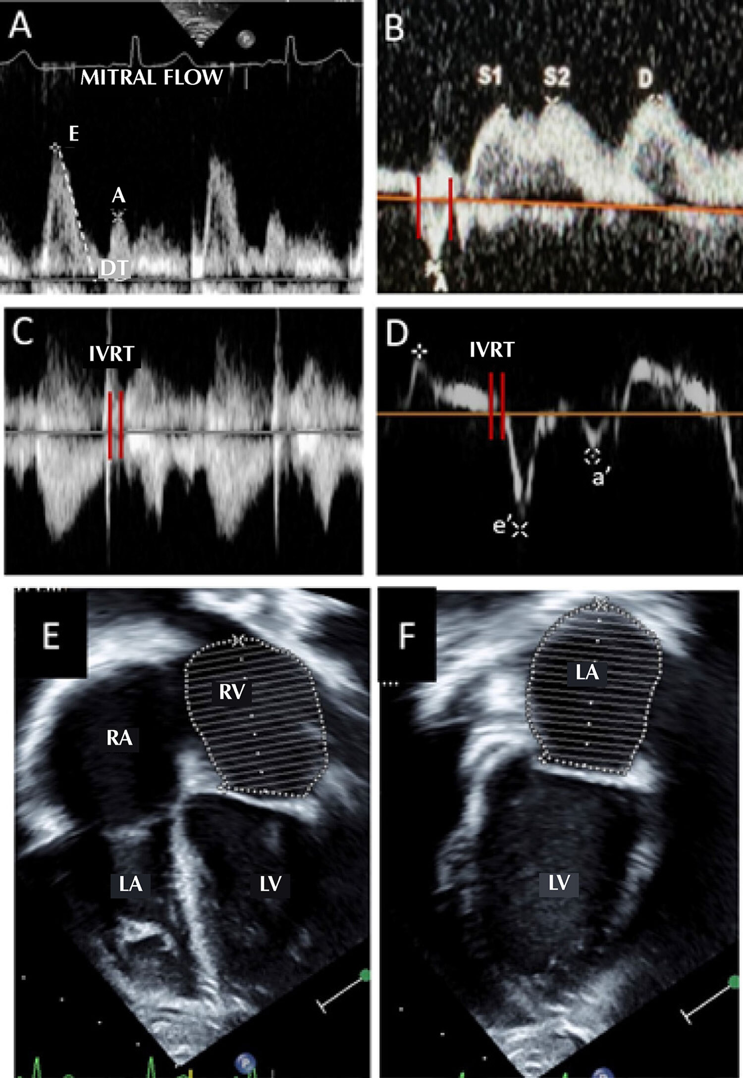
Echocardiography is the primary diagnostic method for assessing ventricular function in children and patients with congenital heart disease; however, it traditionally focuses on systolic function. Despite the growing interest in echocardiographic parameters for assessing diastolic function in children, diastolic dysfunction has been less investigated and, therefore, is less understood.

Some factors have limited this assessment:,

[…]

  • Child
  • Congenital Heart Disease
  • Echocardiography
Read Full Article
Creative Commons Attribution (CC BY) Creative Commons license
23/09/2022 DOI: 10.47593/2675-312X/20223503ecom29
Figure 1
Image

Symptomatic Aortic Mural Thrombus in a Patient With COVID-19

Milton Sérgio Bohatch Júnior et al. Amanda Fernandes Vidal da Silva Marcelo Bellini Dálio Mauricio Serra Ribeiro Edwaldo Edner Joviliano

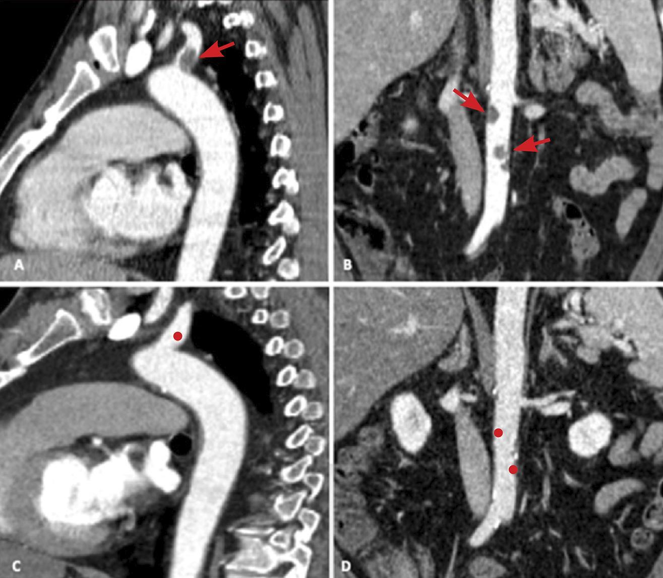
A 56-year-old male former smoker with a history of diabetes mellitus developed flu-like symptoms (cough, fever, and myalgia) that worsened after nine days (dyspnea and asthenia) sought medical attention in an emergency care unit. He was hospitalized with novel coronavirus (sudden acute respiratory syndrome coronavirus 2) infection confirmed by polymerase chain reaction testing. His condition progressed with sudden pain in the left lower limb, toe coldness and paresthesia without motor changes, and pain in the third finger of the left hand associated with non-fixed cyanosis. The patient was referred to the emergency room of Hospital das Clínicas of Ribeirão Preto with acute arterial occlusion. On evaluation, he presented non-fixed cyanosis in the distal phalanx of the third finger of the left hand with no pulse or neurological changes throughout the limb. The left lower limb presented no pulse of or arterial flow in the anterior and posterior tibial arteries. He underwent urgent thrombectomy (Rutherford class 2A) with thrombus removal, and limb perfusion was restored with pulsatile tibial artery flow. The patient remained on anticoagulant therapy with unfractionated heparin. Computed tomography angioplasty used to analyze the embolism demonstrated mural thrombi at the origin of the left subclavian artery and in the infrarenal aorta (, ). The patient progressed with good arterial condition (resolution of left upper limb non-fixed cyanosis and presence of pulse in the left leg), stayed in the intensive care unit for three days, and was discharged after two days of anticoagulant treatment (rivaroxaban [Xarelto®]). In outpatient follow-up, he remained asymptomatic and presented no new embolic events, and the mural thrombi resolved completely after one year of anticoagulant therapy (, ).

[…]

  • Aorta
  • SARS-CoV-2
  • Thrombosis
Read Full Article
Creative Commons Attribution (CC BY) Creative Commons license
17/08/2022 DOI: 10.47593/2675-312X/20223503eabc270
My Approach to Lung Ultrasound to Evaluate Extravascular Lung Water
My Approach To

My Approach to Lung Ultrasound to Evaluate Extravascular Lung Water

Maria Estefânia Bosco Otto et al. Vanessa Andreoli Esmanhoto

Introduction

A clinical history, physical examination, and focused ultrasound can accelerate the appropriate diagnosis and treatment of patients with progressive or acute dyspnea. Extravascular lung water (EVLW) is easily detected with lung ultrasound (LUS). Pivetta et al. reported excellent accuracy detecting EVLW by clinical evaluation and by the presence of “B” lines on LUS heigher than B natriuretic peptide dosage.

The usefulness of LUS for intensive care unit patients was described in the 1980s, later than that of other ultrasound modalities. LUS is useful not only for diagnosing EVLW but also for risk stratification in myocardial infarction and acute congestive heart failure (CHF).

[…]

  • Dyspneia
  • Lung
  • Ultrasonography
Read Full Article
Creative Commons Attribution (CC BY) Creative Commons license
17/08/2022 DOI: 10.47593/2675-312X/20223503ecom22
Figure 1
Case Report

Use of Contrast-Enhanced Ultrasound for the Diagnosis of Endoleak After EVAR: A Case Report

Ana Carolina N. Netto Djaló et al. Glauber Lutterbach de Oliveira Pires Adriana Ferraz Carlos Gustavo Abath … Idalécio Souto Fonseca Filho Milena O. Marques

Introduction

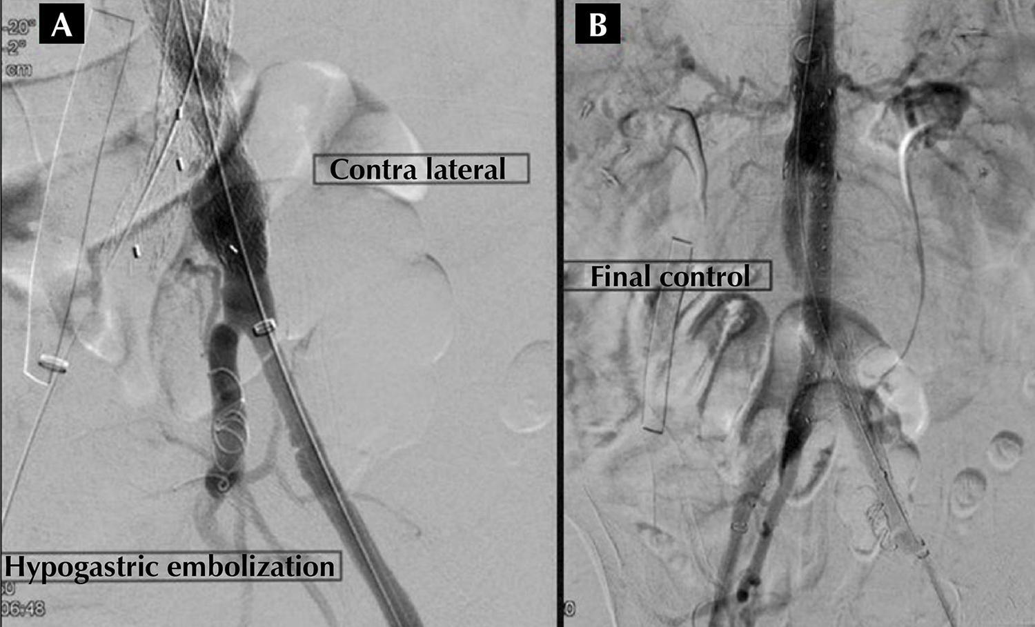
Abdominal aortic aneurysm (AAA), with an estimated prevalence of 4–8% and a higher incidence in men, older people, hypertensive patients, and smokers, can be treated with surgical or endovascular aneurysm repair (EVAR). When well-indicated, EVAR is primarily used since it is less invasive and has lower morbidity and mortality rates. However, due to possible complications, routine clinical and imaging surveillance is necessary to identify or prevent periprosthetic leaks (endoleaks) associated with aneurysmal disease progression.

Accordingly, computed tomography angiography (CTA) has always been considered the gold standard for endoleak surveillance due to its high image quality and usefulness for two- and three-dimensional reconstruction; however, its use of iodinated contrast and exposure to ionizing radiation are disadvantages. Therefore, other post-EVAR control imaging methods such as contrast-enhanced ultrasound (CEUS) have been studied as an endoleak dynamic and high-quality assessment method.,

[…]

  • abdominal
  • Aortic Aneurysm
  • Endoleak
Read Full Article
Creative Commons Attribution (CC BY) Creative Commons license
17/08/2022 DOI: 10.47593/2675-312X/20223503eabc315
Figure 1
Special Article

Pediatric Cardiac Tumors: Echocardiographic Imaging Features

Nathalie Jeanne Magioli Bravo-Valenzuela et al. Eliane Lucas Nadinne Velloso Netto Larissa Vieira da Conceição … Natalia Pinheiro Duque Estrada Maria Respondek-Liberska Edward Araujo Júnior

Abstract

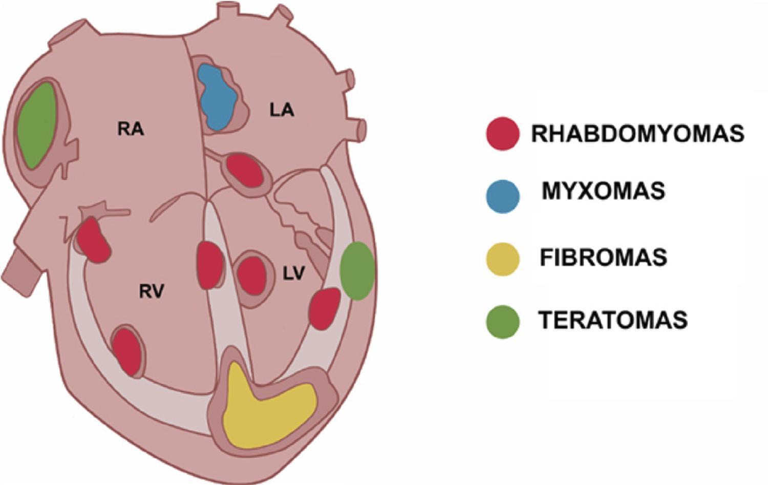
Cardiac tumors (CTs) in children are very rare, but their diagnosis is crucial for patient management. Echocardiography is the most commonly used cardiovascular imaging modality in clinical practice for the initial diagnosis of CTs in pediatric patients. Knowing the echocardiographic characteristics of CTs can enable an increasingly early diagnosis and the identification of its most likely etiology. Primary and benign CTs are the most frequent types in the pediatric population. Among benign CTs, the most frequent in fetuses and neonates are rhabdomyomas and teratomas. In children and adolescents, rhabdomyomas and fibromas are more common. Here we describe the echocardiographic characteristics of the most common CTs in pediatric patients.

  • Cardiac Tumors
  • Echocardiography
  • Imaging
  • Pediatrics
  • screening
Read Full Article
Creative Commons Attribution (CC BY) Creative Commons license
17/08/2022 DOI: 10.47593/2675-312X/20223503eabc302
Figure 1
Case Report

Long-Term Follow-Up of an Asymptomatic Man with a Cardiac Fibroma

Marina Costa Jonas et al. Maria Fernanda Braggion-Santos Marcel Koenigkam-Santos Paulo Henrique Manso … Luiza Frison Dias André Schmidt Fernando Amaral

Abstract

Here we describe a case of a 19-year-old asymptomatic man with a left ventricular fibroma on follow-up for 15 years with no treatment required.

  • Adult
  • Cardiac Neoplasms
  • Fibroma
  • Sudden Death
Read Full Article
Creative Commons Attribution (CC BY) Creative Commons license
07/07/2022 DOI: 10.47593/2675-312X/20223503eabc287 Views 1
Figure 1
Case Report

Three-Dimensional Transesophageal Echocardiography in Native Valve Endocarditis: A Case Report and Similarities Between Echocardiography and Surgical Findings

Giulia Bevilacqua Schmitz et al. Amadeu Antonio Bertuol Filho Solano Vinicius Berger Rodrigo Petersen Saadi … Álvaro Albrecht Angela Barreto Santiago Santos

Abstract

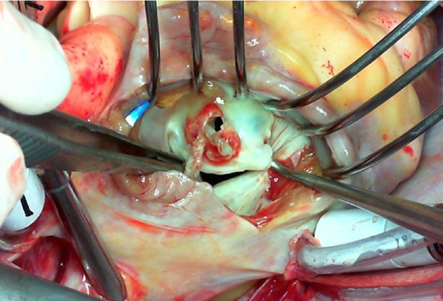
Native valve bacterial endocarditis is an uncommon, complex, and highly morbid disease that requires prolonged clinical treatment and challenging surgical interventions. Transthoracic and transesophageal echocardiography are paramount assessment tools whose findings are included in the diagnostic criteria. Three-dimensional echocardiography shows further realistic imaging details. Here we present a case demonstrating the role of echocardiography in the diagnosis of endocarditis and the identification of its complications to show how advanced imaging techniques may have a remarkable resemblance with in vivo surgical findings.

  • Echocardiography
  • Endocarditis
  • Mitral Valve
  • Three-Dimensional
Read Full Article
Creative Commons Attribution (CC BY) Creative Commons license
07/07/2022 DOI: 10.47593/2675-312X/20223503eabc279 Views 1
Figure 1
Review Article

Mitral Annulus Disjunction: Diagnostic Modalities, Clinical Implications, and Prognostic Progression

Alexandre Costa Souza et al. Marcus Vinicius Silva Freire de Carvalho Marco André Sales Suellen Lacerda Bezerra … Jorge Adinon Torreão Carolina Thé Macêdo

Abstract

Mitral annulus disjunction (MAD) is an abnormal insertion of the flexion line of the mitral annulus into the atrial wall. The annulus presents a separation (disjunction) between the posterior leaflet–atrial wall junction and the left ventricular myocardial crest.1 MAD was first described more than 30 years ago in an autopsy study and is reportedly related to mitral valve prolapse (MVP) in 92% of cases.2 Since then, several studies have been conducted, and reports on the prevalence of MAD in patients with MVP have varied. Ultimately, it may or may not be associated with mitral regurgitation. Transthoracic echocardiography is part of initial MVP assessment, allowing its diagnosis and the assessment of related complications. As new diagnostic methods emerged, cardiac magnetic resonance imaging and transesophageal echocardiography improved the assessment of this pathology in terms of its diagnosis, extension, and location. However, the phenotypic characteristics of MVP that are more closely associated with MAD remain uncertain mainly due to the limited number of patients in classic studies on the subject. Patients with MAD may develop symptoms related to ventricular arrhythmias, configuring the MAD arrhythmic syndrome, which may progress to sudden death. The literature presents conflicting prognostic data among several studies on the subject from clear diagnostic criteria and best imaging method to be used to treatment and prognosis. This review describes MAD characteristics associated (or not) with valve prolapse to improve the diagnosis and management of this important pathology.

  • Diagnosis
  • Mitral Valve Insufficiency
  • Mitral Valve Prolapse
Read Full Article
Creative Commons Attribution (CC BY) Creative Commons license
07/07/2022 DOI: 10.47593/2675-312X/20223503eabc300 Views 1
Figure 1
Review Article

Stress Echocardiography Using Real-Time 3D Imaging

José Roberto Matos-Souza et al. Guilherme De Rossi

Abstract

The need to examine the heart using a three-dimensional (3D) tool is not new. This complex and dynamic organ has always required 3D and real-time understanding. Without this feature, the examiner has to transform two-dimensional images to understand its volume, which requires complex knowledge and approximation interactions. Echocardiography was invented three decades ago, and its improvements resulted in commercial products at the beginning of the century. Some studies demonstrate 3D equivalence with gains in handling the necessary time. We use triplane modalities in our routine, with time gain and less stress on the examiner’s upper limb. Thus, 3D examinations can answer more complex questions and provide a more geometric approach to contraction, with thickening being analyzed in the background.

  • Echocardiography, Stress
  • Echocardiography, Three-Dimensional
  • Imaging, Three-Dimensional
Read Full Article
Creative Commons Attribution (CC BY) Creative Commons license
07/07/2022 DOI: 10.47593/2675-312X/20223503erer_07
Case Report

Cor Triatriatum Sinistrum in an Asymptomatic Adult: A Case Report

Mauro Alves et al. Fernando Antonio de Portugal Morcerf Cynara Silvia Sousa do Amaral

Abstract

Cor triatriatum is a rare congenital heart anomaly often diagnosed in early childhood. This case study features an adult with an incidental finding of cor triatriatum sinistrum. Based on the clinical presentation, the patient was treated conservatively. Cor triatriatum sinistrum echocardiographic image findings of this patient are presented along with a narrative review of the literature about this disease.

  • Congenital
  • Cor Triatriatum sinistrum
  • Echocardiogram
  • Heart Defects
Read Full Article
Creative Commons Attribution (CC BY) Creative Commons license
07/07/2022 DOI: 10.47593/2675-312X/20223503eabc280 Views 1
Figure 1
Case Report

Acquired Pulmonary Stenosis in an Adolescent with Lymphoma

Themissa Helena Voss et al. Flávia Bittar Britto Arantes

Case report

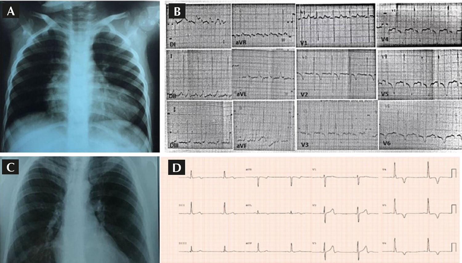
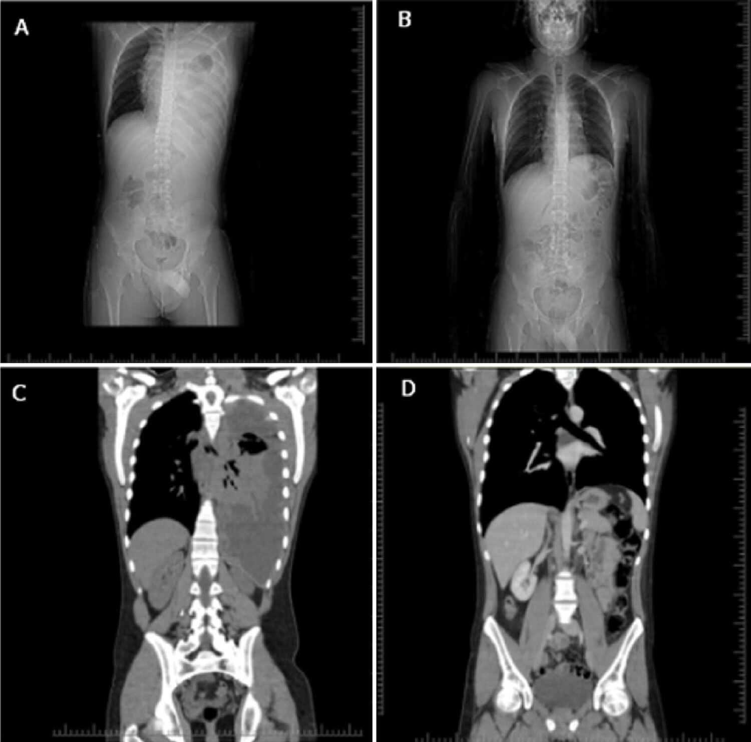
A previously healthy 14-year-old male patient sought emergency care complaining of dyspnea. He was New York Heart Association (NYHA) functional class III and had a 1-month history of orthopnea and night sweats. Auscultation abolished in the left hemithorax, while a chest X-ray revealed extensive opacity in that region. Chest computed tomography (CT) showed a large mediastinal mass with concomitant involvement of the left pleura and retroperitoneal lymph nodes. No other palpable lymph nodes were present on physical examination.

The patient underwent pleuroscopy for pleural biopsy, which identified lymphoid cell proliferation in a “starry sky” pattern compatible with lymphoma and a histochemical pattern compatible with precursor T-cell lymphoblastic non-Hodgkin lymphoma (NHL) (pan-T and TdT, CD4 and CD4 co-expression, and high cell proliferation rate with positive Ki67 test results in 90%). Blast tests in the medulla and cerebrospinal fluid were negative.

[…]

Read Full Article
Creative Commons Attribution (CC BY) Creative Commons license
07/07/2022 DOI: 10.47593/2675-312X/20223503eabc294 Views 2

How to cite this article

EndNote RIS

Electronic Document Format (Vancouver)

Ramirez DG, Hernandez JM. What does the navigator system in interventional procedures in the hybrid room?. ABC Imagem Cardiovascular 2022;35(3):eabc332.

Electronic Document Format (ABNT)

Ramirez, Denisse Guzman; Hernandez, Jose Maria. What does the navigator system in interventional procedures in the hybrid room?. ABC Imagem Cardiovascular, v. 35, n. 3, eabc332, Sep. 2022.

Electronic Document Format (APA)

Ramirez, D. G., & Hernandez, J. M. (2022). What does the navigator system in interventional procedures in the hybrid room?. ABC Imagem Cardiovascular, 35(3), eabc332.

Electronic Document Format (ISO)

Ramirez, Denisse Guzman and Hernandez, Jose Maria. What does the navigator system in interventional procedures in the hybrid room?. ABC Imagem Cardiovascular [online]. 2022, vol. 35, n. 3, [cited 2026-10-14], eabc332. Available from: <https://journals.cardiol.br/en/article/what-does-the-navigator-system-in-interventional-procedures-in-the-hybrid-room/>. ISSN 2318-8219

EndNote RIS

Electronic Document Format (Vancouver)

Brito ATAB, Silva JBM, Rassi DC, Silva DGST, Costa SA, Oliveira DS, Jardim FSV, et al. Correlation Between Echocardiographic and Hemodynamic Measurements in Right Atrium and Right Ventricle Assessments of Patients with Pulmonary Hypertension. ABC Imagem Cardiovascular 2022;35(3):eabc308.

Electronic Document Format (ABNT)

Brito, Ana Tereza Azeredo Bastos; Silva, João Batista Masson; Rassi, Daniela do Carmo; Silva, Daniela Graner Schuwartz Tannus; Costa, Sandra Araújo; Oliveira, Diogo Sampaio de; Jardim, Flavio de Souza Veiga; Freitas, Leandro Zacarias F. de; Freitas Júnior, Aguinaldo F. de; Rassi, Salvador. Correlation Between Echocardiographic and Hemodynamic Measurements in Right Atrium and Right Ventricle Assessments of Patients with Pulmonary Hypertension. ABC Imagem Cardiovascular, v. 35, n. 3, eabc308, Sep. 2022.

Electronic Document Format (APA)

Brito, A. T. A. B., Silva, J. B. M., Rassi, D. C., Silva, D. G. S. T., Costa, S. A., Oliveira, D. S. , Jardim, F. S. V., Freitas, L. Z. F. , Freitas Júnior, A. F. , & Rassi, S. (2022). Correlation Between Echocardiographic and Hemodynamic Measurements in Right Atrium and Right Ventricle Assessments of Patients with Pulmonary Hypertension. ABC Imagem Cardiovascular, 35(3), eabc308.

Electronic Document Format (ISO)

Brito, Ana Tereza Azeredo Bastos and Silva, João Batista Masson and Rassi, Daniela do Carmo and Silva, Daniela Graner Schuwartz Tannus and Costa, Sandra Araújo and Oliveira, Diogo Sampaio de and Jardim, Flavio de Souza Veiga and Freitas, Leandro Zacarias F. de and Freitas Júnior, Aguinaldo F. de and Rassi, Salvador. Correlation Between Echocardiographic and Hemodynamic Measurements in Right Atrium and Right Ventricle Assessments of Patients with Pulmonary Hypertension. ABC Imagem Cardiovascular [online]. 2022, vol. 35, n. 3, [cited 2026-10-14], eabc308. Available from: <https://journals.cardiol.br/en/article/correlation-between-echocardiographic-and-hemodynamic-measurements-in-right-atrium-and-right-ventricle-assessments-of-patients-with-pulmonary-hypertension/>. ISSN 2318-8219

EndNote RIS

Electronic Document Format (Vancouver)

Nastari L. What do cardiologists expect from Echocardiography in the evaluation of non-compacted myocardium. ABC Imagem Cardiovascular 2022;35(3):ecard07.

Electronic Document Format (ABNT)

Nastari, Luciano. What do cardiologists expect from Echocardiography in the evaluation of non-compacted myocardium. ABC Imagem Cardiovascular, v. 35, n. 3, ecard07, Sep. 2022.

Electronic Document Format (APA)

Nastari, L. (2022). What do cardiologists expect from Echocardiography in the evaluation of non-compacted myocardium. ABC Imagem Cardiovascular, 35(3), ecard07.

Electronic Document Format (ISO)

Nastari, Luciano. What do cardiologists expect from Echocardiography in the evaluation of non-compacted myocardium. ABC Imagem Cardiovascular [online]. 2022, vol. 35, n. 3, [cited 2026-10-14], ecard07. Available from: <https://journals.cardiol.br/en/article/what-do-cardiologists-expect-from-echocardiography-in-the-evaluation-of-non-compacted-myocardium/>. ISSN 2318-8219

EndNote RIS

Electronic Document Format (Vancouver)

Rodrigues ACT, Kay FU, Roque M, Gripp TEH, Becker D, Arruda AL, Cerri GG, et al. Impairment of Right Ventricular Function in Patients with Systemic Sclerosis and Interstitial Lung Disease:Tissue Doppler Evaluation. ABC Imagem Cardiovascular 2022;35(3):eabc331.

Electronic Document Format (ABNT)

Rodrigues, Ana Clara Tude; Kay, Fernando U.; Roque, Marina; Gripp, Thais Emerick Henriques; Becker, Derly; Arruda, Ana Lucia; Cerri, Giovanni Guido; Sampaio-Barros, Percival D.. Impairment of Right Ventricular Function in Patients with Systemic Sclerosis and Interstitial Lung Disease:Tissue Doppler Evaluation. ABC Imagem Cardiovascular, v. 35, n. 3, eabc331, Sep. 2022.

Electronic Document Format (APA)

Rodrigues, A. C. T., Kay, F. U., Roque, M., Gripp, T. E. H., Becker, D., Arruda, A. L., Cerri, G. G., & Sampaio-Barros, P. D. (2022). Impairment of Right Ventricular Function in Patients with Systemic Sclerosis and Interstitial Lung Disease:Tissue Doppler Evaluation. ABC Imagem Cardiovascular, 35(3), eabc331.

Electronic Document Format (ISO)

Rodrigues, Ana Clara Tude and Kay, Fernando U. and Roque, Marina and Gripp, Thais Emerick Henriques and Becker, Derly and Arruda, Ana Lucia and Cerri, Giovanni Guido and Sampaio-Barros, Percival D.. Impairment of Right Ventricular Function in Patients with Systemic Sclerosis and Interstitial Lung Disease:Tissue Doppler Evaluation. ABC Imagem Cardiovascular [online]. 2022, vol. 35, n. 3, [cited 2026-10-14], eabc331. Available from: <https://journals.cardiol.br/en/article/impairment-of-right-ventricular-function-in-patients-with-systemic-sclerosis-and-interstitial-lung-diseasetissue-doppler-evaluation/>. ISSN 2318-8219

EndNote RIS

Electronic Document Format (Vancouver)

Hotta VT. Role of Echocardiography in Assessment of Patients with Suspected Ph: Beyond the Peak Tricuspid Regurgitant Velocity. ABC Imagem Cardiovascular 2022;35(3):eed24.

Electronic Document Format (ABNT)

Hotta, Viviane Tiemi. Role of Echocardiography in Assessment of Patients with Suspected Ph: Beyond the Peak Tricuspid Regurgitant Velocity. ABC Imagem Cardiovascular, v. 35, n. 3, eed24, Sep. 2022.

Electronic Document Format (APA)

Hotta, V. T. (2022). Role of Echocardiography in Assessment of Patients with Suspected Ph: Beyond the Peak Tricuspid Regurgitant Velocity. ABC Imagem Cardiovascular, 35(3), eed24.

Electronic Document Format (ISO)

Hotta, Viviane Tiemi. Role of Echocardiography in Assessment of Patients with Suspected Ph: Beyond the Peak Tricuspid Regurgitant Velocity. ABC Imagem Cardiovascular [online]. 2022, vol. 35, n. 3, [cited 2026-10-14], eed24. Available from: <https://journals.cardiol.br/en/article/role-of-echocardiography-in-assessment-of-patients-with-suspected-ph-beyond-the-peak-tricuspid-regurgitant-velocity/>. ISSN 2318-8219

EndNote RIS

Electronic Document Format (Vancouver)

Rosa LM, Morhy SS. My Approach to Managing Left Ventricular Diastolic Function of Children and in Congenital Heart Disease. ABC Imagem Cardiovascular 2022;35(3):ecom29.

Electronic Document Format (ABNT)

Rosa, Laura Mercer; Morhy, Samira Saady. My Approach to Managing Left Ventricular Diastolic Function of Children and in Congenital Heart Disease. ABC Imagem Cardiovascular, v. 35, n. 3, ecom29, Sep. 2022.

Electronic Document Format (APA)

Rosa, L. M., & Morhy, S. S. (2022). My Approach to Managing Left Ventricular Diastolic Function of Children and in Congenital Heart Disease. ABC Imagem Cardiovascular, 35(3), ecom29.

Electronic Document Format (ISO)

Rosa, Laura Mercer and Morhy, Samira Saady. My Approach to Managing Left Ventricular Diastolic Function of Children and in Congenital Heart Disease. ABC Imagem Cardiovascular [online]. 2022, vol. 35, n. 3, [cited 2026-10-14], ecom29. Available from: <https://journals.cardiol.br/en/article/my-approach-to-managing-left-ventricular-diastolic-function-of-children-and-in-congenital-heart-disease/>. ISSN 2318-8219

EndNote RIS

Electronic Document Format (Vancouver)

Bohatch Júnior MS, Silva AFV, Dálio MB, Ribeiro MS, Joviliano EE. Symptomatic Aortic Mural Thrombus in a Patient With COVID-19. ABC Imagem Cardiovascular 2022;35(3):eabc270.

Electronic Document Format (ABNT)

Bohatch Júnior, Milton Sérgio; Silva, Amanda Fernandes Vidal da; Dálio, Marcelo Bellini; Ribeiro, Mauricio Serra; Joviliano, Edwaldo Edner. Symptomatic Aortic Mural Thrombus in a Patient With COVID-19. ABC Imagem Cardiovascular, v. 35, n. 3, eabc270, Aug. 2022.

Electronic Document Format (APA)

Bohatch Júnior, M. S., Silva, A. F. V. , Dálio, M. B., Ribeiro, M. S., & Joviliano, E. E. (2022). Symptomatic Aortic Mural Thrombus in a Patient With COVID-19. ABC Imagem Cardiovascular, 35(3), eabc270.

Electronic Document Format (ISO)

Bohatch Júnior, Milton Sérgio and Silva, Amanda Fernandes Vidal da and Dálio, Marcelo Bellini and Ribeiro, Mauricio Serra and Joviliano, Edwaldo Edner. Symptomatic Aortic Mural Thrombus in a Patient With COVID-19. ABC Imagem Cardiovascular [online]. 2022, vol. 35, n. 3, [cited 2026-10-14], eabc270. Available from: <https://journals.cardiol.br/en/article/symptomatic-aortic-mural-thrombus-in-a-patient-with-covid-19/>. ISSN 2318-8219

EndNote RIS

Electronic Document Format (Vancouver)

Otto MEB, Esmanhoto VA. My Approach to Lung Ultrasound to Evaluate Extravascular Lung Water. ABC Imagem Cardiovascular 2022;35(3):ecom22.

Electronic Document Format (ABNT)

Otto, Maria Estefânia Bosco; Esmanhoto, Vanessa Andreoli. My Approach to Lung Ultrasound to Evaluate Extravascular Lung Water. ABC Imagem Cardiovascular, v. 35, n. 3, ecom22, Aug. 2022.

Electronic Document Format (APA)

Otto, M. E. B., & Esmanhoto, V. A. (2022). My Approach to Lung Ultrasound to Evaluate Extravascular Lung Water. ABC Imagem Cardiovascular, 35(3), ecom22.

Electronic Document Format (ISO)

Otto, Maria Estefânia Bosco and Esmanhoto, Vanessa Andreoli. My Approach to Lung Ultrasound to Evaluate Extravascular Lung Water. ABC Imagem Cardiovascular [online]. 2022, vol. 35, n. 3, [cited 2026-10-14], ecom22. Available from: <https://journals.cardiol.br/en/article/my-approach-to-lung-ultrasound-to-evaluate-extravascular-lung-water/>. ISSN 2318-8219

EndNote RIS

Electronic Document Format (Vancouver)

Djaló ACNN, Pires GLO, Ferraz A, Abath CG, Fonseca Filho IS, Marques MO. Use of Contrast-Enhanced Ultrasound for the Diagnosis of Endoleak After EVAR: A Case Report. ABC Imagem Cardiovascular 2022;35(3):eabc315.

Electronic Document Format (ABNT)

Djaló, Ana Carolina N. Netto; Pires, Glauber Lutterbach de Oliveira; Ferraz, Adriana; Abath, Carlos Gustavo; Fonseca Filho, Idalécio Souto; Marques, Milena O.. Use of Contrast-Enhanced Ultrasound for the Diagnosis of Endoleak After EVAR: A Case Report. ABC Imagem Cardiovascular, v. 35, n. 3, eabc315, Aug. 2022.

Electronic Document Format (APA)

Djaló, A. C. N. N., Pires, G. L. O., Ferraz, A., Abath, C. G., Fonseca Filho, I. S., & Marques, M. O. (2022). Use of Contrast-Enhanced Ultrasound for the Diagnosis of Endoleak After EVAR: A Case Report. ABC Imagem Cardiovascular, 35(3), eabc315.

Electronic Document Format (ISO)

Djaló, Ana Carolina N. Netto and Pires, Glauber Lutterbach de Oliveira and Ferraz, Adriana and Abath, Carlos Gustavo and Fonseca Filho, Idalécio Souto and Marques, Milena O.. Use of Contrast-Enhanced Ultrasound for the Diagnosis of Endoleak After EVAR: A Case Report. ABC Imagem Cardiovascular [online]. 2022, vol. 35, n. 3, [cited 2026-10-14], eabc315. Available from: <https://journals.cardiol.br/en/article/use-of-contrast-enhanced-ultrasound-for-the-diagnosis-of-endoleak-after-evar-a-case-report/>. ISSN 2318-8219

EndNote RIS

Electronic Document Format (Vancouver)

Bravo-Valenzuela NJM, Lucas E, Netto NV, Conceição LV, Estrada NPD, Respondek-Liberska M, Araujo Júnior E, et al. Pediatric Cardiac Tumors: Echocardiographic Imaging Features. ABC Imagem Cardiovascular 2022;35(3):eabc302.

Electronic Document Format (ABNT)

Bravo-Valenzuela, Nathalie Jeanne Magioli; Lucas, Eliane; Netto, Nadinne Velloso; Conceição, Larissa Vieira da; Estrada, Natalia Pinheiro Duque; Respondek-Liberska, Maria; Araujo Júnior, Edward. Pediatric Cardiac Tumors: Echocardiographic Imaging Features. ABC Imagem Cardiovascular, v. 35, n. 3, eabc302, Aug. 2022.

Electronic Document Format (APA)

Bravo-Valenzuela, N. J. M., Lucas, E., Netto, N. V., Conceição, L. V. , Estrada, N. P. D., Respondek-Liberska, M., & Araujo Júnior, E. (2022). Pediatric Cardiac Tumors: Echocardiographic Imaging Features. ABC Imagem Cardiovascular, 35(3), eabc302.

Electronic Document Format (ISO)

Bravo-Valenzuela, Nathalie Jeanne Magioli and Lucas, Eliane and Netto, Nadinne Velloso and Conceição, Larissa Vieira da and Estrada, Natalia Pinheiro Duque and Respondek-Liberska, Maria and Araujo Júnior, Edward. Pediatric Cardiac Tumors: Echocardiographic Imaging Features. ABC Imagem Cardiovascular [online]. 2022, vol. 35, n. 3, [cited 2026-10-14], eabc302. Available from: <https://journals.cardiol.br/en/article/pediatric-cardiac-tumors-echocardiographic-imaging-features/>. ISSN 2318-8219

EndNote RIS

Electronic Document Format (Vancouver)

Jonas MC, Braggion-Santos MF, Koenigkam-Santos M, Manso PH, Dias LF, Schmidt A, Amaral F, et al. Long-Term Follow-Up of an Asymptomatic Man with a Cardiac Fibroma. ABC Imagem Cardiovascular 2022;35(3):eabc287.

Electronic Document Format (ABNT)

Jonas, Marina Costa; Braggion-Santos, Maria Fernanda; Koenigkam-Santos, Marcel; Manso, Paulo Henrique; Dias, Luiza Frison; Schmidt, André; Amaral, Fernando. Long-Term Follow-Up of an Asymptomatic Man with a Cardiac Fibroma. ABC Imagem Cardiovascular, v. 35, n. 3, eabc287, Jul. 2022.

Electronic Document Format (APA)

Jonas, M. C., Braggion-Santos, M. F., Koenigkam-Santos, M., Manso, P. H., Dias, L. F., Schmidt, A., & Amaral, F. (2022). Long-Term Follow-Up of an Asymptomatic Man with a Cardiac Fibroma. ABC Imagem Cardiovascular, 35(3), eabc287.

Electronic Document Format (ISO)

Jonas, Marina Costa and Braggion-Santos, Maria Fernanda and Koenigkam-Santos, Marcel and Manso, Paulo Henrique and Dias, Luiza Frison and Schmidt, André and Amaral, Fernando. Long-Term Follow-Up of an Asymptomatic Man with a Cardiac Fibroma. ABC Imagem Cardiovascular [online]. 2022, vol. 35, n. 3, [cited 2026-10-14], eabc287. Available from: <https://journals.cardiol.br/en/article/long-term-follow-up-of-an-asymptomatic-man-with-a-cardiac-fibroma/>. ISSN 2318-8219

EndNote RIS

Electronic Document Format (Vancouver)

Schmitz GB, Bertuol Filho AA, Berger SV, Saadi RP, Albrecht , Santos ABS. Three-Dimensional Transesophageal Echocardiography in Native Valve Endocarditis: A Case Report and Similarities Between Echocardiography and Surgical Findings. ABC Imagem Cardiovascular 2022;35(3):eabc279.

Electronic Document Format (ABNT)

Schmitz, Giulia Bevilacqua; Bertuol Filho, Amadeu Antonio; Berger, Solano Vinicius; Saadi, Rodrigo Petersen; Albrecht, Álvaro; Santos, Angela Barreto Santiago. Three-Dimensional Transesophageal Echocardiography in Native Valve Endocarditis: A Case Report and Similarities Between Echocardiography and Surgical Findings. ABC Imagem Cardiovascular, v. 35, n. 3, eabc279, Jul. 2022.

Electronic Document Format (APA)

Schmitz, G. B., Bertuol Filho, A. A., Berger, S. V., Saadi, R. P., Albrecht, , & Santos, A. B. S. (2022). Three-Dimensional Transesophageal Echocardiography in Native Valve Endocarditis: A Case Report and Similarities Between Echocardiography and Surgical Findings. ABC Imagem Cardiovascular, 35(3), eabc279.

Electronic Document Format (ISO)

Schmitz, Giulia Bevilacqua and Bertuol Filho, Amadeu Antonio and Berger, Solano Vinicius and Saadi, Rodrigo Petersen and Albrecht, Álvaro and Santos, Angela Barreto Santiago. Three-Dimensional Transesophageal Echocardiography in Native Valve Endocarditis: A Case Report and Similarities Between Echocardiography and Surgical Findings. ABC Imagem Cardiovascular [online]. 2022, vol. 35, n. 3, [cited 2026-10-14], eabc279. Available from: <https://journals.cardiol.br/en/article/three-dimensional-transesophageal-echocardiography-in-native-valve-endocarditis-a-case-report-and-similarities-between-echocardiography-and-surgical-findings/>. ISSN 2318-8219

EndNote RIS

Electronic Document Format (Vancouver)

Souza AC, Carvalho MVSF, Sales MA, Bezerra SL, Torreão JA, Macêdo CT. Mitral Annulus Disjunction: Diagnostic Modalities, Clinical Implications, and Prognostic Progression. ABC Imagem Cardiovascular 2022;35(3):eabc300.

Electronic Document Format (ABNT)

Souza, Alexandre Costa; Carvalho, Marcus Vinicius Silva Freire de; Sales, Marco André; Bezerra, Suellen Lacerda; Torreão, Jorge Adinon; Macêdo, Carolina Thé. Mitral Annulus Disjunction: Diagnostic Modalities, Clinical Implications, and Prognostic Progression. ABC Imagem Cardiovascular, v. 35, n. 3, eabc300, Jul. 2022.

Electronic Document Format (APA)

Souza, A. C., Carvalho, M. V. S. F. , Sales, M. A., Bezerra, S. L., Torreão, J. A., & Macêdo, C. T. (2022). Mitral Annulus Disjunction: Diagnostic Modalities, Clinical Implications, and Prognostic Progression. ABC Imagem Cardiovascular, 35(3), eabc300.

Electronic Document Format (ISO)

Souza, Alexandre Costa and Carvalho, Marcus Vinicius Silva Freire de and Sales, Marco André and Bezerra, Suellen Lacerda and Torreão, Jorge Adinon and Macêdo, Carolina Thé. Mitral Annulus Disjunction: Diagnostic Modalities, Clinical Implications, and Prognostic Progression. ABC Imagem Cardiovascular [online]. 2022, vol. 35, n. 3, [cited 2026-10-14], eabc300. Available from: <https://journals.cardiol.br/en/article/mitral-annulus-disjunction-diagnostic-modalities-clinical-implications-and-prognostic-progression/>. ISSN 2318-8219

EndNote RIS

Electronic Document Format (Vancouver)

Matos-Souza JR, Rossi GD. Stress Echocardiography Using Real-Time 3D Imaging. ABC Imagem Cardiovascular 2022;35(3):erer_07.

Electronic Document Format (ABNT)

Matos-Souza, José Roberto; Rossi, Guilherme De. Stress Echocardiography Using Real-Time 3D Imaging. ABC Imagem Cardiovascular, v. 35, n. 3, erer_07, Jul. 2022.

Electronic Document Format (APA)

Matos-Souza, J. R., & Rossi, G. D. (2022). Stress Echocardiography Using Real-Time 3D Imaging. ABC Imagem Cardiovascular, 35(3), erer_07.

Electronic Document Format (ISO)

Matos-Souza, José Roberto and Rossi, Guilherme De. Stress Echocardiography Using Real-Time 3D Imaging. ABC Imagem Cardiovascular [online]. 2022, vol. 35, n. 3, [cited 2026-10-14], erer_07. Available from: <https://journals.cardiol.br/en/article/stress-echocardiography-using-real-time-3d-imaging/>. ISSN 2318-8219

EndNote RIS

Electronic Document Format (Vancouver)

Alves M, Morcerf FAP, Amaral CSS. Cor Triatriatum Sinistrum in an Asymptomatic Adult: A Case Report. ABC Imagem Cardiovascular 2022;35(3):eabc280.

Electronic Document Format (ABNT)

Alves, Mauro; Morcerf, Fernando Antonio de Portugal; Amaral, Cynara Silvia Sousa do. Cor Triatriatum Sinistrum in an Asymptomatic Adult: A Case Report. ABC Imagem Cardiovascular, v. 35, n. 3, eabc280, Jul. 2022.

Electronic Document Format (APA)

Alves, M., Morcerf, F. A. P., & Amaral, C. S. S. (2022). Cor Triatriatum Sinistrum in an Asymptomatic Adult: A Case Report. ABC Imagem Cardiovascular, 35(3), eabc280.

Electronic Document Format (ISO)

Alves, Mauro and Morcerf, Fernando Antonio de Portugal and Amaral, Cynara Silvia Sousa do. Cor Triatriatum Sinistrum in an Asymptomatic Adult: A Case Report. ABC Imagem Cardiovascular [online]. 2022, vol. 35, n. 3, [cited 2026-10-14], eabc280. Available from: <https://journals.cardiol.br/en/article/cor-triatriatum-sinistrum-in-an-asymptomatic-adult-a-case-report/>. ISSN 2318-8219

EndNote RIS

Electronic Document Format (Vancouver)

Voss TH, Arantes FBB. Acquired Pulmonary Stenosis in an Adolescent with Lymphoma. ABC Imagem Cardiovascular 2022;35(3):eabc294.

Electronic Document Format (ABNT)

Voss, Themissa Helena; Arantes, Flávia Bittar Britto. Acquired Pulmonary Stenosis in an Adolescent with Lymphoma. ABC Imagem Cardiovascular, v. 35, n. 3, eabc294, Jul. 2022.

Electronic Document Format (APA)

Voss, T. H., & Arantes, F. B. B. (2022). Acquired Pulmonary Stenosis in an Adolescent with Lymphoma. ABC Imagem Cardiovascular, 35(3), eabc294.

Electronic Document Format (ISO)

Voss, Themissa Helena and Arantes, Flávia Bittar Britto. Acquired Pulmonary Stenosis in an Adolescent with Lymphoma. ABC Imagem Cardiovascular [online]. 2022, vol. 35, n. 3, [cited 2026-10-14], eabc294. Available from: <https://journals.cardiol.br/en/article/acquired-pulmonary-stenosis-in-an-adolescent-with-lymphoma/>. ISSN 2318-8219

Sede – São Paulo
Rua Barata Ribeiro, 380 cj 54 Bela Vista
São Paulo, Brasil – CEP: 01308-000
Tel.: +55 (11) 3120-3363
Telefax: +55 (11) 3259-2988
E-mail: depeco@cardiol.br

Sede – Rio de Janeiro
Av. Marechal Câmara, 160
3º andar – Sala: 330 – Centro
Rio de Janeiro – CEP: 20020-907
Tel.: +55 (21) 3478-2700
ou: +55 (21) 99386-3961
E-mail: abcimaging@cardiol.br

  • About the Journal
  • Internal Regulation
  • Editors Section
  • Editorial Board
  • SBC Award

  • Guidelines for Publication
  • Editorial Policy
  • Ethics Policy
  • Policy on the Use of Artificial Intelligence
  • Mandatory Publication Form

Issues

SBC Guidelines