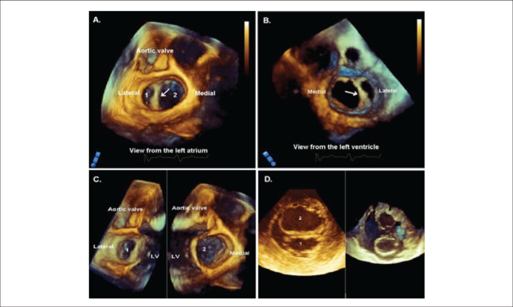
Real-time 3D-transesophageal echocardiogram „en face” view from the left atrium („surgeon’s view”, Panel A) and from the LV (Panel B) revealing two asymmetrical orifices, a smaller lateral (1) and a bigger medial (2) mitral orifice, separated by fibrous tissue (arrow). Panel C- achieved after using a sagittal crosssectional plane to “split” the valve and the LV in two for better view of the orifices and unrestricted opening of the mitral valve. Panel D. 2D (left) and 3D-zoom mode (right) transgastric view of the two orifices. LV, left ventricle.
Comments are moderated by the editorial team before publication. Discussions should be evidence-based and use scientific language. Conflicts of interest should be declared.
Comments are moderated by the editorial team before publication. Discussions should be evidence-based and use scientific language. Conflicts of interest should be declared.