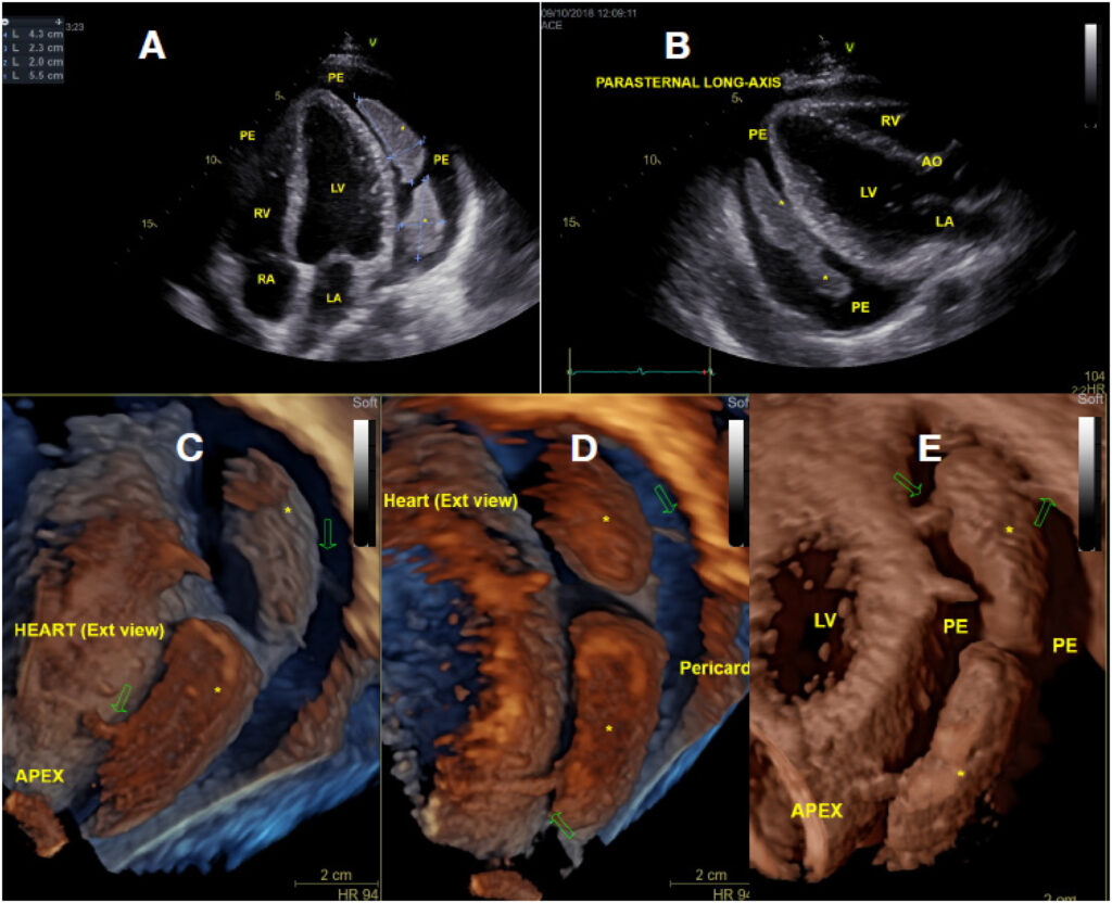
Transthoracic echocardiogram (TTE), showing large pericardial efusion with two intrapericardial masses (*). Two discoid masses were detected by 2D TTE, measuring 5.5 × 2.0cm and 4.3 × 2.3cm in its major diameters, from apical (A) and parasternal views (B). Rendered 3D images were obtained by post processing of datasets obtained by 3DTTE. Oblique 3D views in C, D and E show morphologic details of the masses, which are attached to visceral and parietal pericardial layers by fibrinous strands (arrows), floating inside the pericardial fluid, without invasion of surrounding tissues. LV: Left ventricle; RV: Right ventricle; LA: Left atrium; RA: Right atrium; PE: Pericardial effusion.
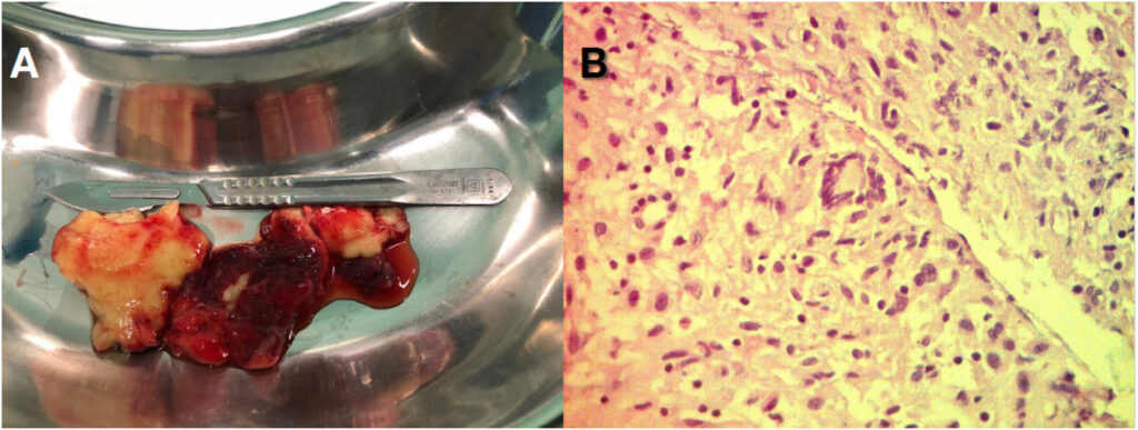
Histopathologic analysis of the intrapericardial masses. Macroscopically (A) the masses were disc-shaped and constituted by a lobulated yellowish soft tissue. In B, histopathological analysis with hematoxylin/eosin stain (magnification 40x) shows a pattern of chronic granulomatous inflammation with necrosis, consistent with tuberculosis granuloma.
Comments are moderated by the editorial team before publication. Discussions should be evidence-based and use scientific language. Conflicts of interest should be declared.