Dynamic three-dimensional reconstruction of the heart by transesophageal echocardiography
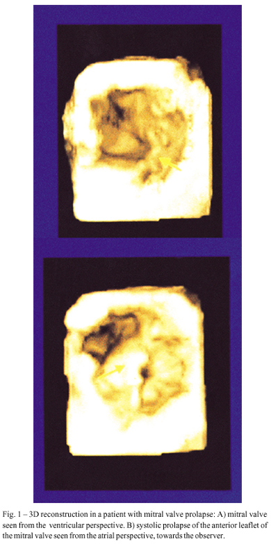
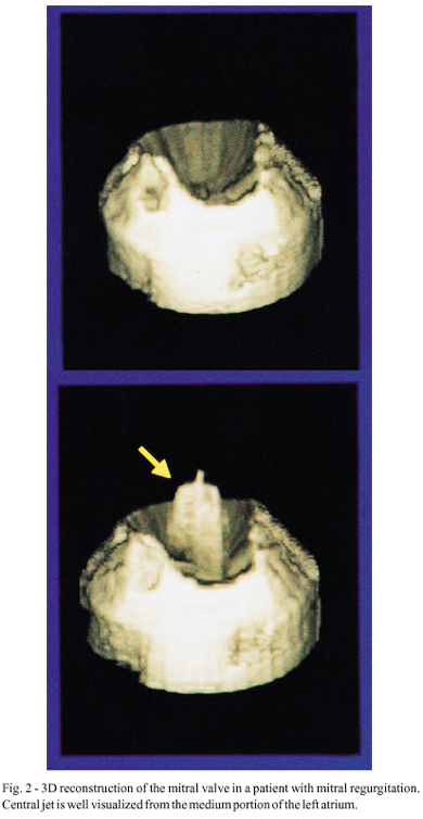
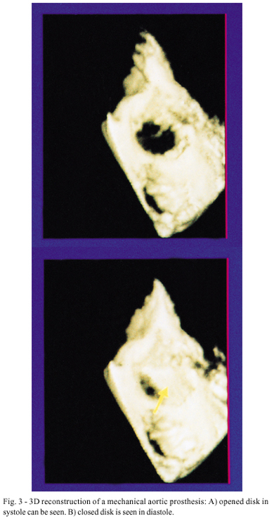
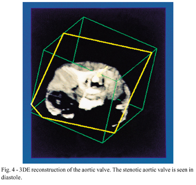
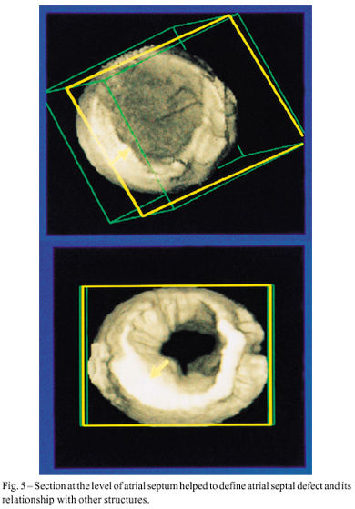
OBJECTIVE: To evaluate echocardiography accuracy in performing and obtaining images for dynamical three-dimensional (3D) reconstruction. METHODS: Three-dimensional (3D) image reconstruction was obtained in 20 consecutive patients who underwent transesophageal echocardiography. A multiplanar 5 MHz transducer was used for 3D reconstruction. RESULTS: Twenty patients were studied consecutively. The following cardiac diseases were present: valvar prostheses–6 (2 mitral, 2 aortic and 2 mitral and aortic); mitral valve prolapse– 3; mitral and aortic disease – 2; aortic valve disease– 5; congenital heart disease– 3 (2 atrial septal defect– ASD – and 1 transposition of the great arteries -TGA); arteriovenous fistula– 1. In 7 patients, color Doppler was also obtained and used for 3D flow reconstruction. Twenty five cardiac structures were acquired and 60 reconstructions generated (28 of mitral valves, 14 of aortic valves, 4 of mitral prostheses, 7 of aortic prostheses and 7 of the ASD). Fifty five of 60 (91.6%) reconstructions were considered of good quality by 2 independent observers. The 11 reconstructed mitral valves/prostheses and the 2 reconstructed ASDs provided more anatomical information than two dimensional echocardiography (2DE) alone. CONCLUSION: 3D echocardiography using a transesophageal transducer is a feasible technique, which improves detection of anatomical details of cardiac structures, particularly of the mitral valve and atrial septum.
Images
Image gallery fullscreen viewer
Comments are moderated by the editorial team before publication. Discussions should be evidence-based and use scientific language. Conflicts of interest should be declared.