| ABCIC
ABC Family Arquivos Brasileiros de Cardiologia International Journal of Cardiovascular Sciences ABC Heart Failure & Cardiomyopathy ABC Imagem Cardiovascular
Português English
ABC Family | ABCIC
Português English
  • About
    • About the Journal
    • Internal Regulation
    • Editors Section
    • Editorial Board
    • SBC Award
  • Instructions to Authors
    • Guidelines for Publication
    • Editorial Policy
    • Ethics Policy
    • Policy on the Use of Artificial Intelligence
    • Mandatory Publication Form
  • Issues
  • SBC Guidelines
  • About
    • About the Journal
    • Internal Regulation
    • Editors Section
    • Editorial Board
    • SBC Award
  • Instructions to Authors
    • Guidelines for Publication
    • Editorial Policy
    • Ethics Policy
    • Policy on the Use of Artificial Intelligence
    • Mandatory Publication Form
  • Issues
  • SBC Guidelines

ABC Family

  • ABC Family
  • Arquivos Brasileiros de Cardiologia
  • International Journal of Cardiovascular Sciences
  • ABC Heart Failure & Cardiomyopathy
  • ABC Imagem Cardiovascular
Search in

No content found.

Journals
Ordering
Article types
Others

  1. Home
  2. ABCIC
  3. Issues
  4. 2022, Volume 35, Number 1
Figure 1
Original Article

Tricuspid Annular Plane Systolic Excursion and Mitral Annular Systolic Excursion: Correlation with Left Ventricular Diastolic Function

André Américo Bedenko Martins et al. Leonardo Yoshida Osaku Ana Cristina Camarozano Cintia Rocha Fortes de Sá … Daniela de Castro Carmo Jerônimo Antonio Fortunato Rubens Zenóbio Darwich Liz Andréa Villela Baroncini

Abstract

Introduction:

Tricuspid annular plane systolic excursion and mitral annular systolic excursion are parameters used to assess the systolic function of the right ventricle and left ventricle, respectively. Little is known about its relationship with left ventricular diastolic function.

Objective:

To assess whether the values of mitral annular systolic excursion and tricuspid annular plane systolic excursion correlate with parameters used in the evaluation of left ventricular diastolic function.

Method:

Observational cross-sectional study. Two hundred nine individuals were selected, 116 women, with both ventricles normal systolic function. The analyzes were performed for men and women, through Pearson correlation coefficient and Sperman correlation coefficient. Tricuspid annular plane systolic excursion, mitral annular systolic excursion, atrial volumes and left ventricular diastolic function parameters on transthoracic echocardiogram were obtained.

Results:

In women, mitral annular systolic excursion was positively correlated with lateral e ‘(Sperman correlation coefficient of 0.22; p=0.016) and tricuspid annular plane systolic excursion was positively correlated with E / A ratio (Sperman correlation coefficient of 0.23; p=0.037), lateral e’ (Sperman correlation coefficient of 0.28; p=0.012), and septal e’ (Sperman correlation coefficient of 0.28; p=0.012), and negatively with the E/e’ ratio (Pearson correlation coefficient of -0.27; p=0.018), and A wave (Pearson correlation coefficient of -0.29; p=0.009). In men, only mitral annular systolic excursion correlated positively with E wave (Pearson correlation coefficient of 0.21; p=0.037), lateral e’ (Sperman correlation coefficient of 0.34; p <0.001) and the septal e’ (Sperman correlation coefficient of 0.26; p=0.008). There was no correlation between mitral annular systolic excursion and tricuspid annular plane systolic excursion and atrial volumes. Hypertension and diabetes mellitus influenced tricuspid annular plane systolic excursion and mitral annular systolic excursion values correlated to E and A waves, E/A ratio, septal and lateral e’ waves, and E/e’ ratio.

Conclusion:

In the present study, mitral annular systolic excursion and tricuspid annular plane systolic excursion values showed a significant correlation with some parameters of left ventricular diastolic function, with stronger evidence on female sex.
  • Cardiac function
  • Cardiovascular disease
  • COVID-19
  • Echocardiography
  • SARS-CoV-2
Read Full Article
Creative Commons Attribution (CC BY) Creative Commons license
30/03/2022 DOI: 10.47593/2675-312X/20223501eabc245 Views 1
Original Article

Factors Associated With Abnormalities On Myocardial Perfusion Scintigraphy In Diabetic Patients

Alfredo Lírio da Cruz et al. Álisson Carvalho de Freitas Bianca Naomi Sada Watanabe Helena Bigarella Nascimento … Heloisa Porath Luiz Antonio Fruet Bettini Luciane Moreira Marcos Germano Dopheide

Abstract

Diabetes mellitus (DM) is the greatest risk factor for coronary artery disease. In addition to a long duration of diabetes, the presence of peripheral arterial disease and smoking are strong predictors of abnormalities on myocardial perfusion scintigraphy (MPS). This study aimed to assess the impact of risk factors in diabetic patients on MPS results and compare them with those of non-diabetic patients in a nuclear medicine clinic. A retrospective cross-sectional study was performed through the analysis of the medical records of patients who underwent MPS in 2010–2019. A total of 34,736 medical records were evaluated. Analyzing the stress phase of MPS, DM patients required two-fold more pharmacological stimulation than non-diabetic patients for MPS. Factors that negatively impact the MPS results were also evaluated, and DM (33.6%), insulin therapy (18.1%), systemic arterial hypertension (69.9%), dyslipidemia (53%), sedentary lifestyle (83.1%), use of pharmacological stress (50.6%), typical chest pain (8.5%), and limiting angina during the test (1.7%) were significantly associated (p < 0.001) with test abnormalities.

  • Diabetes Mellitus
  • Pharmacological Stress
  • Ventilation-Perfusion Scan
Read Full Article
Creative Commons Attribution (CC BY) Creative Commons license
30/03/2022 DOI: 10.47593/2675-312X/20223501eabc274 Views 1
Figure 1
My Approach To

My Approach To Echocardiographic Assessment In Pulmonary Hypertension

João Batista Masson-Silva et al. Cloves Geraldino da Silva Júnior

Echocardiographic assessment of PH probability

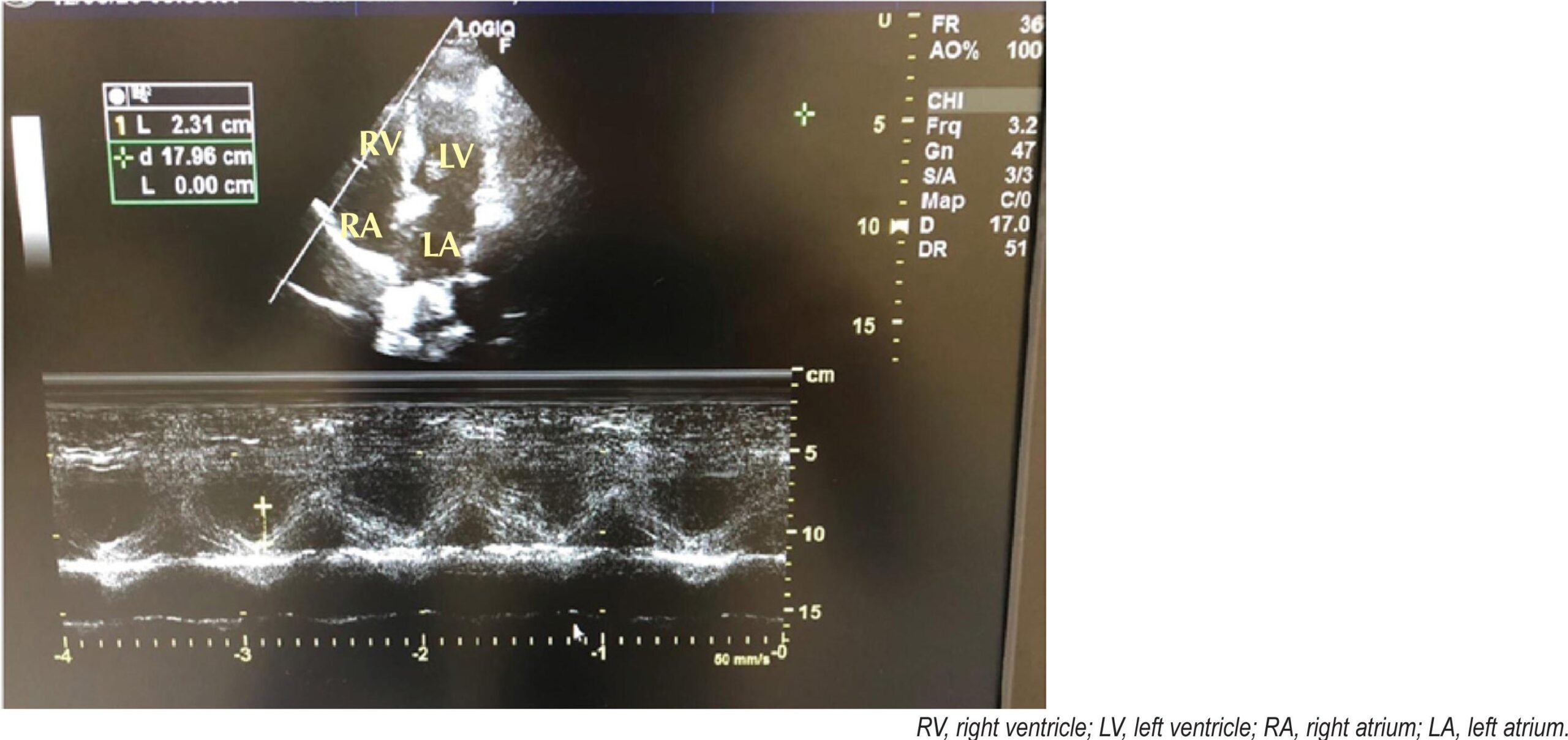
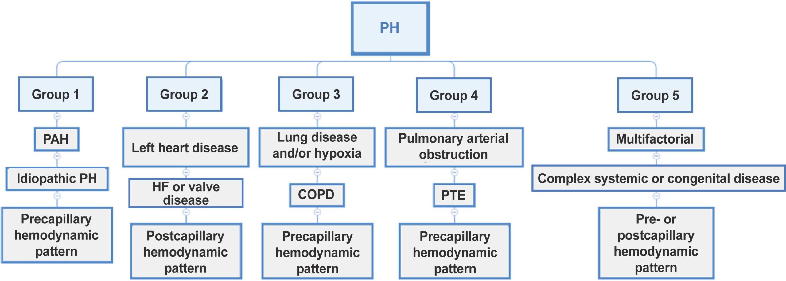
Echocardiographic PH probability can be classified as low, intermediate, or high. This categorization is based on the assessment of peak tricuspid regurgitation (TR) velocity and the presence of indirect signs suggestive of increased pulmonary vascular bed pressure ( ). ,

Peak TR velocity is the key criterion and should be measured by continuous-wave Doppler during smooth respiration according to the following recommendations: , – ( ).

[…]

  • Diagnosis
  • Echocardiography
  • Hypertension
  • Pulmonary
Read Full Article
Creative Commons Attribution (CC BY) Creative Commons license
30/03/2022 DOI: 10.47593/2675-312X/20223501ecom18
Figure 1
Case Report

Coronary Flow Velocity Reserve and Myocardial Contractility Under Stress in the Post-Infarction Ischemic Memory Dilemma

Marilia Esther Benevides de Abreu et al. Tereza Cristina Diógenes Pinheiro Antônio Augusto Lima Guimarães José Sebastião de Abreu

Introduction

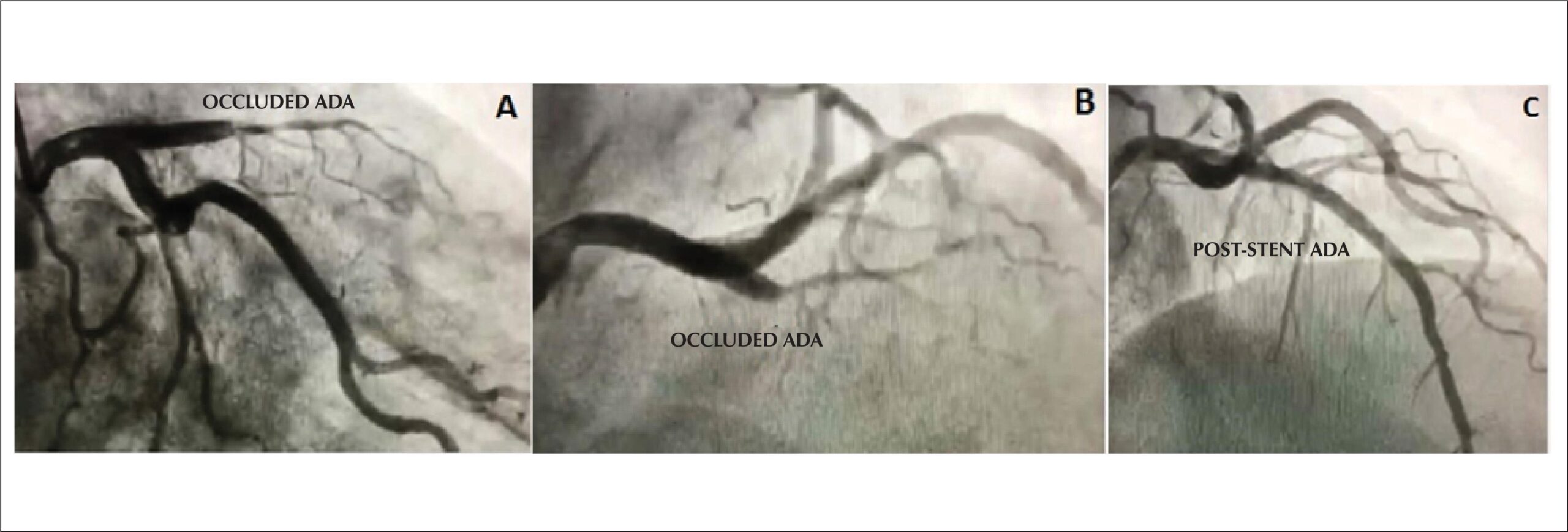
After acute myocardial infarction (AMI), some cases progress with deep T wave inversions on electrocardiogram (ECG). This electrocardiographic abnormality can continue for a prolonged period, even if a coronary intervention within a short time of AMI progression results in successful myocardial reperfusion. However, these deep T wave inversions can raise management questions when the patient is already being followed up in an outpatient clinic, particularly in the absence of relevant symptoms to support the resurgence of an ischemic process. In this context, a safe noninvasive assessment such as dobutamine stress echocardiography (DSE) associated with the analysis of coronary flow velocity reserve (CFVR) may be an option for this dilemma between myocardial ischemia and the possible post-infarction ischemic memory. –

[…]

  • Dobutamine
  • Myocardial Infarction
  • Stress echocardiography
Read Full Article
Creative Commons Attribution (CC BY) Creative Commons license
30/03/2022 DOI: 10.47593/2675-312X/20223501eabc221 Views 1
Figure 1
My Approach To

My Approach to Aortic Stenosis Assessment with Discrepant Quantification

Adenalva Lima de Souza Beck et al. Luiz Carlos Madruga Ribeiro

Introduction

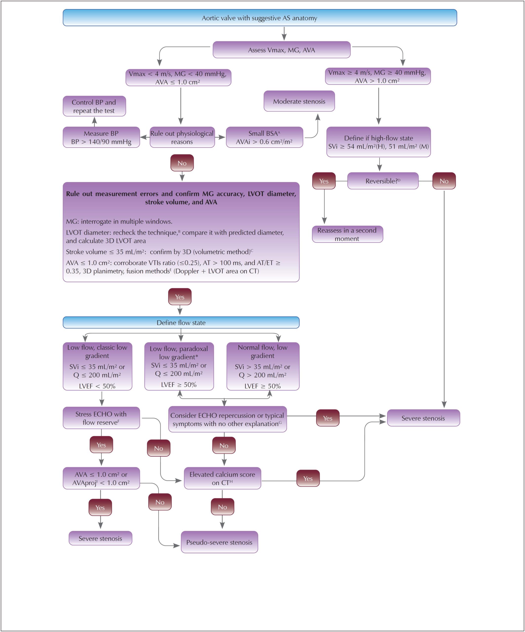
Aortic stenosis (AS) is the most common primary valve disease and an important cause of morbidity and mortality worldwide. Echocardiography is the first-line method for its diagnosis, quantification of severity, prognosis, and determination of intervention timing.

The association of maximum velocity (Vmax) ≥ 4m/s, aortic valve area (AVA) ≤ 1.0 cm², and mean gradient (MG) ≥ 40 mmHg defines the diagnosis of classic severe AS. However, MG and AVA are discrepant (AVA ≤ 1.0 cm², MG < 40 mmHg) in 30–40% of the population with severe AS, leading to low-gradient AS (LGAS), the focus of this article. Less frequently, high-gradient AS can also occur in cases of MG ≥ 40 mmHg and AVA > 1.0 cm² due to high flow situations.

[…]

  • Aortic Stenosis
  • Diagnosis
  • Echocardiography
Read Full Article
Creative Commons Attribution (CC BY) Creative Commons license
30/03/2022 DOI: 10.47593/2675-312X/20223501ecom21
Figure 1
My Approach To

My approach to diagnosis of and echocardiographic follow-up for muscular dystrophies

Raphael Henrique Déa Cirino et al. Renata Dal-Prá Ducci

Introduction

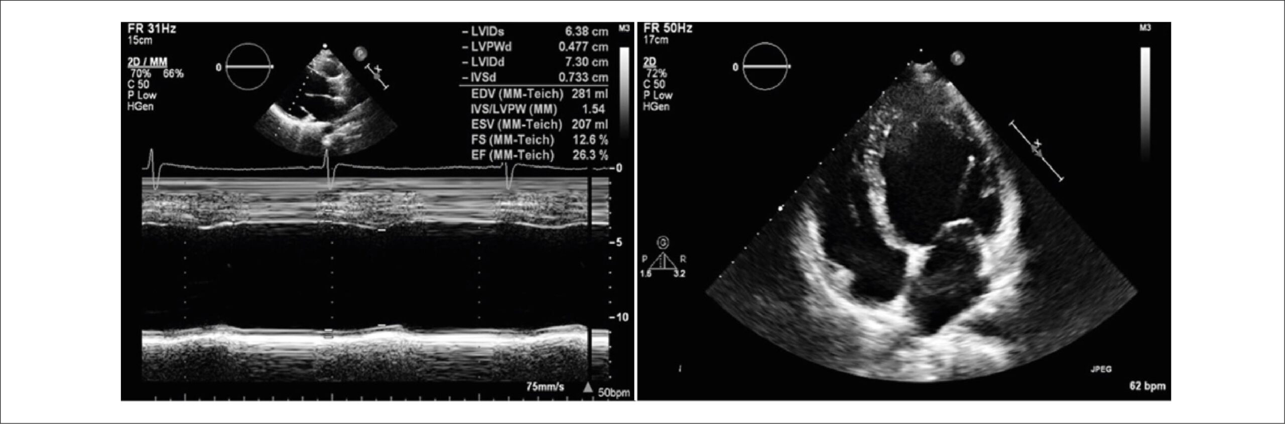
Muscular dystrophies (MD) are a group of heterogeneous diseases genetically determined and characterized by progressive muscle atrophy and weakness. Mutations in genes encoding muscle fiber proteins lead to the development of MD. MD types vary by deficient protein, each with its natural history and characteristic clinical presentation. As with other clinical aspects, cardiac involvement is also quite variable. ,

This article will review the most important aspects of the clinical-echocardiographic follow-up of MD types frequently associated with cardiomyopathies, including dystrophinopathy, limb-girdle muscular dystrophy (LGMD), myotonic dystrophy, and Emery-Dreifuss muscular dystrophy (EDMD).

[…]

  • Cardiomyopathy, Dilated
  • Echocardiography
  • Muscular dystrophies
Read Full Article
Creative Commons Attribution (CC BY) Creative Commons license
30/03/2022 DOI: 10.47593/2675-312X/20223501eabc292 Views 1
Figure 1
Image

Intermittent Aortic Intraprosthetic Failure

Luciano H Melato et al. Daniela do Carmo Rassi Rogério G Furtado Ana Caroline R Oliveira

I

n

[…]

  • Echocardiography
  • Heart Valve Prosthesis
  • Prosthesis Failure
Read Full Article
Creative Commons Attribution (CC BY) Creative Commons license
30/03/2022 DOI: 10.47593/2675-312X/20223501eabc284 Views 1
Figure 1
My Approach To

My Approach to the Echocardiographic Assessment of Mitral Annular Calcification: Challenges and Particularities

Antonio Carlos Leite de Barros Filho et al. Minna Moreira Dias Romano

Introduction

Mitral annular calcification (MAC) is a chronic degenerative process that affects the fibrous structure supporting the mitral valve. It is present in 8–15% of the general population; however, its prevalence can reach 40% in older people and 50% in patients with chronic kidney disease. Although it was previously classified as a degenerative pathophysiology associated with age or calcium metabolism, other etiological factors have been suggested, such as atherosclerotic disease and its risk factors (hypertension, obesity, dyslipidemia, diabetes mellitus, smoking), exposure to radiation therapy, and conditions that increase mitral valve stress, such as aortic valve stenosis, hypertrophic cardiomyopathy, and mitral valve prolapse. –

MAC is related to an increased risk of a number of adverse cardiac events, such as acute myocardial infarction, stroke, arrhythmias (conduction disorders and atrial fibrillation), and mitral valve disease (mitral stenosis, mitral regurgitation, and infective endocarditis). – It is also associated with a higher risk of complications and mortality in patients undergoing mitral valve surgery. Although most cases are not associated with mitral valve dysfunction, some may progress with an increased mitral transvalvular gradient. ,

[…]

  • Calcinosis
  • Echocardiography
  • Mitral Valve
Read Full Article
Creative Commons Attribution (CC BY) Creative Commons license
30/03/2022 DOI: 10.47593/2675-312X/20223501ecom19 Views 1
Figure 1
Review Article

Myocardial Involvement in Chagas Disease: From the Perspective of Cardiovascular Magnetic Ressonance Assessment

Henrique Turin Moreira et al. Gustavo Jardim Volpe André Schmidt

Abstract

Chagas disease represents an important public health problem, especially in endemic countries in Latin America. Chronic cardiomyopathy is its most frequent clinical presentation. Myocardial involvement has a multifactorial pathogenesis and can lead to heart failure, thromboembolic events, arrhythmias, and sudden death. In this context, cardiovascular magnetic resonance imaging (CMR) is an excellent noninvasive method for investigating myocardial damage and understanding the mechanisms and consequences of these injuries. CMR has high spatial resolution and tissue characterization capacity, enabling a highly reliable morphofunctional analysis and the identification of risk markers for adverse events in patients with Chagas disease. This exam is very useful for the diagnosis and follow-up of these patients in the routine clinical setting.

  • Chagas disease
  • Magnetic Resonance Imaging
  • Ventricular Dysfunction
Read Full Article
Creative Commons Attribution (CC BY) Creative Commons license
30/03/2022 DOI: 10.47593/2675-312X/20223501eabc285
Figure 1
Artigo Original

Right Ventricular Outflow Tract Acceleration Time: Correlation with Left Ventricular Diastolic Function, Sex, And Age

Renan Macedo Coimbra et al. Ana Cristina Camarozano Cintia Rocha Fortes de Sá Daniela de Castro Carmo … Jerônimo Antonio Fortunato Rubens Zenóbio Darwich Liz Andréa Villela Baroncini

Abstract

Background:

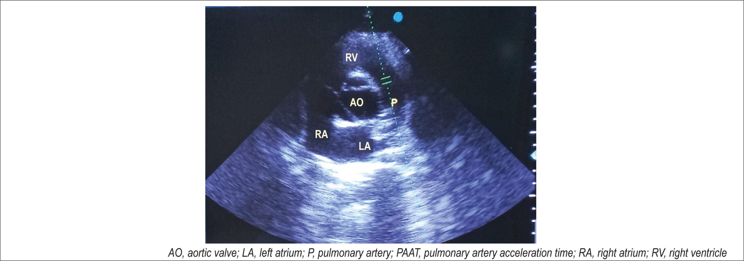
Pulmonary artery acceleration time (PAAT) can be used as a parameter in the evaluation of pulmonary hypertension and aids left ventricular diastolic function (LVDF) analyses.

Objective:

To assess whether there is a correlation between PAAT and LVDF parameters in individuals with a preserved left ventricular systolic function and by sex, age, and cardiovascular risk factors.

Methods:

Observational cross-sectional study. One hundred nineteen patients were selected (59 women [49.6%]). The subjects underwent transthoracic echocardiography including measurements of PAAT, E and A waves and E/A ratio, e’ septal and e’ lateral waves and E/e’ ratio, pulmonary artery systolic pressure (PASP), and left atrial volume.

Results:

In female patients, a positive correlation (Spearman’s correlation coefficient – Spearman correlation coefficient [SCC]) was found between the PAAT value and the lateral e’ (SCC, 0.47; p = 0.002), with the E/A ratio (SCC, 0.32; p = 0.04), and with septal e’ (SCC, 0.36; p = 0.023), and a negative correlation between PAAT and PASP (SCC, -0.43; p = 0.034). In men, no correlation was found between PAAT and any parameters. Lower PAAT values were found in women with systemic arterial hypertension (hypertension) than in women without hypertension (0.13 ± 0.03 s versus 0.16 ± 0.03 s; p = 0.015).

Conclusion:

The present study showed a significant correlation between PAAT and some LVDF parameters in female patients only. Hypertension was correlated with lower PAAT values in women.

  • Cardiac function
  • Cardiovascular disease
  • COVID-19
  • Echocardiography
  • SARS-CoV-2
Read Full Article
Creative Commons Attribution (CC BY) Creative Commons license
30/03/2022 DOI: 10.47593/2675-312X/20223501eabc242 Views 1
Figure 1
Case Report

Role of Transthoracic Echocardiography in the Diagnosis of Double Aortic Arch

Guilherme Gomes de Souza et al. Adriana Furletti Machado Guimarães Ricardo Wang Sandra Regina Tolentino Castilho … Henrique de Assis Fonseca Tonelli Fátima Derlene da Rocha Araújo

Introduction

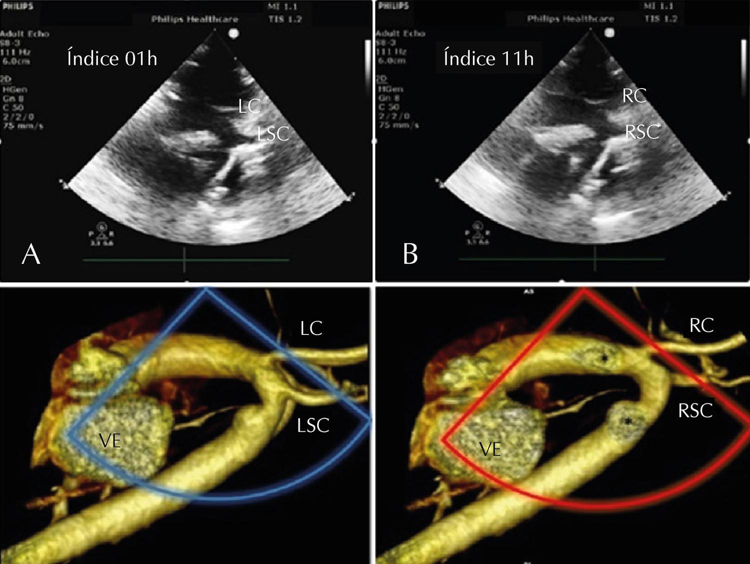
Double aortic arch is the main cause of vascular ring in cardiovascular surgical case series records. This rare anomaly, caused by the non-involution of one of the embryonic aortic arches, is a challenge for the pediatric echocardiographer, often requiring other imaging methods such as computed tomography or cardiac magnetic resonance. This case report aimed to systematize echocardiographic findings to help diagnose double aortic arch.

[…]

  • Congenital
  • Echocardiography
  • Heart Defects
  • Vascular ring
Read Full Article
Creative Commons Attribution (CC BY) Creative Commons license
04/03/2022 DOI: 10.47593/2675-312X/20223501eabc267 Views 1
Figure 1
Case Report

Libman-Sacks endocarditis in a male patient with systemic lupus erythematosus

Álvaro Augusto Vedana Filho et al. Vivien Ritchie Bittencourt Thaíssa Antunes Mattar Valente Fernando Silva Botelho Marco Stephan Lofrano Alves

Introduction

Libman-Sacks endocarditis (LSE), also known as marantic or verrucous endocarditis, is a form of non-bacterial thrombotic endocarditis (NBTE) that involves the presence of sterile vegetations in the heart valves. These vegetations may be associated with malignancy, systemic lupus erythematosus (SLE), or antiphospholipid syndrome (APS), and although they are more commonly found in the mitral and aortic valves, other valves may be involved. Diagnosis requires high clinical suspicion when evaluating specific populations.

[…]

  • Echocardiogram
  • Endocarditis
  • Lupus Erythematosus, Systemic
Read Full Article
Creative Commons Attribution (CC BY) Creative Commons license
04/03/2022 DOI: 10.47593/2675-312X/20223501eabc224
Figure 1
Case Report

Cardiac Fibroma: Long-Term Follow-up by Cardiac Magnetic Resonance

Fernanda Sayuri Oshiro et al. Raul Serra Valério Alfredo Augusto Eyer Rodrigues Gilberto Szarf … Marly Uellendahl Maria Eduarda Menezes de Siqueira

Abstract

Background

Primary cardiac neoplasms are rare, and a correct diagnosis is essential to planning the most appropriate treatment.

Objective

To demonstrate the role of cardiac magnetic resonance imaging (CMR) in the assessment, diagnosis, and follow-up of cardiac fibroma.

Case report

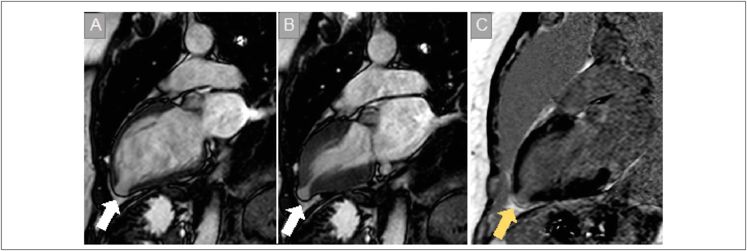
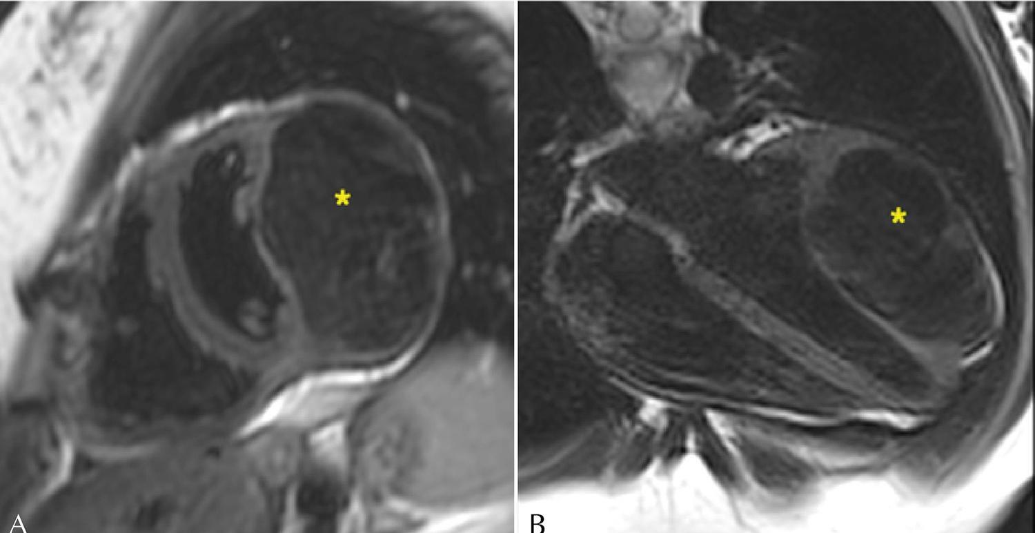
Female 21-year-old patient with a myocardial mass detected on echocardiogram. CMR confirmed a diagnosis of cardiac fibroma. The patient chose to be followed up, and her condition remained stable after six years.

Discussion

Cardiac fibromas are the second most common neoplasm in children and young people. On CMR, it is characterized by intense and homogeneous delayed enhancement. CMR plays an important role in the assessment of cardiac masses.

  • Fibromas
  • Heart Neoplasms
  • Magnetic Resonance Imaging
Read Full Article
Creative Commons Attribution (CC BY) Creative Commons license
04/03/2022 DOI: 10.47593/2675-312X/20223501eabc263 Views 1
Figure 1
Image

Incidental Finding of Iliac Vein Compression Syndrome

Marcelo Bellini Dalio et al. Milton Sérgio Bohatch Júnior Tércio Ferreira de Oliveira Maurício Serra Ribeiro Edwaldo Edner Joviliano

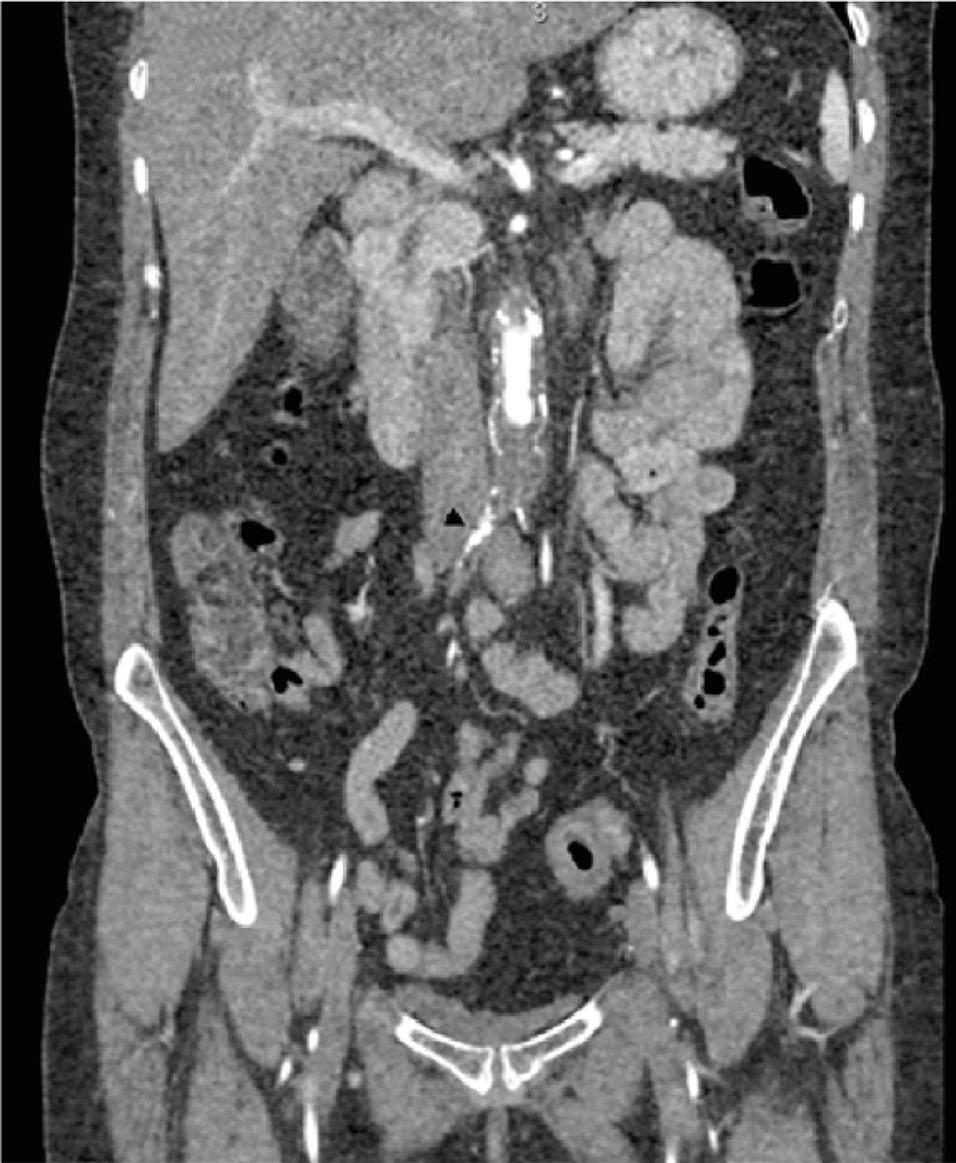
A 49-year-old woman presented with a progressive history of bilateral buttock and calf intermittent claudication, reaching a walking distance of 10 m. She was a smoker and had hypertension. She had no history of venous thrombosis, leg edema, varicose veins, or signs of chronic venous disease. Her femoral pulses were absent. Computed tomography angiography showed occlusion of the infrarenal aorta with recanalization of both iliac arteries. Interestingly, the right common iliac artery compressed the left common iliac vein, which was enlarged ( ). Left-sided dilated pelvic collaterals were observed: uterine and gonadal veins ( ). There was no compression on the left renal vein.

[…]

  • Asymptomatic Diseases
  • Cockett
  • Iliac vein compression
  • syndrome, May-Thurner
Read Full Article
Creative Commons Attribution (CC BY) Creative Commons license
04/03/2022 DOI: 10.47593/2675-312X/20223501eabc225 Views 2

How to cite this article

EndNote RIS

Electronic Document Format (Vancouver)

Martins AAB, Osaku LY, Camarozano AC, Sá CRF, Carmo DC, Fortunato JA, Darwich RZ, et al. Tricuspid Annular Plane Systolic Excursion and Mitral Annular Systolic Excursion: Correlation with Left Ventricular Diastolic Function. ABC Imagem Cardiovascular 2022;35(1):eabc245.

Electronic Document Format (ABNT)

Martins, André Américo Bedenko; Osaku, Leonardo Yoshida; Camarozano, Ana Cristina; Sá, Cintia Rocha Fortes de; Carmo, Daniela de Castro; Fortunato, Jerônimo Antonio; Darwich, Rubens Zenóbio; Baroncini, Liz Andréa Villela. Tricuspid Annular Plane Systolic Excursion and Mitral Annular Systolic Excursion: Correlation with Left Ventricular Diastolic Function. ABC Imagem Cardiovascular, v. 35, n. 1, eabc245, Mar. 2022.

Electronic Document Format (APA)

Martins, A. A. B., Osaku, L. Y., Camarozano, A. C., Sá, C. R. F. , Carmo, D. C., Fortunato, J. A., Darwich, R. Z., & Baroncini, L. A. V. (2022). Tricuspid Annular Plane Systolic Excursion and Mitral Annular Systolic Excursion: Correlation with Left Ventricular Diastolic Function. ABC Imagem Cardiovascular, 35(1), eabc245.

Electronic Document Format (ISO)

Martins, André Américo Bedenko and Osaku, Leonardo Yoshida and Camarozano, Ana Cristina and Sá, Cintia Rocha Fortes de and Carmo, Daniela de Castro and Fortunato, Jerônimo Antonio and Darwich, Rubens Zenóbio and Baroncini, Liz Andréa Villela. Tricuspid Annular Plane Systolic Excursion and Mitral Annular Systolic Excursion: Correlation with Left Ventricular Diastolic Function. ABC Imagem Cardiovascular [online]. 2022, vol. 35, n. 1, [cited 2026-10-14], eabc245. Available from: <https://journals.cardiol.br/en/article/tricuspid-annular-plane-systolic-excursion-and-mitral-annular-systolic-excursion-correlation-with-left-ventricular-diastolic-function/>. ISSN 2318-8219

EndNote RIS

Electronic Document Format (Vancouver)

Cruz AL, Freitas C, Watanabe BNS, Nascimento HB, Porath H, Bettini LAF, Moreira L, et al. Factors Associated With Abnormalities On Myocardial Perfusion Scintigraphy In Diabetic Patients. ABC Imagem Cardiovascular 2022;35(1):eabc274.

Electronic Document Format (ABNT)

Cruz, Alfredo Lírio da; Freitas, Álisson Carvalho de; Watanabe, Bianca Naomi Sada; Nascimento, Helena Bigarella; Porath, Heloisa; Bettini, Luiz Antonio Fruet; Moreira, Luciane; Dopheide, Marcos Germano. Factors Associated With Abnormalities On Myocardial Perfusion Scintigraphy In Diabetic Patients. ABC Imagem Cardiovascular, v. 35, n. 1, eabc274, Mar. 2022.

Electronic Document Format (APA)

Cruz, A. L. , Freitas, C. , Watanabe, B. N. S., Nascimento, H. B., Porath, H., Bettini, L. A. F., Moreira, L., & Dopheide, M. G. (2022). Factors Associated With Abnormalities On Myocardial Perfusion Scintigraphy In Diabetic Patients. ABC Imagem Cardiovascular, 35(1), eabc274.

Electronic Document Format (ISO)

Cruz, Alfredo Lírio da and Freitas, Álisson Carvalho de and Watanabe, Bianca Naomi Sada and Nascimento, Helena Bigarella and Porath, Heloisa and Bettini, Luiz Antonio Fruet and Moreira, Luciane and Dopheide, Marcos Germano. Factors Associated With Abnormalities On Myocardial Perfusion Scintigraphy In Diabetic Patients. ABC Imagem Cardiovascular [online]. 2022, vol. 35, n. 1, [cited 2026-10-14], eabc274. Available from: <https://journals.cardiol.br/en/article/factors-associated-with-abnormalities-on-myocardial-perfusion-scintigraphy-in-diabetic-patients/>. ISSN 2318-8219

EndNote RIS

Electronic Document Format (Vancouver)

Masson-Silva JB, Silva Júnior CG. My Approach To Echocardiographic Assessment In Pulmonary Hypertension. ABC Imagem Cardiovascular 2022;35(1):ecom18.

Electronic Document Format (ABNT)

Masson-Silva, João Batista; Silva Júnior, Cloves Geraldino da. My Approach To Echocardiographic Assessment In Pulmonary Hypertension. ABC Imagem Cardiovascular, v. 35, n. 1, ecom18, Mar. 2022.

Electronic Document Format (APA)

Masson-Silva, J. B., & Silva Júnior, C. G. (2022). My Approach To Echocardiographic Assessment In Pulmonary Hypertension. ABC Imagem Cardiovascular, 35(1), ecom18.

Electronic Document Format (ISO)

Masson-Silva, João Batista and Silva Júnior, Cloves Geraldino da. My Approach To Echocardiographic Assessment In Pulmonary Hypertension. ABC Imagem Cardiovascular [online]. 2022, vol. 35, n. 1, [cited 2026-10-14], ecom18. Available from: <https://journals.cardiol.br/en/article/my-approach-to-echocardiographic-assessment-in-pulmonary-hypertension/>. ISSN 2318-8219

EndNote RIS

Electronic Document Format (Vancouver)

Abreu MEB, Pinheiro TCD, Guimarães AAL, Abreu JS. Coronary Flow Velocity Reserve and Myocardial Contractility Under Stress in the Post-Infarction Ischemic Memory Dilemma. ABC Imagem Cardiovascular 2022;35(1):eabc221.

Electronic Document Format (ABNT)

Abreu, Marilia Esther Benevides de; Pinheiro, Tereza Cristina Diógenes; Guimarães, Antônio Augusto Lima; Abreu, José Sebastião de. Coronary Flow Velocity Reserve and Myocardial Contractility Under Stress in the Post-Infarction Ischemic Memory Dilemma. ABC Imagem Cardiovascular, v. 35, n. 1, eabc221, Mar. 2022.

Electronic Document Format (APA)

Abreu, M. E. B. , Pinheiro, T. C. D., Guimarães, A. A. L., & Abreu, J. S. (2022). Coronary Flow Velocity Reserve and Myocardial Contractility Under Stress in the Post-Infarction Ischemic Memory Dilemma. ABC Imagem Cardiovascular, 35(1), eabc221.

Electronic Document Format (ISO)

Abreu, Marilia Esther Benevides de and Pinheiro, Tereza Cristina Diógenes and Guimarães, Antônio Augusto Lima and Abreu, José Sebastião de. Coronary Flow Velocity Reserve and Myocardial Contractility Under Stress in the Post-Infarction Ischemic Memory Dilemma. ABC Imagem Cardiovascular [online]. 2022, vol. 35, n. 1, [cited 2026-10-14], eabc221. Available from: <https://journals.cardiol.br/en/article/coronary-flow-velocity-reserve-and-myocardial-contractility-under-stress-in-the-post-infarction-ischemic-memory-dilemma/>. ISSN 2318-8219

EndNote RIS

Electronic Document Format (Vancouver)

Beck ALS, Ribeiro LCM. My Approach to Aortic Stenosis Assessment with Discrepant Quantification. ABC Imagem Cardiovascular 2022;35(1):ecom21.

Electronic Document Format (ABNT)

Beck, Adenalva Lima de Souza; Ribeiro, Luiz Carlos Madruga. My Approach to Aortic Stenosis Assessment with Discrepant Quantification. ABC Imagem Cardiovascular, v. 35, n. 1, ecom21, Mar. 2022.

Electronic Document Format (APA)

Beck, A. L. S., & Ribeiro, L. C. M. (2022). My Approach to Aortic Stenosis Assessment with Discrepant Quantification. ABC Imagem Cardiovascular, 35(1), ecom21.

Electronic Document Format (ISO)

Beck, Adenalva Lima de Souza and Ribeiro, Luiz Carlos Madruga. My Approach to Aortic Stenosis Assessment with Discrepant Quantification. ABC Imagem Cardiovascular [online]. 2022, vol. 35, n. 1, [cited 2026-10-14], ecom21. Available from: <https://journals.cardiol.br/en/article/my-approach-to-aortic-stenosis-assessment-with-discrepant-quantification/>. ISSN 2318-8219

EndNote RIS

Electronic Document Format (Vancouver)

Cirino RHD, Ducci RD. My approach to diagnosis of and echocardiographic follow-up for muscular dystrophies. ABC Imagem Cardiovascular 2022;35(1):eabc292.

Electronic Document Format (ABNT)

Cirino, Raphael Henrique Déa; Ducci, Renata Dal-Prá. My approach to diagnosis of and echocardiographic follow-up for muscular dystrophies. ABC Imagem Cardiovascular, v. 35, n. 1, eabc292, Mar. 2022.

Electronic Document Format (APA)

Cirino, R. H. D., & Ducci, R. D. (2022). My approach to diagnosis of and echocardiographic follow-up for muscular dystrophies. ABC Imagem Cardiovascular, 35(1), eabc292.

Electronic Document Format (ISO)

Cirino, Raphael Henrique Déa and Ducci, Renata Dal-Prá. My approach to diagnosis of and echocardiographic follow-up for muscular dystrophies. ABC Imagem Cardiovascular [online]. 2022, vol. 35, n. 1, [cited 2026-10-14], eabc292. Available from: <https://journals.cardiol.br/en/article/my-approach-to-diagnosis-of-and-echocardiographic-follow-up-for-muscular-dystrophies/>. ISSN 2318-8219

EndNote RIS

Electronic Document Format (Vancouver)

Melato LH, Rassi DC, Furtado RG, Oliveira ACR. Intermittent Aortic Intraprosthetic Failure. ABC Imagem Cardiovascular 2022;35(1):eabc284.

Electronic Document Format (ABNT)

Melato, Luciano H; Rassi, Daniela do Carmo; Furtado, Rogério G; Oliveira, Ana Caroline R. Intermittent Aortic Intraprosthetic Failure. ABC Imagem Cardiovascular, v. 35, n. 1, eabc284, Mar. 2022.

Electronic Document Format (APA)

Melato, L. H., Rassi, D. C., Furtado, R. G., & Oliveira, A. C. R. (2022). Intermittent Aortic Intraprosthetic Failure. ABC Imagem Cardiovascular, 35(1), eabc284.

Electronic Document Format (ISO)

Melato, Luciano H and Rassi, Daniela do Carmo and Furtado, Rogério G and Oliveira, Ana Caroline R. Intermittent Aortic Intraprosthetic Failure. ABC Imagem Cardiovascular [online]. 2022, vol. 35, n. 1, [cited 2026-10-14], eabc284. Available from: <https://journals.cardiol.br/en/article/intermittent-aortic-intraprosthetic-failure/>. ISSN 2318-8219

EndNote RIS

Electronic Document Format (Vancouver)

Barros Filho ACL, Romano MMD. My Approach to the Echocardiographic Assessment of Mitral Annular Calcification: Challenges and Particularities. ABC Imagem Cardiovascular 2022;35(1):ecom19.

Electronic Document Format (ABNT)

Barros Filho, Antonio Carlos Leite de; Romano, Minna Moreira Dias. My Approach to the Echocardiographic Assessment of Mitral Annular Calcification: Challenges and Particularities. ABC Imagem Cardiovascular, v. 35, n. 1, ecom19, Mar. 2022.

Electronic Document Format (APA)

Barros Filho, A. C. L. , & Romano, M. M. D. (2022). My Approach to the Echocardiographic Assessment of Mitral Annular Calcification: Challenges and Particularities. ABC Imagem Cardiovascular, 35(1), ecom19.

Electronic Document Format (ISO)

Barros Filho, Antonio Carlos Leite de and Romano, Minna Moreira Dias. My Approach to the Echocardiographic Assessment of Mitral Annular Calcification: Challenges and Particularities. ABC Imagem Cardiovascular [online]. 2022, vol. 35, n. 1, [cited 2026-10-14], ecom19. Available from: <https://journals.cardiol.br/en/article/my-approach-to-the-echocardiographic-assessment-of-mitral-annular-calcification-challenges-and-particularities/>. ISSN 2318-8219

EndNote RIS

Electronic Document Format (Vancouver)

Moreira HT, Volpe GJ, Schmidt A. Myocardial Involvement in Chagas Disease: From the Perspective of Cardiovascular Magnetic Ressonance Assessment. ABC Imagem Cardiovascular 2022;35(1):eabc285.

Electronic Document Format (ABNT)

Moreira, Henrique Turin; Volpe, Gustavo Jardim; Schmidt, André. Myocardial Involvement in Chagas Disease: From the Perspective of Cardiovascular Magnetic Ressonance Assessment. ABC Imagem Cardiovascular, v. 35, n. 1, eabc285, Mar. 2022.

Electronic Document Format (APA)

Moreira, H. T., Volpe, G. J., & Schmidt, A. (2022). Myocardial Involvement in Chagas Disease: From the Perspective of Cardiovascular Magnetic Ressonance Assessment. ABC Imagem Cardiovascular, 35(1), eabc285.

Electronic Document Format (ISO)

Moreira, Henrique Turin and Volpe, Gustavo Jardim and Schmidt, André. Myocardial Involvement in Chagas Disease: From the Perspective of Cardiovascular Magnetic Ressonance Assessment. ABC Imagem Cardiovascular [online]. 2022, vol. 35, n. 1, [cited 2026-10-14], eabc285. Available from: <https://journals.cardiol.br/en/article/myocardial-involvement-in-chagas-disease-from-the-perspective-of-cardiovascular-magnetic-ressonance-assessment/>. ISSN 2318-8219

EndNote RIS

Electronic Document Format (Vancouver)

Coimbra RM, Camarozano AC, Sá CRF, Carmo DC, Fortunato JA, Darwich RZ, Baroncini LAV, et al. Right Ventricular Outflow Tract Acceleration Time: Correlation with Left Ventricular Diastolic Function, Sex, And Age. ABC Imagem Cardiovascular 2022;35(1):eabc242.

Electronic Document Format (ABNT)

Coimbra, Renan Macedo; Camarozano, Ana Cristina; Sá, Cintia Rocha Fortes de; Carmo, Daniela de Castro; Fortunato, Jerônimo Antonio; Darwich, Rubens Zenóbio; Baroncini, Liz Andréa Villela. Right Ventricular Outflow Tract Acceleration Time: Correlation with Left Ventricular Diastolic Function, Sex, And Age. ABC Imagem Cardiovascular, v. 35, n. 1, eabc242, Mar. 2022.

Electronic Document Format (APA)

Coimbra, R. M., Camarozano, A. C., Sá, C. R. F. , Carmo, D. C., Fortunato, J. A., Darwich, R. Z., & Baroncini, L. A. V. (2022). Right Ventricular Outflow Tract Acceleration Time: Correlation with Left Ventricular Diastolic Function, Sex, And Age. ABC Imagem Cardiovascular, 35(1), eabc242.

Electronic Document Format (ISO)

Coimbra, Renan Macedo and Camarozano, Ana Cristina and Sá, Cintia Rocha Fortes de and Carmo, Daniela de Castro and Fortunato, Jerônimo Antonio and Darwich, Rubens Zenóbio and Baroncini, Liz Andréa Villela. Right Ventricular Outflow Tract Acceleration Time: Correlation with Left Ventricular Diastolic Function, Sex, And Age. ABC Imagem Cardiovascular [online]. 2022, vol. 35, n. 1, [cited 2026-10-14], eabc242. Available from: <https://journals.cardiol.br/en/article/right-ventricular-outflow-tract-acceleration-time-correlation-with-left-ventricular-diastolic-function-sex-and-age/>. ISSN 2318-8219

EndNote RIS

Electronic Document Format (Vancouver)

Souza GG, Guimarães AFM, Wang R, Castilho SRT, Tonelli HAF, Araújo FDR. Role of Transthoracic Echocardiography in the Diagnosis of Double Aortic Arch. Arquivos Brasileiros de Cardiologia Imagem cardiovascular 2022;35(1):eabc267.

Electronic Document Format (ABNT)

Souza, Guilherme Gomes de; Guimarães, Adriana Furletti Machado; Wang, Ricardo; Castilho, Sandra Regina Tolentino; Tonelli, Henrique de Assis Fonseca; Araújo, Fátima Derlene da Rocha. Role of Transthoracic Echocardiography in the Diagnosis of Double Aortic Arch. Arquivos Brasileiros de Cardiologia Imagem cardiovascular, v. 35, n. 1, eabc267, Mar. 2022.

Electronic Document Format (APA)

Souza, G. G. , Guimarães, A. F. M., Wang, R., Castilho, S. R. T., Tonelli, H. A. F., & Araújo, F. D. R. (2022). Role of Transthoracic Echocardiography in the Diagnosis of Double Aortic Arch. Arquivos Brasileiros de Cardiologia Imagem cardiovascular, 35(1), eabc267.

Electronic Document Format (ISO)

Souza, Guilherme Gomes de and Guimarães, Adriana Furletti Machado and Wang, Ricardo and Castilho, Sandra Regina Tolentino and Tonelli, Henrique de Assis Fonseca and Araújo, Fátima Derlene da Rocha. Role of Transthoracic Echocardiography in the Diagnosis of Double Aortic Arch. Arquivos Brasileiros de Cardiologia Imagem cardiovascular [online]. 2022, vol. 35, n. 1, [cited 2026-10-14], eabc267. Available from: <https://journals.cardiol.br/en/article/role-of-transthoracic-echocardiography-in-the-diagnosis-of-double-aortic-arch/>. ISSN 2318-8219

EndNote RIS

Electronic Document Format (Vancouver)

Vedana Filho A, Bittencourt VR, Valente TAM, Botelho FS, Alves MSL. Libman-Sacks endocarditis in a male patient with systemic lupus erythematosus. Arquivos Brasileiros de Cardiologia Imagem cardiovascular 2022;35(1):eabc224.

Electronic Document Format (ABNT)

Vedana Filho, Álvaro Augusto; Bittencourt, Vivien Ritchie; Valente, Thaíssa Antunes Mattar; Botelho, Fernando Silva; Alves, Marco Stephan Lofrano. Libman-Sacks endocarditis in a male patient with systemic lupus erythematosus. Arquivos Brasileiros de Cardiologia Imagem cardiovascular, v. 35, n. 1, eabc224, Mar. 2022.

Electronic Document Format (APA)

Vedana Filho, A., Bittencourt, V. R., Valente, T. A. M., Botelho, F. S., & Alves, M. S. L. (2022). Libman-Sacks endocarditis in a male patient with systemic lupus erythematosus. Arquivos Brasileiros de Cardiologia Imagem cardiovascular, 35(1), eabc224.

Electronic Document Format (ISO)

Vedana Filho, Álvaro Augusto and Bittencourt, Vivien Ritchie and Valente, Thaíssa Antunes Mattar and Botelho, Fernando Silva and Alves, Marco Stephan Lofrano. Libman-Sacks endocarditis in a male patient with systemic lupus erythematosus. Arquivos Brasileiros de Cardiologia Imagem cardiovascular [online]. 2022, vol. 35, n. 1, [cited 2026-10-14], eabc224. Available from: <https://journals.cardiol.br/en/article/libman-sacks-endocarditis-in-a-male-patient-with-systemic-lupus-erythematosus/>. ISSN 2318-8219

EndNote RIS

Electronic Document Format (Vancouver)

Oshiro FS, Valério RS, Rodrigues AAE, Szarf G, Uellendahl M, Siqueira MEM. Cardiac Fibroma: Long-Term Follow-up by Cardiac Magnetic Resonance. Arquivos Brasileiros de Cardiologia Imagem cardiovascular 2022;35(1):eabc263.

Electronic Document Format (ABNT)

Oshiro, Fernanda Sayuri; Valério, Raul Serra; Rodrigues, Alfredo Augusto Eyer; Szarf, Gilberto; Uellendahl, Marly; Siqueira, Maria Eduarda Menezes de. Cardiac Fibroma: Long-Term Follow-up by Cardiac Magnetic Resonance. Arquivos Brasileiros de Cardiologia Imagem cardiovascular, v. 35, n. 1, eabc263, Mar. 2022.

Electronic Document Format (APA)

Oshiro, F. S., Valério, R. S., Rodrigues, A. A. E., Szarf, G., Uellendahl, M., & Siqueira, M. E. M. (2022). Cardiac Fibroma: Long-Term Follow-up by Cardiac Magnetic Resonance. Arquivos Brasileiros de Cardiologia Imagem cardiovascular, 35(1), eabc263.

Electronic Document Format (ISO)

Oshiro, Fernanda Sayuri and Valério, Raul Serra and Rodrigues, Alfredo Augusto Eyer and Szarf, Gilberto and Uellendahl, Marly and Siqueira, Maria Eduarda Menezes de. Cardiac Fibroma: Long-Term Follow-up by Cardiac Magnetic Resonance. Arquivos Brasileiros de Cardiologia Imagem cardiovascular [online]. 2022, vol. 35, n. 1, [cited 2026-10-14], eabc263. Available from: <https://journals.cardiol.br/en/article/cardiac-fibroma-long-term-follow-up-by-cardiac-magnetic-resonance/>. ISSN 2318-8219

EndNote RIS

Electronic Document Format (Vancouver)

Dalio MB, Bohatch Júnior MS, Oliveira TF, Ribeiro MS, Joviliano EE. Incidental Finding of Iliac Vein Compression Syndrome. Arquivos Brasileiros de Cardiologia Imagem cardiovascular 2022;35(1):eabc225.

Electronic Document Format (ABNT)

Dalio, Marcelo Bellini; Bohatch Júnior, Milton Sérgio; Oliveira, Tércio Ferreira de; Ribeiro, Maurício Serra; Joviliano, Edwaldo Edner. Incidental Finding of Iliac Vein Compression Syndrome. Arquivos Brasileiros de Cardiologia Imagem cardiovascular, v. 35, n. 1, eabc225, Mar. 2022.

Electronic Document Format (APA)

Dalio, M. B., Bohatch Júnior, M. S., Oliveira, T. F. , Ribeiro, M. S., & Joviliano, E. E. (2022). Incidental Finding of Iliac Vein Compression Syndrome. Arquivos Brasileiros de Cardiologia Imagem cardiovascular, 35(1), eabc225.

Electronic Document Format (ISO)

Dalio, Marcelo Bellini and Bohatch Júnior, Milton Sérgio and Oliveira, Tércio Ferreira de and Ribeiro, Maurício Serra and Joviliano, Edwaldo Edner. Incidental Finding of Iliac Vein Compression Syndrome. Arquivos Brasileiros de Cardiologia Imagem cardiovascular [online]. 2022, vol. 35, n. 1, [cited 2026-10-14], eabc225. Available from: <https://journals.cardiol.br/en/article/incidental-finding-of-iliac-vein-compression-syndrome/>. ISSN 2318-8219

Sede – São Paulo
Rua Barata Ribeiro, 380 cj 54 Bela Vista
São Paulo, Brasil – CEP: 01308-000
Tel.: +55 (11) 3120-3363
Telefax: +55 (11) 3259-2988
E-mail: depeco@cardiol.br

Sede – Rio de Janeiro
Av. Marechal Câmara, 160
3º andar – Sala: 330 – Centro
Rio de Janeiro – CEP: 20020-907
Tel.: +55 (21) 3478-2700
ou: +55 (21) 99386-3961
E-mail: abcimaging@cardiol.br

  • About the Journal
  • Internal Regulation
  • Editors Section
  • Editorial Board
  • SBC Award

  • Guidelines for Publication
  • Editorial Policy
  • Ethics Policy
  • Policy on the Use of Artificial Intelligence
  • Mandatory Publication Form

Issues

SBC Guidelines