Cardiac Lipoma as an Incidental Finding in Cardiovascular Imaging Exam
Introduction
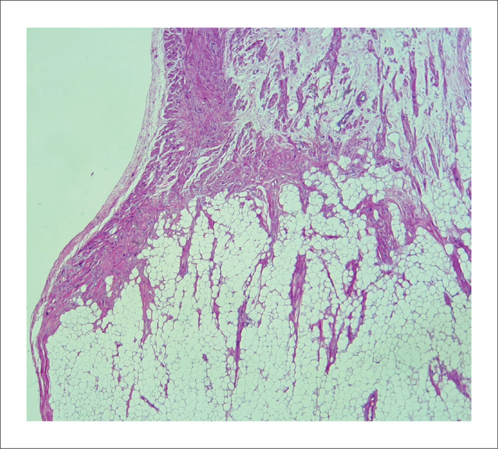
The advent of cardiac imaging has ushered in an era where incidental findings can be detected using less invasive methods, even in asymptomatic patients. The clinical presentation of cardiac masses, characterized by their varying growth rates and diverse locations within the heart, poses a formidable challenge to cardiologists. Therefore, paramount importance lies in reducing the clinical burden and costs, considering the potential of cardiac imaging to optimize the clinical pathway. The most common cardiac masses in the right atrium include thrombi, vegetations, and neoplasia. Approximately 75% of primary tumors are benign, with myxomas comprising 50% of benign cases. Other benign tumors that usually may arise in the right atrium are rhabdomyomas, fibromas, fibroelastomas, and lipomas. Cardiac lipomas are rare, accounting for 2.9% to 8% of all benign cardiac tumors. They rank third in frequency after myxomas and papillary fibroelastomas. The literature suggests that 25% of cardiac lipomas are intramyocardial; 25% are extracavitary of epicardial origin, and 50% are intracavitary of subendocardial origin. Lipomas are well-encapsulated and homogeneous masses composed of mature fat. Although the etiology of cardiac lipomas is unknown, they can originate from any of the three cardiac tissues: subendocardial (the most common), pericardial, or myocardial.
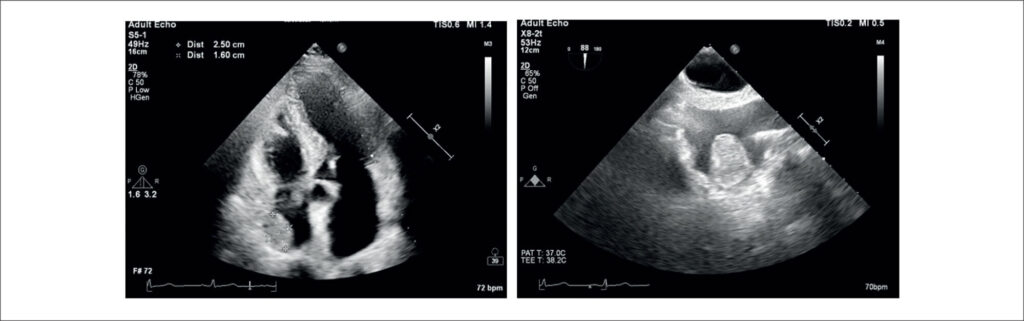
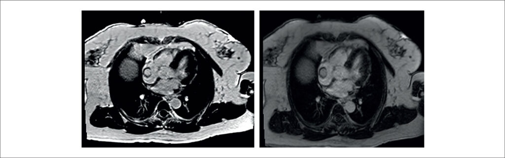
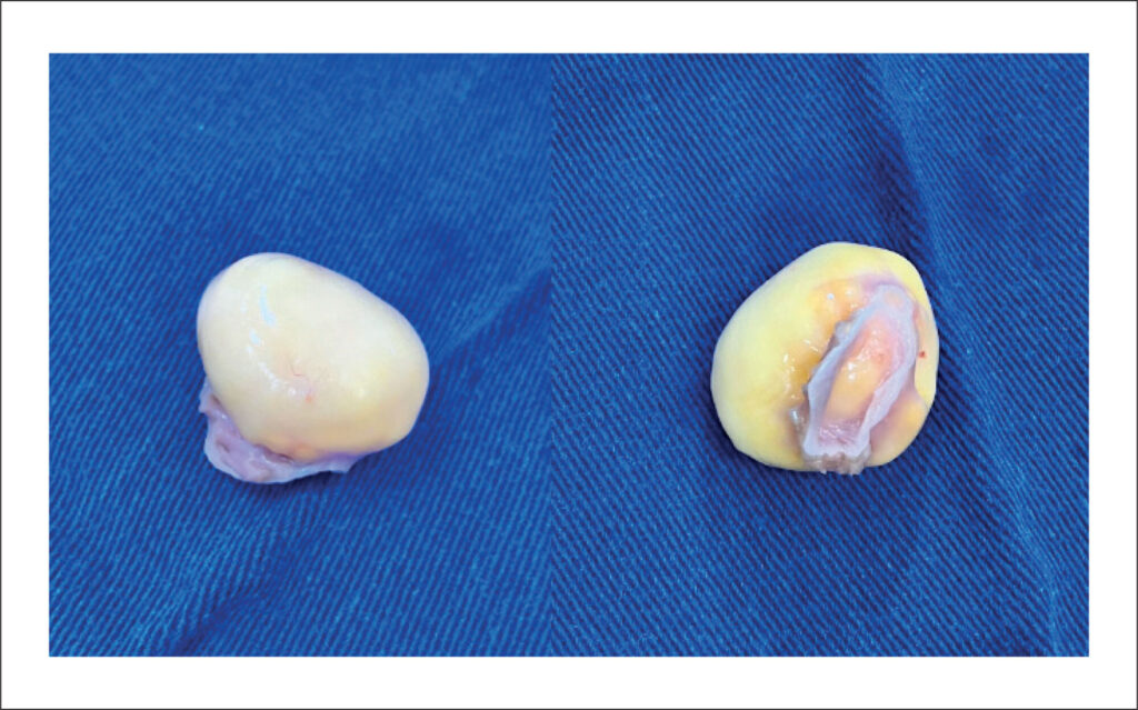
The aim of this report is to present a case of an asymptomatic right atrial lipoma and to discuss the etiology, natural history, and optimal management strategies for right atrial lipomas.
[…]
Images
Image gallery fullscreen viewer
Comments are moderated by the editorial team before publication. Discussions should be evidence-based and use scientific language. Conflicts of interest should be declared.