My Approach to Imaging the Atrial Septal Defect by Three-Dimensional Echocardiography
Pre-procedural assessment
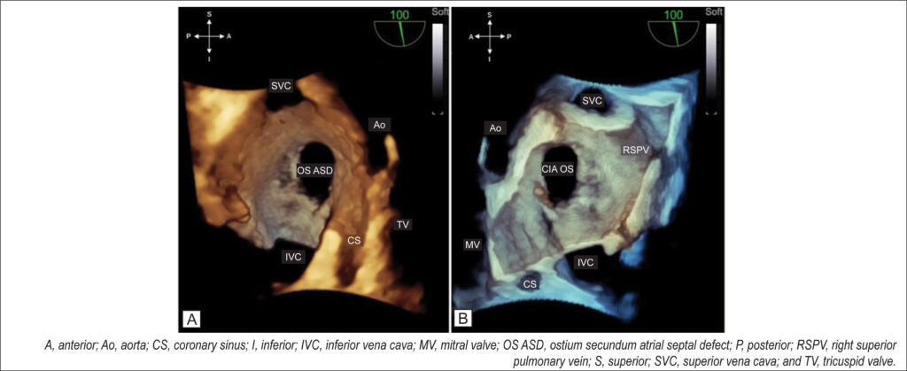
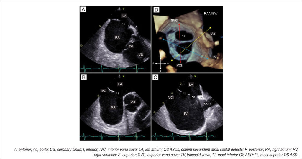
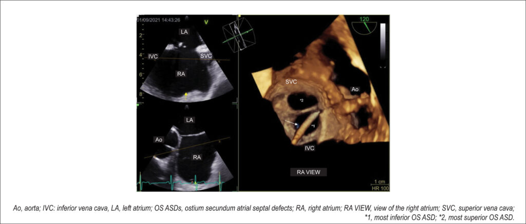
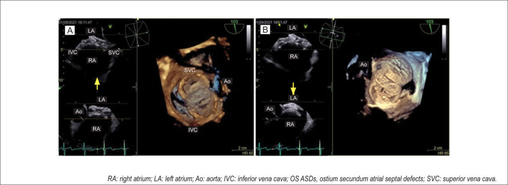
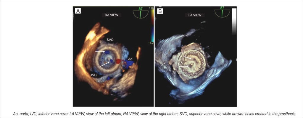
At the end of a detailed 2D TEE with a full septal analysis from the four-chamber view to bicaval section and color mapping, we commence the 3D image acquisition. The en-face projection of the ASD and the defect’s positioning in the anatomic position provide valuable information from a single image: size, shape, location in the interatrial septum, characteristics of the defect’s borders, and the presence of additional fenestration.
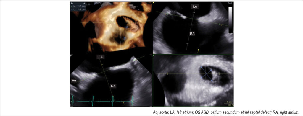
I usually start the 3D evaluation from the perspective of the right atrium to facilitate the identification of anatomical markers. I acquire a medium angle block (3D or 4D zoom) or a large block (full-volume using the two-section tool) starting from an initial bicaval section and proceeding with approximate 90° rotation in the z-plane so that the superior and inferior vena cava are aligned in the vertical plane (i.e., superior vena cava at the 11 o’clock position) and the aorta in the anterior and superior region. I prefer not to use the narrow-angle mode for acquisition (live 3D or bird’s eye view) or very high magnification due to a loss of anatomical references adjacent to the defect, which can interfere with this fundamental initial analysis. From this reconstruction (), I can characterize the defect as unique, the OS type, having an oval shape, being central, being medium-sized, and having adequate borders. shows the same ASD and allows assessment of the occurrence of dynamic variation in its dimension with the cardiac cycle.
[…]
Images
Image gallery fullscreen viewer
Comments are moderated by the editorial team before publication. Discussions should be evidence-based and use scientific language. Conflicts of interest should be declared.