Focal Arterialization and Neoatherosclerosis of a Saphenous Vein Graft. Improving our Understanding of Late Graft Failures
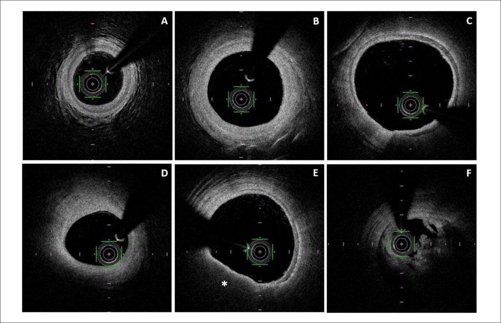
A 76 year-old man with a past medical history of type 2 Diabetes, stage 3 chronic kidney disease and previous multivessel coronary artery bypass surgery 10 years before was admitted with an acute coronary syndrome. Coronary angiogram showed an occluded saphenous vein graft (SVG) to first diagonal. After restoring flow by means of thrombus aspiration, optical coherence tomography (OCT) runs were required. Localized and widely patent segments of the graft showed a clear three-layer configuration, suggestive of SVG focal arterialization. Proximally, predominantly red thrombus determined a critical stenosis and a thin cap fibroatheroma was evident in an arterialized segment of the graft contiguous to the thrombotic lesion. Conversely, the rest of the SVG kept its venous appearance and had three different areas of stenosis ().
[…]
Images
Image gallery fullscreen viewer
Comments are moderated by the editorial team before publication. Discussions should be evidence-based and use scientific language. Conflicts of interest should be declared.