My Approach To Manage Valve Prosthetic Dysfunction: What is the Role of Tomography?
Abstract
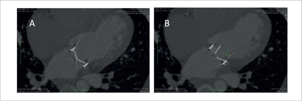
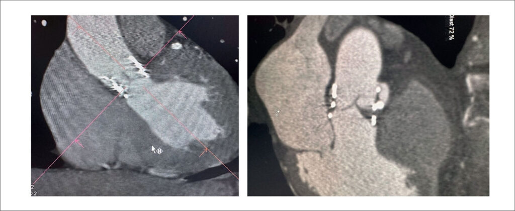
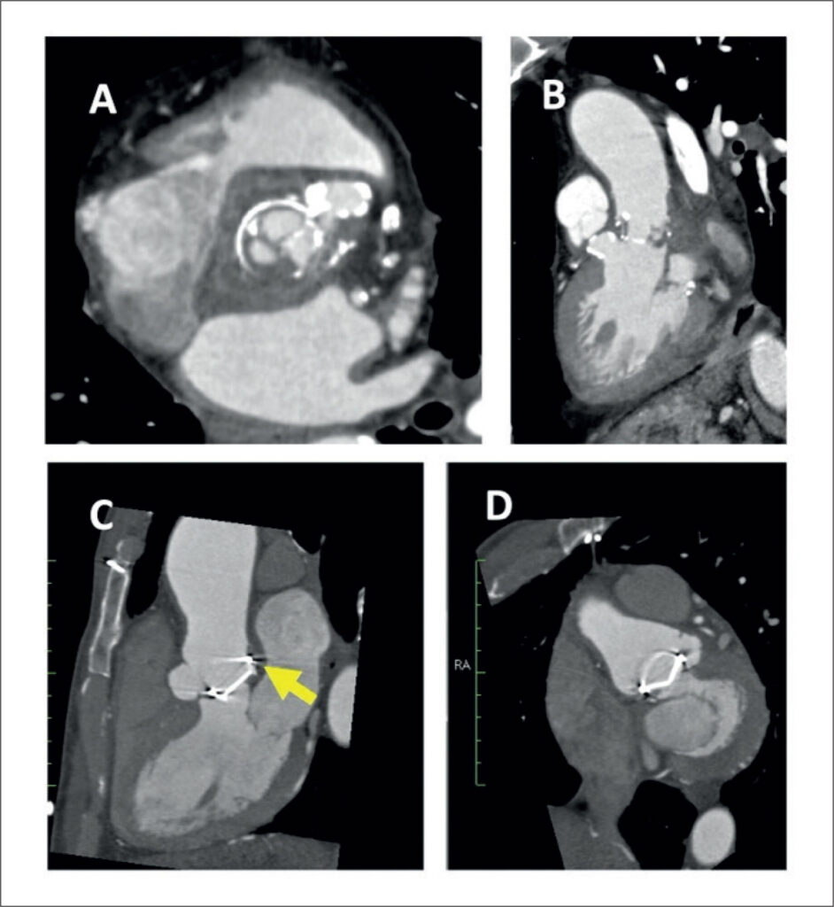
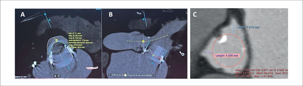
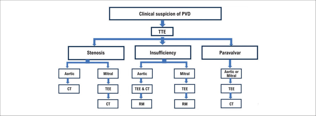
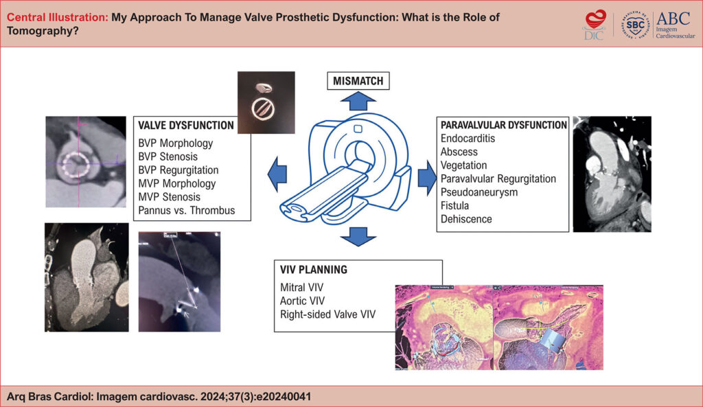
The prevalence of valvular heart disease remains significant worldwide, as does the resulting use of valve replacement devices. Currently, mechanical valve prostheses (MVP), biological valve prostheses (BVP), and transcatheter valve prostheses are available. Computer tomography (CT) provides excellent spatial resolution and plays a critical role in the diagnostic process for prosthetic valve dysfunction (PVD). CT allows proper visualization of leaflet calcification/thickening (biological) or leaflet motion and angulation (mechanical). CT plays a fundamental role in the diagnosis of pannus and/or thrombus and in the characterization of paravalvular complications (e.g., dehiscence, fistula, leak, abscess, and pseudoaneurysm). Finally, CT has an established role as a necessary tool for planning percutaneous (valve-in-valve [VIV]) or surgical interventions to replace dysfunctional biological prostheses (Central Figure).
Images
Image gallery fullscreen viewer
Comments are moderated by the editorial team before publication. Discussions should be evidence-based and use scientific language. Conflicts of interest should be declared.