Case 1/2014 – 24-Year-Old Man with Left Single Ventricle in Chronic Hypoxia
Complementary tests
Electrocardiogram showed sinus rhythm and signs of biatrial and left ventricle overloads. P wave was peaked in I, V2-5, and extended in II, F, V5-6, with negative deflection in V1 and V2. The QRS complex was of RS morphology in V1 and Rs in V6. AQRS: +120º, AT: -60º, AP: +40º.
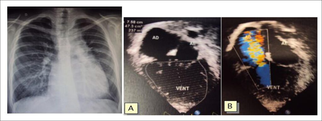
Chest radiography showed an enlarged heart of mild degree (cardiothoracic ratio: 0.54) with left long ventricular arch and also a long left medium arch. The pulmonary vascular bed was increased ().
[…]
Images
Image gallery fullscreen viewer
Comments are moderated by the editorial team before publication. Discussions should be evidence-based and use scientific language. Conflicts of interest should be declared.