A Very Complicated Inferior Myocardial Infarction: The Role of Multimodality Imaging Approach
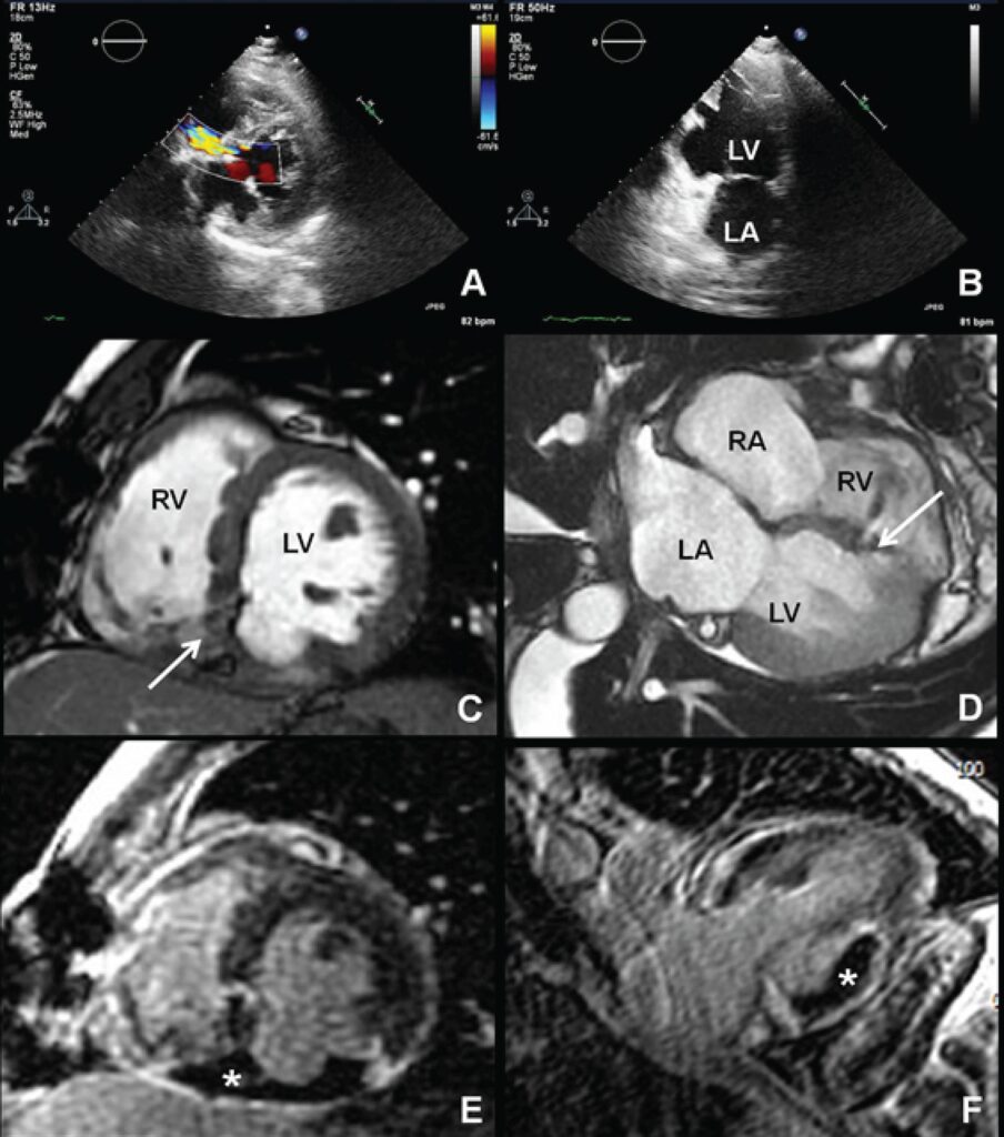
A 47-year-old male presented with a subacute inferior myocardial infarction (MI) complicated by inferobasal ventricular septal rupture (VSR) and underwent emergency surgical repair of VSR with a pericardial patch. In the postoperative period he remained hypotensive, with low exercise tolerance and a holosystolic murmur was present. Echocardiography showed persistence of VSR with left-right shunt () and revealed an aneurysmatic region at the inferobasal wall of the left ventricle (LV), suggestive of a true aneurysm due to its large “neck” and smooth transition from normal myocardium to the aneurysm ( and ). Cardiac magnetic resonance confirmed the VSR ( and ) with significant shunt (Qp/Qs of 2.3), showed transmural necrosis of the basal and mid segments of the inferior and free wall of the right ventricle with moderate systolic dysfunction and also revealed that the aneurysmatic inferobasal region of LV was actually a pseudoaneurysm, without myocardial tissue but with a sessile and adherent thrombus within it ( and ).
[…]
Images
Image gallery fullscreen viewer
Comments are moderated by the editorial team before publication. Discussions should be evidence-based and use scientific language. Conflicts of interest should be declared.