ABC Family Arquivos Brasileiros de Cardiologia International Journal of Cardiovascular Sciences ABC Heart Failure & Cardiomyopathy ABC Imagem Cardiovascular
Português English
ABC Family
Português English
  • About
    • About ABC Family
    • Internal Regulation
    • Editorial Board
    • Brazilian Society of Cardiology (SBC)
  • Issues
  • SBC Guidelines
  • Content
    • Videos
    • Podcasts
    • News
  • About
    • About ABC Family
    • Internal Regulation
    • Editorial Board
    • Brazilian Society of Cardiology (SBC)
  • Issues
  • SBC Guidelines
  • Content
    • Videos
    • Podcasts
    • News

ABC Family

  • ABC Family
  • Arquivos Brasileiros de Cardiologia
  • International Journal of Cardiovascular Sciences
  • ABC Heart Failure & Cardiomyopathy
  • ABC Imagem Cardiovascular
Search in

No content found.

Journals
Ordering
Article types
Others

  1. Home
  2. Keyword: ventricular septal defect
Figure 1
ABC
Clinicoradiological Session

Case 2/2015 A 33-Year-Old Woman with Double Right Ventricular Chamber and Ventricular Septal Defect

Edmar Atik

Laboratory tests

Electrocardiogram showed normal sinus rhythm and signs of right ventricular (RV) overload. Low-voltage QRS complex with qR morphology in V1, RS in V6 with final conduction disturbance through the right bundle branch. QRSA: -70º, TA: + 30º.

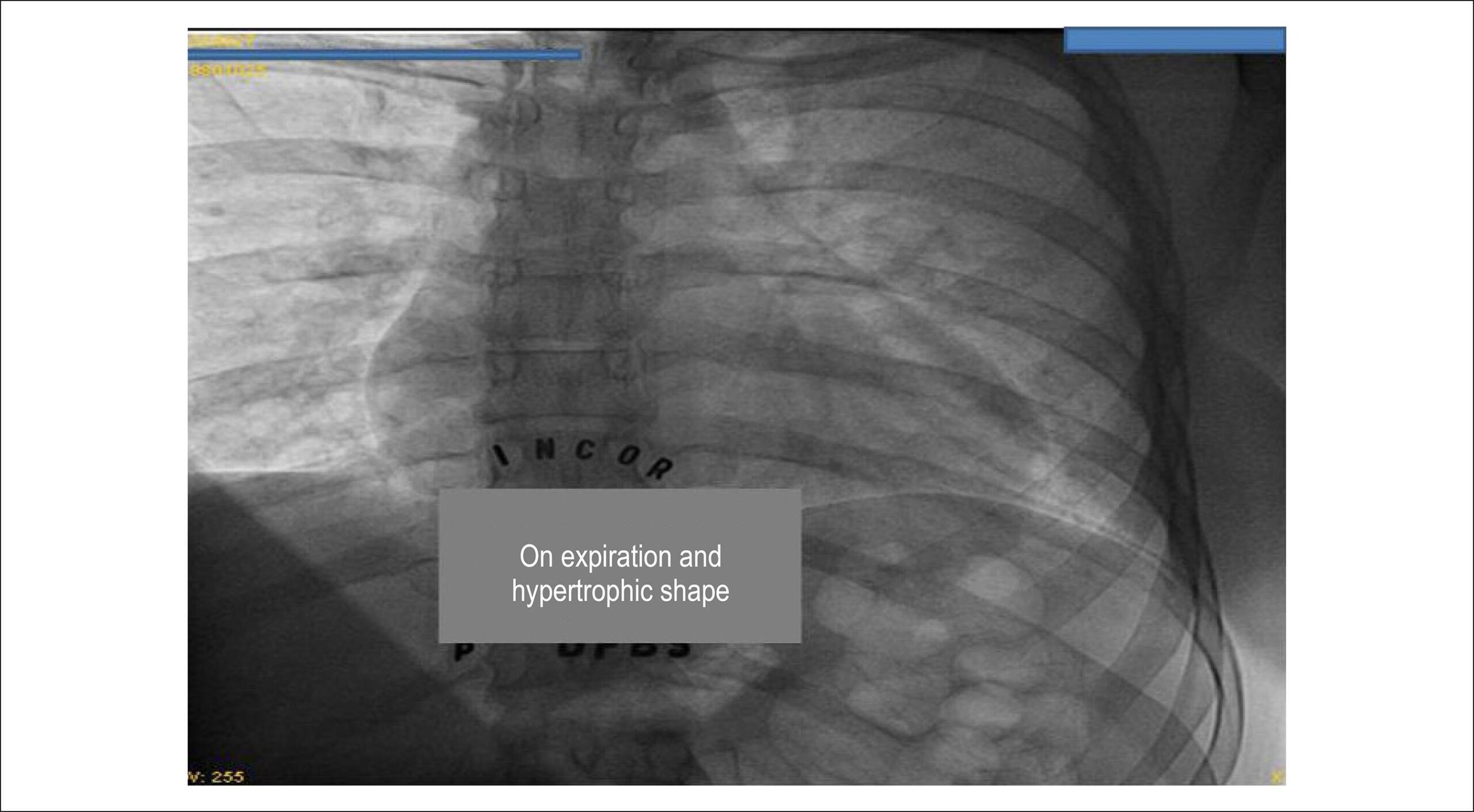
Chest radiograph showed normal cardiac silhouette (cardiothoracic ratio of 0.46). The pulmonary vascular network was normal and the arch of the pulmonary artery was concave ().

[…]

  • Double Chambered Right Ventricle
  • hipertrophy of right ventricle
  • right ventricle inflow tract stenosis
  • ventricular septal defect
Read Full Article
Creative Commons Attribution (CC BY) Creative Commons license
01/02/2015 DOI: 10.5935/abc.20140218 Views 1
Small ventricular septal defect: long-term expectant clinical management
ABC
Artigo Original

Small ventricular septal defect: long-term expectant clinical management

Edmar Atik

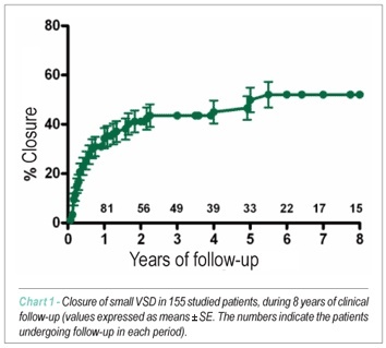
BACKGROUND: The small ventricular septal defect (VSD) usually presents good clinical evolution, even at long-term follow-up. OBJECTIVE: To verify the clinical evolution of patients with small VSD in order to determine the continuation or not of the expectant conduct, considering the low operative risk, which results in a more liberal indication for surgery. METHODS: From October 1976 to December 2007, 187 cases of small VSD (diameter < 3 mm at the echocardiogram) were evaluated and 155 of them were assessed at long-term follow-up. Time of the clinical manifestation of the murmur and evolution aspects such as the spontaneous closure of the defect (group I) – 64 cases, persistence of the initial size (group II – 74 cases and decrease in the size of the defect (group III) – 17 cases, in addition to clinical complications, were studied. RESULTS: The clinical manifestation of the murmur occurred, in the majority of cases, during the first month of life, corresponding to 48 (75%), 54 (72.9%) and 12 (70.5%) patients, in the three groups, respectively and after the first year of life in 11 (5.8%) patients. Spontaneous closure occurred in the first year of life in 48 cases (75%), mean of 7.6 months and from 1 to 5.5 years in 15 patients (23.4%), with a maximum follow-up of 18 years. The persistence of the defect until 40 years of age was observed. The decrease in the size of the defect occurred on a mean of 15 months, followed for up to 9 years. The probability of VSD closure by the actuarial curve was 34.38% in 1 year and 49.89% in 5 years. There were no clinical complications. CONLCUSION: A favorable evolution of the small VSD at long-term follow-up does not require surgical intervention, with concerns regarding the strict antibiotic prophylaxis.

  • clinical evalution
  • Heart Defects, Congenital
  • ventricular septal defect
Read Full Article
Creative Commons Attribution (CC BY) Creative Commons license
20/07/2009 DOI: 10.1590/S0066-782X2009000600003
Subaortic stenosis associated with perimembranous ventricular septal defect: clinical follow-up of 36 patients
ABC
Artigo Original

Subaortic stenosis associated with perimembranous ventricular septal defect: clinical follow-up of 36 patients

Maria da Gloria Cruvinel Horta et al. Carlos Alberto Franco Faria Dilermando Fazito Rezende Tereza Lucia Masci … Cathia Costa C. Rabelo Tamara Katina Marly de Oliveira Luciana Paulino Oliveira

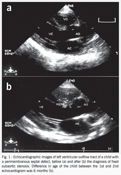
OBJECTIVE: To study the clinical pattern of subaortic stenosis associated with perimembranous ventricular septal defect. METHODS: From January 1979 to June 2000, 36 children with perimembranous ventricular septal defect and fixed subaortic stenosis were followed-up regarding anatomic characteristics, evolvement, and clinical events. RESULTS: Age at diagnosis of subaortic stenosis ranged from 6 months to 170 months, and it was less than 1 year in only 2 children. Regarding sex, the distribution was 2:1 with a greater predominance of males. Ventricular septal defect was small in 61.0% of cases, medium in 30.56%, and large in 8.40%; the size of the septal defect decreased during follow-up in 30.56% (11 cases). In all patients, subaortic stenosis was membranous and fixed. During follow-up, 23 patients experienced evolvement of the stenosis. Surgical treatment was performed in 21 cases, and one patient underwent surgery for restenosis. Infectious endocarditis occurred in 2 patients; one of the patients died. CONCLUSION: Subaortic stenosis occurs in the natural history of ventricular septal defect usually after the first year of life, and it is progressive and requires surgery in most cases.

  • Congenital Heart Disease
  • Endocarditis
  • subaortic stenosis
  • ventricular septal defect
Read Full Article
Creative Commons Attribution (CC BY) Creative Commons license
09/03/2005 DOI: 10.1590/S0066-782X2005000200002
Assessment of the pulmonary vascular blood supply in patients with pulmonary atresia, ventricular septal defect, and aortopulmonary collateral arteries
ABC
Artigo Original

Assessment of the pulmonary vascular blood supply in patients with pulmonary atresia, ventricular septal defect, and aortopulmonary collateral arteries

Ulisses Alexandre Croti et al. Miguel Lorenzo Barbero Marcial Carla Tanamati Marcelo Biscegli Jatene … Domingo Marcolino Braile Sergio Almeida de Oliveira

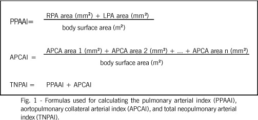
OBJECTIVE: To study the morphometric characteristics of the central pulmonary arteries and aortopulmonary collateral arteries by assessing the morphology of the pulmonary vascular blood supply, to determine their significance in surgical treatment. METHOD: From January 1990 to June 2001, 40 patients were studied. Those who had the complete cineangiocardiographic study prior to the first surgical intervention were included in the study. The morphometric characteristics of the central pulmonary arteries (PPAA) and aortopulmonary collateral arteries (APCA) were analyzed, as was the distribution of blood irrigation to the lungs. The following indices were calculated: pulmonary arterial index (PPAAI), aortopulmonary collateral arterial index (APCAI), and total neopulmonary arterial index (TNPAI = PPAAI + APCAI). The surgical treatment was considered palliative (PT), palliative definitive (PDT), and definitive (DT). RESULTS: The palliative treatment predominated. No statistically significant differences were observed in the patients undergoing PT, PDT, and DT, in regard to PPAAI, APCAI, and TNPAI. Comparing PPAAI and APCAI, no difference was observed for DT (P=0.4309); APCAI was greater than PPAAI for PT (P=0.0176); and APCAI was descriptively greater for PDT. The TNPAI of patients undergoing DT was greater than that of patients undergoing PT (P=0.0959). Five morphologically similar subgroups were identified and designated as B1, B2, B3, B4, and B5. Overall mortality was 17.5%. CONCLUSION: The morphometric characteristics are important, but the morphology of the pulmonary vascular blood supply of the PPAA and APCA proved to be better for guiding the surgical treatment. Independently of the didactical division into subgroups, PT predominated. Mortality was not correlated with the morphometric characteristics.

  • aortopulmonary collateral arteries
  • Pulmonary Atresia
  • ventricular septal defect
Read Full Article
Creative Commons Attribution (CC BY) Creative Commons license
14/02/2005 DOI: 10.1590/S0066-782X2005000100002

How to cite this article

EndNote RIS

Electronic Document Format (Vancouver)

Atik E. Case 2/2015 A 33-Year-Old Woman with Double Right Ventricular Chamber and Ventricular Septal Defect. Arquivos Brasileiros de Cardiologia 2015;104(2):e12-4.

Electronic Document Format (ABNT)

Atik, Edmar. Case 2/2015 A 33-Year-Old Woman with Double Right Ventricular Chamber and Ventricular Septal Defect. Arquivos Brasileiros de Cardiologia, v. 104, n. 2, p. e12-e14, Jan. 2015.

Electronic Document Format (APA)

Atik, E. (2015). Case 2/2015 A 33-Year-Old Woman with Double Right Ventricular Chamber and Ventricular Septal Defect. Arquivos Brasileiros de Cardiologia, 104(2), e12-e14.

Electronic Document Format (ISO)

Atik, Edmar. Case 2/2015 A 33-Year-Old Woman with Double Right Ventricular Chamber and Ventricular Septal Defect. Arquivos Brasileiros de Cardiologia [online]. 2015, vol. 104, n. 2, [cited 2026-10-14], pp. e12-e14. Available from: <https://journals.cardiol.br/en/article/case-2-2015-a-33-year-old-woman-with-double-right-ventricular-chamber-and-ventricular-septal-defect/>. ISSN 0066-782X

EndNote RIS

Electronic Document Format (Vancouver)

Atik E. Small ventricular septal defect: long-term expectant clinical management. Arquivos Brasileiros de Cardiologia 2009;92(6):429-32.

Electronic Document Format (ABNT)

Atik, Edmar. Small ventricular septal defect: long-term expectant clinical management. Arquivos Brasileiros de Cardiologia, v. 92, n. 6, p. 429-432, Jul. 2009.

Electronic Document Format (APA)

Atik, E. (2009). Small ventricular septal defect: long-term expectant clinical management. Arquivos Brasileiros de Cardiologia, 92(6), 429-432.

Electronic Document Format (ISO)

Atik, Edmar. Small ventricular septal defect: long-term expectant clinical management. Arquivos Brasileiros de Cardiologia [online]. 2009, vol. 92, n. 6, [cited 2026-10-14], pp. 429-432. Available from: <https://journals.cardiol.br/en/article/small-ventricular-septal-defect-long-term-expectant-clinical-management/>. ISSN 0066-782X

EndNote RIS

Electronic Document Format (Vancouver)

Horta MGC, Faria CAF, Rezende DF, Masci TL, Rabelo CCC, Katina T, Oliveira M, et al. Subaortic stenosis associated with perimembranous ventricular septal defect: clinical follow-up of 36 patients. Arquivos Brasileiros de Cardiologia 2005;84(2):103-7.

Electronic Document Format (ABNT)

Horta, Maria da Gloria Cruvinel; Faria, Carlos Alberto Franco; Rezende, Dilermando Fazito; Masci, Tereza Lucia; Rabelo, Cathia Costa C.; Katina, Tamara; Oliveira, Marly de; Oliveira, Luciana Paulino. Subaortic stenosis associated with perimembranous ventricular septal defect: clinical follow-up of 36 patients. Arquivos Brasileiros de Cardiologia, v. 84, n. 2, p. 103-107, Mar. 2005.

Electronic Document Format (APA)

Horta, M. G. C., Faria, C. A. F., Rezende, D. F., Masci, T. L., Rabelo, C. C. C., Katina, T., Oliveira, M. , & Oliveira, L. P. (2005). Subaortic stenosis associated with perimembranous ventricular septal defect: clinical follow-up of 36 patients. Arquivos Brasileiros de Cardiologia, 84(2), 103-107.

Electronic Document Format (ISO)

Horta, Maria da Gloria Cruvinel and Faria, Carlos Alberto Franco and Rezende, Dilermando Fazito and Masci, Tereza Lucia and Rabelo, Cathia Costa C. and Katina, Tamara and Oliveira, Marly de and Oliveira, Luciana Paulino. Subaortic stenosis associated with perimembranous ventricular septal defect: clinical follow-up of 36 patients. Arquivos Brasileiros de Cardiologia [online]. 2005, vol. 84, n. 2, [cited 2026-10-14], pp. 103-107. Available from: <https://journals.cardiol.br/en/article/subaortic-stenosis-associated-with-perimembranous-ventricular-septal-defect-clinical-follow-up-of-36-patients/>. ISSN 0066-782X

EndNote RIS

Electronic Document Format (Vancouver)

Croti UA, Marcial MLB, Tanamati C, Jatene MB, Braile DM, Oliveira SA. Assessment of the pulmonary vascular blood supply in patients with pulmonary atresia, ventricular septal defect, and aortopulmonary collateral arteries. Arquivos Brasileiros de Cardiologia 2005;84(1):3-9.

Electronic Document Format (ABNT)

Croti, Ulisses Alexandre; Marcial, Miguel Lorenzo Barbero; Tanamati, Carla; Jatene, Marcelo Biscegli; Braile, Domingo Marcolino; Oliveira, Sergio Almeida de. Assessment of the pulmonary vascular blood supply in patients with pulmonary atresia, ventricular septal defect, and aortopulmonary collateral arteries. Arquivos Brasileiros de Cardiologia, v. 84, n. 1, p. 3-9, Feb. 2005.

Electronic Document Format (APA)

Croti, U. A., Marcial, M. L. B., Tanamati, C., Jatene, M. B., Braile, D. M., & Oliveira, S. A. (2005). Assessment of the pulmonary vascular blood supply in patients with pulmonary atresia, ventricular septal defect, and aortopulmonary collateral arteries. Arquivos Brasileiros de Cardiologia, 84(1), 3-9.

Electronic Document Format (ISO)

Croti, Ulisses Alexandre and Marcial, Miguel Lorenzo Barbero and Tanamati, Carla and Jatene, Marcelo Biscegli and Braile, Domingo Marcolino and Oliveira, Sergio Almeida de. Assessment of the pulmonary vascular blood supply in patients with pulmonary atresia, ventricular septal defect, and aortopulmonary collateral arteries. Arquivos Brasileiros de Cardiologia [online]. 2005, vol. 84, n. 1, [cited 2026-10-14], pp. 3-9. Available from: <https://journals.cardiol.br/en/article/assessment-of-the-pulmonary-vascular-blood-supply-in-patients-with-pulmonary-atresia-ventricular-septal-defect-and-aortopulmonary-collateral-arteries/>. ISSN 0066-782X

  • About ABC Family
  • Internal Regulation
  • Editorial Board
  • Brazilian Society of Cardiology (SBC)

Issues

SBC Guidelines

  • Videos
  • Podcasts
  • News