Unusual Presentation of Takayasu’s Disease
Unusual presentation of Takayasu’s disease
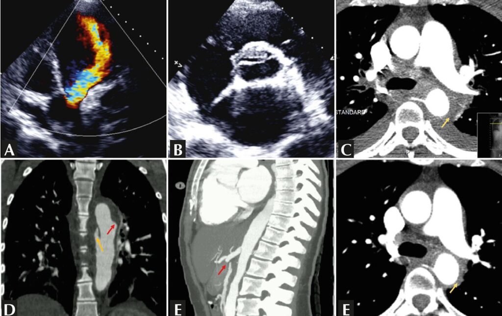
A 21-year-old woman was admitted with heart failure and chest pain. Echocardiography revealed a tricuspid aortic valve with severe regurgitation (A), a thickened and deformed right coronary cusp with a linear image (B), and severe left ventricular dilation. Angio-computed tomography showed continuous and concentric thickening of the aortic wall (C) with a penetrating ulcer and dissection of the descending aorta (D) and superior mesenteric and left renal arterial occlusion (E). Takayasu aortitis type V was assumed for which immunosuppression was commenced. Significant improvement was noted (F). After achieving remission, the patient underwent endovascular surgery of the aorta and aortic valve replacement. No episodes of relapses were seen during a 3-year follow-up period.
[…]
Images
Image gallery fullscreen viewer
Comments are moderated by the editorial team before publication. Discussions should be evidence-based and use scientific language. Conflicts of interest should be declared.