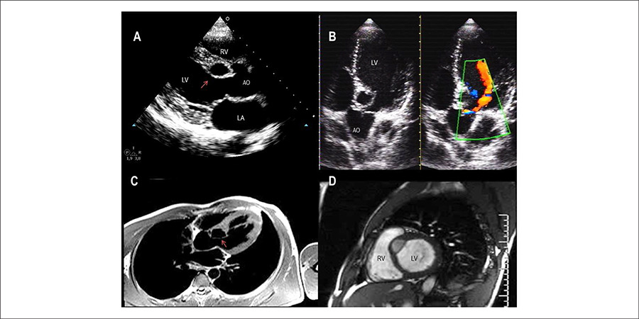
A) Transthoracic echocardiogram, parasternal longitudinal view, showing the diverticulum in the interventricular septum (arrow) adjacent to the right coronary cusp of aortic valve; B) apical five-chamber view showing the aortic regurgitant jet and apparent structural communication with the LV cavity (Doppler flow); C) Cardiac MRI, axial view in dark blood sequence, and T1-weighted image, confirms the continuity solution (arrow); D) short basal axis view sequence of cine-MRI (perfusion) shows isointense structure (muscular) and hypointense content.
Comments are moderated by the editorial team before publication. Discussions should be evidence-based and use scientific language. Conflicts of interest should be declared.
Comments are moderated by the editorial team before publication. Discussions should be evidence-based and use scientific language. Conflicts of interest should be declared.