Muhieddine Chokr et al. Lucas G. de Moura Italo Bruno dos Santos Sousa Cristiano Faria Pisani … Carina Abigail Hardy Sissy Lara de Melo Arnobio Dias da Ponte Filho Ieda Prata Costa Ronaldo Vasconcelos Tavora Luciana Sacilotto Tan Chen Wu Francisco Carlos da Costa Darrieux Denise Tessariol Hachul Vera Demarchi Aiello Mauricio Scanavacca
Abstract
Background
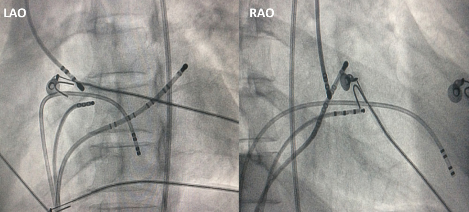
Atrial tachycardia (AT) ablation with earliest activation site close to the His-Bundle is a challenge due to the risk of complete AV block by its proximity to His-Purkinje system (HPS). An alternative to minimize this risk is to position the catheter on the non-coronary cusp (NCC), which is anatomically contiguous to the para-Hisian region.
Objectives
The aim of this study was to perform a literature review and evaluate the electrophysiological characteristics, safety, and success rate of catheter-based radiofrequency (RF) delivery in the NCC for the treatment of para-Hisian AT in a case series.
Methods
This study performed a retrospective evaluation of ten patients (Age: 36±10 y-o) who had been referred for SVT ablation and presented a diagnosis of para-Hisian focal AT confirmed by classical electrophysiological maneuvers. For statistical analysis, a p-value of <0.05 was considered statistically significant.
Results
The earliest atrial activation at the His position was 28±12ms from the P wave and at the NCC was 3±2ms earlier than His position, without evidence of His potential in all patients. RF was applied on the NCC (4-mm-tip catheter; 30W, 55ºC), and the tachycardia was interrupted in 5±3s with no increase in the PR interval or evidence of junctional rhythm. Electrophysiological tests did not reinduce tachycardia in 9/10 of patients. There were no complications in all procedures. During the 30 ± 12 months follow-up, no patient presented tachycardia recurrence.
Conclusion
The percutaneous treatment of para-Hisian AT through the NCC is an effective and safe strategy, which represents an interesting option for the treatment of this complex arrhythmia. (Arq Bras Cardiol. 2021; 116(1):119-126)