Positron Emission Tomography Identification of Cardiovascular Implantable Electronic Device Infection
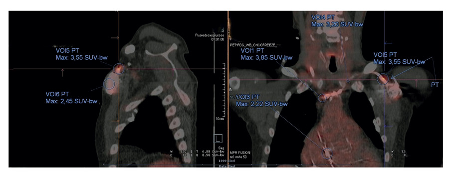
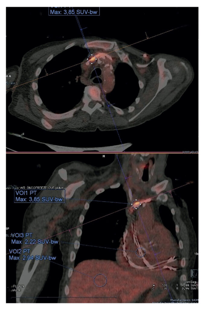
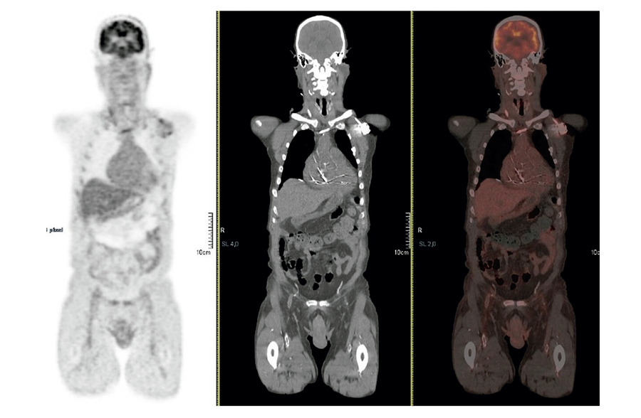
An 84-year-old man with hypertension, dyslipidemia, chronic kidney disease, permanent atrial fibrillation, and a pacemaker implanted for brady-tachy syndrome, with generator and lead replacement one year prior, presented with a 15-day fever, malaise, and fatigue. Physical examination revealed a holosystolic murmur. The skin overlying the pacemaker pocket showed no signs of inflammation. Laboratory testing showed leukocytosis with neutrophilia, C-reactive protein of 9.5 mg/dL, and procalcitonin of 1.3 ng/mL. The rapid respiratory viral panel was negative, and urinalysis was unremarkable. A pair of blood cultures yielded methicillin-susceptible Staphylococcus aureus (MSSA). Transthoracic and transesophageal echocardiography showed no valvular or lead involvement. 18F-fluorodeoxyglucose positron emission tomography /computed tomography (PET/CT) demonstrated increased uptake along the pacemaker lead and generator ( ), confirming cardiac implantable electronic device (CIED) infection. Complete system extraction was performed, and lead cultures confirmed MSSA. Three follow-up blood cultures were obtained, two before extraction and one after extraction, all of which were negative. He received 3 weeks of IV cefazolin pre-extraction and 3 weeks of IV flucloxacillin post-extraction, completing a total of 6 weeks of directed antibiotic therapy. Monitoring revealed permanent atrial fibrillation without pauses, and device reimplantation was not required.
[…]
Images
Image gallery fullscreen viewer
Comments are moderated by the editorial team before publication. Discussions should be evidence-based and use scientific language. Conflicts of interest should be declared.